Implications
The study conducted on weaned pigs challenged with enterotoxigenic Escherichia coli F4 allowed us to consider the possible implications of the use of a well-studied human-derived probiotic, which, however, has not been well-documented in pigs. Thus, we support the need for the in vivo assessment whenever the use of a probiotic has to be extended to the swine species; this has also economic implications, like the increased cost for the experimental dossiers to be produced for the inclusion in the list of in-feed probiotics approved for pigs.
Our research also indicates that lactobacilli administered with a diet can have unpredictable interaction with local microflora, particularly during phases of combined stress. More research is needed regarding this point.
Introduction
After studies with laboratory animal models, in vitro tests and clinical tests, there is an overall consensus about the notion that the addition of certain bacterial strains can enhance growth and health. For animal production, there is rising interest regarding probiotics since the European Union ban on the use of growth-promoting in-feed antibiotics, particularly for subjects during the weaning and growth phase.
However, from early tests to in-farm applications, many steps should be involved in order to document the qualities of a probiotic and its practical relevance. It would be very useful if knowledge of strains, which could be defined as probiotics for one species, could also be used in other species. A recent study indicates that Lactobacillus plantarum 299v specifically is able to adapt its metabolic capacity in the human intestine for carbohydrate acquisition and expression of exopolysaccharide and proteinaceous cell surface compounds (Marco et al., Reference Marco, de Vries, Wels, Molenaar, Mangell, Ahrne, de Vos, Vaughan and Kleerebezem2010); this finding gives an important evidence about the dynamic metabolism of the bacteria, suggesting that the intestinal environment can modify the interplay among the bacteria and the host. There are evidences that in mammals, some gut microbes have diversified into host-adapted lineages, originated by a long-term evolutionary process, and that led to the development of a highly specialized symbiosis (Oh et al., Reference Oh, Benson, Peterson, Patil, Moriyama, Roos and Walter2010). The probiotic species-specificity was reviewed by Saarela et al. (Reference Saarela, Mogensen, Fondén, Mättö and Mattila-Sandholm2000) reporting that in humans, the most current successful probiotic strains are indicated to be of human origin; thus the author speculates that a probiotic strain can function better in an environment similar to that where it was originally isolated from. Conversely, Vinderola et al. (Reference Vinderola, Medici and Perdigón2004), showed that the immunomodulating effect of different Lactobacillus strains was independent of the host specificity, notwithstanding that a greater immune activation was achieved by using indigenous rather than exogenous strains. The author concluded anyway that the effectiveness of exogenous bacteria in the activation of the immune system is undeniable. The most studied lactic acid bacteria for probiotic purposes is probably Lactobacillus rhamnosus GG (LGG) and evidence of the positive effect of the GG strain on human health is relevant. In particular, its positive effect on the humoral immune response in pediatrics (Saavedra, Reference Saavedra2007) and on the prevention and treatment of diarrhea (Kaila et al., Reference Kaila, Isolauri, Saxelin, Arvilommi and Vesikari1995; Majamaa et al., Reference Majamaa, Isolauri, Saxelin and Vesikari1995; Yan and Polk, Reference Yan and Polk2006) has been shown. In the human-derived Caco-2 intestinal cells, LGG inhibited the attachment of the pig-specific enterotoxigenic Escherichia coli F4 (ETEC) and counteracted the pathogen-induced upregulation of the inflammatory cytokines IL-8, IL-1b and TNF-α. Thus, the interest in testing the use of LGG in young pigs is not only motivated by its possible use as a study model of the impact of not species-specific bacteria, but also as an occasion to assess its value for the animal practice.
Therefore, our aim was to assess the effect of a well-defined probiotic, such as LGG, on growth performance, ETEC excretion, diarrhoea score, small intestine morphology and humoral immunity in newly weaned pigs orally challenged with ETEC.
Material and methods
Bacterial strains
As a source of LGG ATCC 53103, we used a commercial product called Dicoflor 60 (Dicofarm S.p.A., Rome, Italy). Each commercial dose contains 6 × 10⁹ CFU of lyophilised bacteria. During the trial, we administered one dose/pig per day. Before administration, LGG ATCC 53103 was suspended in 5 ml of heated water (37°C) in order to reactivate the lyophilised bacteria. The inoculum was added directly to the trough together with the morning meal.
The E. coli O149:F4ac inoculum was isolated from pigs with colibacillosis, was provided by the Istituto Zooprofilattico Sperimentale della Lombardia e dell'Emilia, was grown in Luria broth and further prepared as described by Bosi et al. (Reference Bosi, Casini, Finamore, Gremokolini, Merialdi, Trevisi, Nobili and Mengheri2004a).
Animals and diets
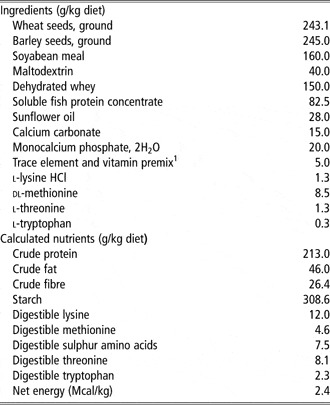
The experiment was conducted in three consecutive runs using 36 crossbred piglets (Landrace × Large-White) weaned at 21 days of age. The pigs were housed in weaning rooms with controlled temperature and ventilation. Continuous access to feed and water was allowed throughout the trial. The pigs were individually penned in cages, except for the first 2 days when they were kept in groups of three in order to stimulate a quick resumption of feed intake after weaning. For the entire experimental period (14 days), the pigs were fed two different diets: one half received a standard pre-starter basal diet (B) without antimicrobials or zinc oxide, and the other half received a basal diet supplemented with LGG ATCC 53103 (LB; Table 1).
Ingredient composition and calculated nutrient content of the basal diet (as-fed)

1Provided per kg of diet: vitamin A, 26 500 IU; vitamin D3, 2400 IU; vitamin E, 35 mg; vitamin B1, 10 mg; vitamin B2, 8 mg; vitamin B6, 6 mg; vitamin B12, 0.04 mg; niacin, 55 mg; biotin, 0.15 mg; d-pantothenic acid, 30 mg; folacin, 1 mg; iron, 200 mg; zinc, 100 mg; copper, 150 mg; manganese, 80 mg; iodine, 2.5 mg; selenium, 0.2 mg.
During the first 2 days of the experiment, all pigs received 5 ml of a water solution with or without the prefixed doses of LGG directly per os in order to ensure that each piglet received the entire bacterial daily dose. After this period, the probiotic supplementation was administered directly in the individual trough.
Experimental procedure
All the procedures complied with Italian law pertaining to experimental animals and were approved by the Ethic-Scientific Committee for Experiments on Animals of the University of Bologna. For each run, that lasted 14 days, the pigs were divided into two groups of six subjects each, balanced for litter and body weight (BW). On day 7, all the piglets were orally infected with a 1.5 ml suspension containing 1010 CFU of E. coli F4ac O149/ml. To determine the severity of the diarrhoea, faecal scores were recorded daily by visual appraisal of each subject using a five-point scoring system (1 to 5): 1 = hard, 5 = watery faeces. The piglets were killed on days 12 and 14. The animals were deeply anaesthetised with sodium thiopenthal (10 mg/kg of BW) and killed by an intracardiac injection of a commercial solution (0.5 ml/kg of BW) with three components (embutramide, mebenzonium iodine and tetracaine hydrochloride; Tanax; Intervet Italia, Peschiera Borromeo, Italy).
Experimental observations and measurements
The pigs were weighed individually at the start of the trial, at day 7 (pre-challenge) and when slaughtered. The individual feed intakes of each pig were recorded, and daily live weight gain and feed efficiency were calculated. On days 7 and 10, an individual sample of saliva was collected and stored at −20°C. Moreover, on days 0, 7 and 11, blood samples from the vena cava were taken from each piglet. The blood was centrifuged at 3000 × g for 10 min and the serum was removed. The serum was incubated at 56°C for 30 min and stored at −20°C until analysis. The saliva and blood serum were analysed using the ELISA test in order to determine the total immunoglobulin A (IgA) concentration, as described in Bosi et al. (Reference Bosi, Sarli, Casini, De Filippi, Trevisi, Mazzoni and Merialdi2007), and the ETEC-specific IgA titres, according to Van den Broeck et al. (Reference Van den Broeck, Cox and Goddeeris1999), using F4 fimbriae isolated from ETEC cultures as reported by Bosi et al. (Reference Bosi, Casini, Finamore, Gremokolini, Merialdi, Trevisi, Nobili and Mengheri2004a). On day 10 (3 days after challenge), the faeces were individually collected and the concentrations of total E. coli and ETEC were calculated as described in Bosi et al. (Reference Bosi, Casini, Finamore, Gremokolini, Merialdi, Trevisi, Nobili and Mengheri2004a and Reference Bosi, Gremokolini, Trevisi, Mazzoni, Bonilauri, Sarli and Casini2004b).
At slaughter, the small intestine was removed, its length measured and empty weight recorded. The pHs of the ileum, colon and cecum were noted. Whole thickness tissue from the duodenum, jejunum and ileum of ∼1 cm2 was removed and fixed in Carlson's formalin pH 7.3. Morphometry measurement of the villus and crypt was performed on these samples using the procedure described by Bosi et al. (Reference Bosi, Sarli, Casini, De Filippi, Trevisi, Mazzoni and Merialdi2007). Two additional samples from the distal jejunum were collected: one to determine the phenotype for adhesion of the ETEC to the intestinal villi, as described in Trevisi et al. (Reference Trevisi, Melchior, Mazzoni, Casini, De Filippi, Minieri, Lalatta-Costerbosa and Bosi2009) and one to measure the concentration of total intestinal secretory IgA and ETEC-specific intestinal secretory IgA (Evans et al., Reference Evans, Newby, Stokes, Patel and Bourne1980). Samples of the colon contents were immediately stored at +2°C and processed within 2 h.
Enterobacteriaceae were counted on VRBGA (Oxoid, Basingstoke, UK) incubated for 24 h at 37°C, lactic acid bacteria were cultured on MRS Agar (Oxoid) at 30°C for 96 h under anaerobic conditions, yeasts were counted on Sabouroud Agar (Oxoid) and incubated at 25°C for 72 h. Three replicates were carried out for each microbial count.
Statistical analysis
The individual data were analysed by ANOVA using the GLM procedure (SAS Institute Inc., Cary, NC, USA), For all the data the initial model included the treatment and the litter. For the factor sensitivity of the intestinal villus to ETEC adhesion, we divided the pigs of each of the two challenged groups into two different subgroups: positive or negative based on the results of the in vitro adhesion test, as suggested by Geenen et al. (Reference Geenen, Van der Meulen, Bouma, Engel, Heesterbeek and De Jong2007). We observed that the ETEC susceptibility factor was not statistically significant; therefore, we decided not to use this parameter in the final statistical analysis. We excluded also the effect of the different runs, because it was almost totally absorbed by the inclusion of the litter in the model. The data of intestinal morphometry and of IgA quantifications were analysed using the MIXED procedure for repeated measurements, adding the effect of the different intestinal section or the effect of time of sampling to the other two factors.
Results
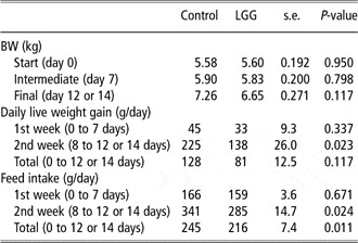
The effect of LGG on growth performance is presented in Table 2. Daily live weight gain and feed intake were not affected in the first week while, in the following week after the challenge with ETEC, both were reduced (−39% and −16%, respectively, P < 0.05). Overall daily live weight gain and feed intake were also reduced (P < 0.05) as a result of the LGG supplementation.
Effect of the dietary addition of LGG on growth and feed intake of pigs orally challenged with E. coli F4 at day 7 post-weaning

LGG = L. rhamnosus GG.
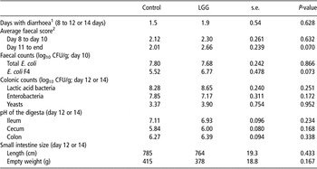
The effect of the addition of LGG to the diet on the faecal score, bacteria counts in the faeces and colic digesta, intestinal pHs and small intestine size is presented in Table 3. Days with diarrhoea were not changed by LGG; the average faecal score tended to worsen from day 11 to the end of the trial (P = 0.07), but not in the first days after the ETEC challenge. The shedding of total E. coli in the faeces was not affected but, with the addition of LGG, a trend of increased concentration of ETEC in the faeces was seen (P = 0.07). The counts of lactic acid bacteria, enterobacteria and yeasts in the colic digesta were not affected. The pHs in the digesta from the ileum, colon and cecum, and small intestine size were also unchanged.
Effect of the addition of LGG to the diet on the faecal score, bacteria counts in the faeces and colonic digesta, intestinal pH and small intestine size of pigs orally challenged with E. coli F4 at day 7 post-weaning

LGG = L. rhamnosus GG.
1Counts of days with faecal score >2.
2Faecal scores were recorded daily by visual appraisal of each subject using a five-point scoring system (1–5): 1 = hard, 5 = watery faeces.
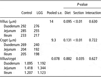
The effect of the addition of LGG to the diet on the morphometry of the different tracts of the small intestinal mucosa is shown in Table 4. No statistically significant interaction of the dietary treatment with the site of measurement was observed for villus height, crypt depth and villus to crypt ratio. Regardless of the site of measurement (duodenum, jejunum or ileum), a trend of decreased villus height was seen with LGG supplementation (P = 0.10). Crypt depth and villus to crypt ratio were unchanged by the diet. All the morphometry parameters differed for the site of sampling (P < 0.05).
Effect of the addition of LGG to the diet on the morphometry of small intestinal mucosa of pigs orally challenged with E. coli F4 at day 7 post-weaning

LGG = L. rhamnosus GG.
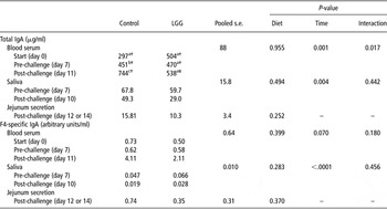
The effect of the addition of LGG to the diet on total and F4-specific IgA content in the blood serum, saliva and jejunum secretion is shown in Table 5. An interaction between the diet and the time of sampling was observed for total IgA in the blood serum. LGG supplementation did not affect the measurements before the challenge, but reduced the IgA values after the challenge (P < 0.05), as compared with the control diet. In the control group the values increased after 1 week of trial, and then increased after the challenge (P < 0.05), but no increase was seen in the treated group. No other interaction was seen for the other IgA quantifications. Total IgA in the saliva and in the jejunum secretion were not affected. The F4-specific IgA activity was not affected by the diet.
Effect of the addition of LGG to the diet on total and F4-specific IgA content in the blood serum, saliva and jejunum secretion of pigs orally challenged with E. coli F4 at day 7 post-weaning

LGG = L. rhamnosus GG.
Test on the means of diet × time interaction: values on the same column with different letters, or on the same row with different symbols, differ for P < 0.05.
Discussion
For probiosis, the use of bacterial strains isolated from the same animal species being treated is often recommended (Mäyrä-Mäkinen et al., Reference Mäyrä-Mäkinen, Manninen and Gyllenberg1983). However, Rinkinen et al. (Reference Rinkinen, Westermarck, Salminen and Ouwehand2003) observed that the adhesion traits of beneficial lactic acid bacteria may be dictated more by the bacterial properties rather than the host species. Consequently, the decision to use a probiotic in a species where it has not been tested could depend more on the demonstrated safety and on legal approval for its use. The absence of DNA from the LGG in the cecum contents of the control pigs used in our trial demonstrates that LGG is not normally present in swine (Casini et al., Reference Casini, Kostantinov, Coloretti, De Filippi, Mazzoni, Trevisi and Bosi2007). Here we show that, after oral stimulation with ETEC, the inclusion of LGG in the diet for weaned piglets impaired growth performance, feed intake and health status as compared with a standard diet.
In our experiment, we used this strain isolated in humans, based on in vitro studies with Caco-2 cells stimulated with ETEC (Roselli et al., Reference Roselli, Finamore, Britti and Mengheri2006). The authors observed that LGG partially blocked the adhesion of ETEC to the surface of Caco-2 cells and that this action was not due to a bactericidal effect. This probiotic did not totally prevent ETEC adhesion or reduction of the trans-epithelial resistance which damaged cell junctions (Roselli et al., Reference Roselli, Finamore, Britti and Mengheri2003), but was able to reduce neutrophil transmigration and the increased expression of inflammatory cytokines induced by ETEC (Roselli et al., Reference Roselli, Finamore, Britti and Mengheri2006). Therefore, we should have expected a reduction of ETEC excretion and, at least, health and performance were not negatively affected. This contrasts with our observation of a trend of increase in ETEC excretion after the challenge with this strain in the LGG-treated pigs. Zhang et al. (Reference Zhang, Xu, Liu, Lai, Ma, Wang and Zhu2010) evaluated the supplementation with LGG in a similar trial with piglets challenged with ETEC; they did not assess ETEC faecal excretion but found a reduction in total coliforms in the faeces as a result of the probiotic supplementation. This observation was consistent with the observed reduction of the incidence of diarrhoea in the treated pigs. Interestingly, in their trial, LGG feeding was directly related to an increased number of Lactobacilli in the faeces after the challenge, whereas we did not observe any effect on counts of lactic acid bacteria in the cecum contents after treatment with LGG.
Alander et al. (Reference Alander, Satokari, Korpela, Saxelin, Vilpponen-Salmela, Mattila-Sandholm and von Wright1999) observed that LGG is able to adhere to the human intestinal wall and then to persist in the intestinal tract, even after suspending its administration; however, the adhering properties of LGG on swine mucosa have not been documented. In addition, we do not know about interaction with the microbiota normally present in the pig gut. In both trials carried out on piglets, the administration of LGG started at the moment of weaning, when the gut microbiota was adapting to the new feeding and environmental conditions; therefore, we cannot exclude the possibility that the LGG interacted differently with already established bacteria.
In our in vivo study, the daily live weight gain was lower in subjects fed an LGG diet v. the controls. This result stems from a slower growth in the post-stimulation phase and lower feed consumption of subjects in the LGG group. This may indicate that subjects fed with LGG were affected due to the greater amount of stress caused by the challenge, stating that LGG not only failed to act as a barrier against pathogens but, in some way, negatively interacted with some aspects of the intestinal barrier. Recently, some research has shed additional light on the possible two-faced behaviour of LGG in the gut. On Caco-2 cells it has also been reported that high doses of LGG (1010 CFU/L) can be pro-inflammatory (more IL-8 production), whereas the opposite is seen at lower doses or in the presence of TNF-α (Zhang et al., Reference Zhang, Li, Caicedo and Neu2005). Some evidence also exists that LGG can exacerbate acute colitis (but not chronical colitis) induced by dextran sulfate sodium in rats (Moon et al., Reference Moon, Myung, Jeong, Yang, Cho, Lee, Chang, Byeon, Lee, Lee, Hong, Kim, Min and Kim2004; Geier et al., Reference Geier, Butler, Giffard and Howarth2007; Mileti et al., Reference Mileti, Matteoli, Iliev and Rescigno2009). In our trial we used LGG in a lower concentration than in the trial carried out by Zhang et al. (Reference Zhang, Xu, Liu, Lai, Ma, Wang and Zhu2010); thus, the different response to LGG cannot be simply explained by the concentration. However, we cannot exclude that the viability of the probiotic product was different. Moreover, we should consider that the challenge models with certain pathogens have some difficulties related, for example, to the sanitary and immunitary background of the animals subjected to the infection or that the modality of administration of the pathogen can affect the response (Madec et al., Reference Madec, Bridoux, Bounaix, Cariolet, Duval-Iflah, Hampson and Jestin2000). Finally, a more complex mechanism should be also investigated in relationship to the reported properties of LGG. In particular, there is evidence that LGG can activate the phenotype of dendritic cells, the key cells locally involved in antigen capture and presentation, and thus affect T cell polarization in a way similar to a pathogen (Mileti et al., Reference Mileti, Matteoli, Iliev and Rescigno2009).
Villus height and its ratio with crypt depth are considered an indicator of the health of the mucosa and of the local availability of nutrients, particularly at the time of weaning. The lower feed intake observed with the probiotic may have caused a minor trophism of the villus, as has previously been observed in studies where the consumption of feed was varied (McCracken et al., Reference McCracken, Spurlock, Roos, Zuckermann and Gaskins1999; Spreeuwenberg et al., Reference Spreeuwenberg, Verdonk, Gaskins and Verstegen2001). This can partially explain the trend of reduction of villus height with LGG supplementation, which was constant all along the small intestine, as indicated by the absence of interaction of the diet with the site of measurement. However, one cannot exclude a direct effect of LGG and a relationship with the increased ETEC shedding and the impaired faecal score in the final days. In a model of acute colitis in rats, LGG impaired the histological score of the colic mucosa (Geier et al., Reference Geier, Butler, Giffard and Howarth2007).
In general, after weaning, the piglet experiences new encounters with environmental microflora, due to mixing with pigs from other litters and a transfer to new spaces. This is relevant for the activation of the local mucosal immune system, and the commensal microflora also stimulates IgA production (Macpherson et al., Reference Macpherson, Gatto, Sainsbury, Harriman, Hengartner and Zinkernagel2000). Thus, a rise in circulating IgA during the first weeks after weaning, as observed in the control pigs, can be considered normal and is also a consequence of the progressive maturation of the gut-associated lymphoid tissue. We were surprised by the absence of such a rise in the case of the LGG group, which led to a reduced IgA content in blood serum at the final sampling. One could be tempted to hypothesise that this is, in some way, related to an immune response against a new microorganism added to the diet in high concentrations. We assessed the anti-LGG IgA activity in the blood serum of the piglets in this experiment (Casini et al., Reference Casini, Kostantinov, Coloretti, De Filippi, Mazzoni, Trevisi and Bosi2007), and we observed that it was present at each sampling time, but we also observed that the activity in proportion to the total IgA rose after 1 week and then stabilised. This could indicate that the immune system was also engaged in the response against the new commensal. However, we also found IgA activity in the control pigs (without the presence of LGG); this was explained by the presence of poly-reactive IgA or by the cross-reactivity with other Lactobacilli (Casini et al., Reference Casini, Kostantinov, Coloretti, De Filippi, Mazzoni, Trevisi and Bosi2007). Total IgA intestinal secretion was not significantly reduced by LGG; as compared to the data on the blood serum, this could be considered more a picture of the final adaptation due to a more rapid turnover in the gut content than in the blood. After ETEC challenge, Zhang et al. (Reference Zhang, Xu, Liu, Lai, Ma, Wang and Zhu2010) found a rise in total secretory IgA recovered in the supernatant from the jejunum and the ileum of pigs supplemented with LGG as compared with the control pigs. This could be because of the different impact of LGG on the gut, which caused a rise of TNF-α in those piglets, which could have stimulated the expression of the immunoglobulin secretory component and IgA transcytosis. Pohjavuori et al. (Reference Pohjavuori, Viljanen, Korpela, Kuitunen, Tiittanen, Vaarala and Savilahti2004) observed that L. rhamnosus GG induces the production of interferon-γ in infants, which stimulates the secretion of polymeric IgA (Sollid et al., Reference Sollid, Kvale, Brandtzaeg, Markussen and Thorsby1987).
The relative activity of F4-specific IgA in the blood, the saliva and the intestinal secretion was not affected. The effect of LGG on these parameters has never before been presented; however, if we also consider that the total circulating IgA was reduced by LGG, this result confirms the fact that this dietary treatment was not able to guarantee an increase in the intestinal barrier.
Conclusions
Our data show that LGG ATCC 53103 administered in feed is not capable of improving growth performance and the health status of weaned pigs after oral challenge with ETEC. Furthermore, there was some evidence that the efficiency of the intestinal barrier was impaired while the overall humoral immune response was depressed with the probiotic supplementation. The reasons for discrepancies with other trials carried out under similar conditions show the need for further studies and, as a first step, a different interaction with different local pre-existent microbiota should be investigated.
Acknowledgements
The authors wish to thank the European Union (EU) for its financial support of the project HEALTHYPIGUT (Contract no. QLK5-CT 2000-00522). The authors are solely responsible for this text, which does not represent the opinion of the EU, and the EU is not responsible for the information provided.