Case report
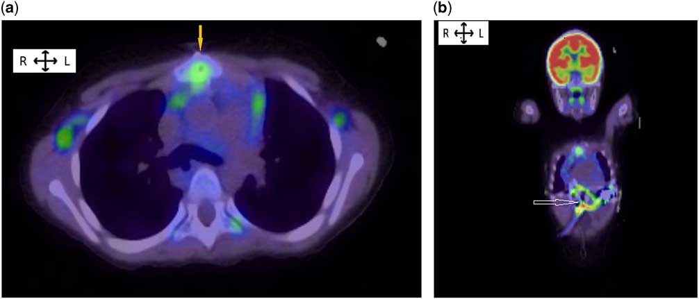
Two children underwent left ventricular assist device implantation (HVAD; HeartWare device, Framingham, Massachusetts, United States of America) for refractory end-stage heart failure caused by dilated cardiomyopathy. Patient 1, a 5-year-old girl, had a history of a suspected culture-negative device-related infection approximately 6 months after device placement and was treated empirically with 6 weeks of vancomycin, cefepime, and gentamicin. A period of 4 weeks after completion of this therapy, the patient presented to the cardiology clinic with epigastric pain, intermittent fever for 2 weeks, elevated serum C-reactive protein concentration (22 mg/dl), and elevated white blood cell count (24×109 cells/L). Initial investigations were performed, including culture of drainage from the percutaneous exit site, three sets of blood cultures, chest radiography, echocardiography, and a CT scan of the chest, abdomen, and pelvis with contrast, but they failed to identify any source of infection. A F-fluorodeoxyglucose-positive emission tomography/CT from head to mid-thigh was then performed. Abnormal F-fluorodeoxyglucose uptake was observed around the outflow tract of the HVAD device (see Fig 1a and b). Empirical intravenous antibiotic therapy, using vancomycin and cefepime, was initiated for culture-negative device-related infection. After antibiotic initiation, the patient became afebrile and her C-reactive protein concentration normalised. She underwent an uneventful heart transplant during her 6th week of antimicrobial therapy.
(a) FDG PET/CT shows increased FDG uptake which extends along the connector between the LVAD “and the aortic outflow graft (yellow arrow)”. Mild FDG uptake is seen in some bilateral axillary lymph nodes. (b) FDG PET/CT shows increased FDG uptake which extends along the connector between the LVAD and outflow graft (white arrow).

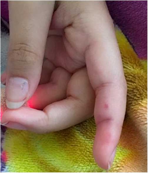
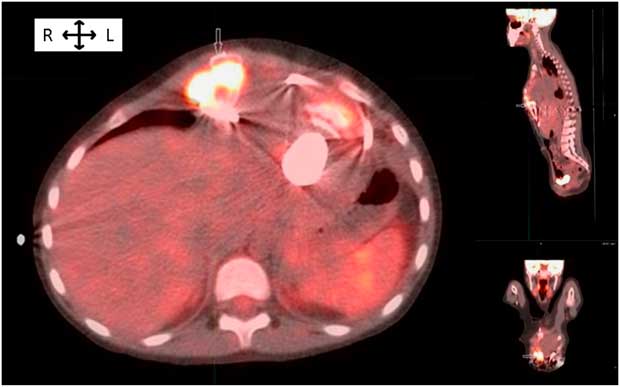
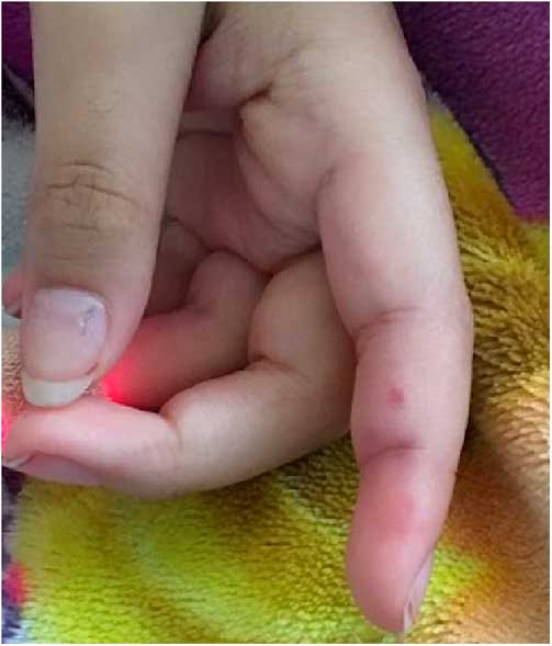
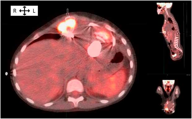
Patient 2, an 8-year-old girl with dilated cardiomyopathy with refractory end-stage heart failure, was hospitalised 10 months after left ventricular assist device implantation with fever, fatigue, and poor oral intake. Laboratory revealed a mild leucocytosis (14×109 cells/L) and elevated C-reactive protein concentration (24 mg/dl). Janeway lesions (see Fig 2) and splenomegaly were noted on physical examination, and blood cultures grew methicillin-sensitive Staphylococcus aureus. Similar to the previous patient, echocardiography and CT scan with contrast failed to localise the source of infection, and a F-fluorodeoxyglucose-positive emission tomography/CT scan was then performed. Abnormal fluorodeoxyglucose uptake was observed around the outflow tract of the device (see Fig 3), consistent with infection. Her inflammatory markers normalised and her condition improved after starting treatment with nafcillin. She completed a 6-week course of nafcillin and continued on chronic suppressive therapy with cephalexin until she successfully underwent cardiac transplantation. The tissue cultures taken at the time of explant for both cases were negative.
Janeway lesions.
FDG PET/CT shows focal intense hypermetabolic activity surrounding the LVAD outflow cannula “in the retrosternal space at the level of the inferior sternum (white arrow)”. There is mild adjacent reactive lymphadenopathy.
Left ventricular assist devices are important therapeutic options for children with end-stage heart failure. Infections are well-known complications of ventricular assist devices and increase the risk of morbidity and mortality associated with device placement. Early detection and accurate diagnosis of infection is critical for prompt initiation of appropriate management in patients with ventricular assist devices; early and appropriate management provides the best opportunity to improve outcomes in these patients.
F-fluorodeoxyglucose-positive emission tomography/CT is highly sensitive for the detection of an inflammatory response. Some adult centres have reported their experiences using positive emission tomography/CT to diagnose infections in ventricular assist device patients.Reference Kim, Feller, Chen and Dilsizian 1 – Reference Akin, Muslem and Constaninescu 3 In a recent meta-analysis of the use of F-fluorodeoxyglucose-positive emission tomography/CT for detecting infections associated with implantable cardiac devices, the pooled sensitivity of F-fluorodeoxyglucose-positive emission tomography-CT was 87% and the pooled specificity was 94%.Reference Juneau, Golfam and Hazra 4 The area under the curve of the receiver operating characteristics analysis showed good accuracy at 0.952. Among ventricular assist device patients, a study of 31 adults showed a sensitivity of 100% and a specificity of 80% for identifying infections.Reference Dell’Aquilla, Mastrobuoni and Alles 5
Other imaging modalities have been used to diagnose infection in ventricular assist device patients. A transthoracic or transesophageal echocardiogram can identify vegetations within the heart, suggestive of endocarditis, but has limited utility beyond that. Abdominal ultrasound and standard CT scan of the chest and abdomen can identify nonspecific fluid collections around the driveline and pump pocket. The use of F-fluorodeoxyglucose-positive emission tomography/CT, however, may provide a more precise location of an inflammatory process and may allow for earlier and more accurate detection.Reference Trachtenberg, Cordero-Reyes, Elias and Loebe 6 One potential limitation of this imaging modality is that patients should receive a low-carbohydrate diet for 24 hours before the study to optimise images by reducing physiological myocardial uptake.Reference Akin, Muslem and Constaninescu 3 Despite this limitation, positive emission tomography/CT appears to be a helpful imaging modality for adult and now paediatric ventricular assist device patients with suspected infectious complications.
Acknowledgements
The authors would like to acknowledge the contribution of Dr Antonio Cabrera to this work.
Financial Support
This research received no specific grant from any funding agency or from commercial or not-for-profit sectors.
Conflicts of Interest
None.
Ethical Standards
The authors assert that all procedures contributing to this work comply with the ethical standards of the relevant national guides and has been approved by the institutional committee of involved institutions.