Introduction
Actinomycosis is a bacterial disease caused by actinomyces species. Actinomyces are anaerobic or facultatively anaerobic Gram-positive bacteria from the actinobacteria genus. The disease commonly presents with granulomatous abscesses of the oral-cervicofacial region, but other sites including the thorax and abdomen have been described.Reference Wong, Turmezei and Weston1
The involvement of the temporal bone and middle ear is far less common. To date, approximately 43 cases of tympanomastoid actinomycosis have been presented in the literature. These cases have been distributed across the world; however, prior to the current paper, a case had not been reported in the UK for over 60 years.
Case report
A nine-year-old female was referred for a second opinion because of a history of intermittent left otorrhoea since early childhood. She had been seen in another ENT department, and had received numerous courses of topical and systemic antibiotics without a definitive diagnosis. Her past medical history was unremarkable.
At the initial assessment her left ear was discharging, with granulation tissue visible on the tympanic membrane. An audiogram revealed a moderate conductive hearing loss in the affected ear and normal hearing in her right ear. In view of these findings, an examination of her left ear was undertaken under general anaesthetic. This revealed an inflammatory polyp arising from an area of myringitis on the pars tensa. The remaining tympanic membrane was featureless.
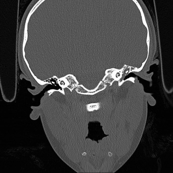
A myringotomy was performed and thick, glue-like secretions were aspirated. At this time there appeared to be a white plaque visible within the middle ear, which raised the possibility of a cholesteatoma. The polyp was removed and sent for histological examination, and was reported as ‘an inflammatory aural polyp’. A swab taken at the time of the examination cultured an antibiotic-sensitive Staphylococcus aureus. A computed tomography scan revealed complete opacification of the middle-ear cleft, with no evidence of ossicular or scutum erosion (Figure 1).
Coronal (high-resolution, unenhanced) computed tomography image showing complete opacification of the left hypo-, meso- and epi-tympanum.

In view of these findings, a combined approach tympanoplasty was performed. This was conducted under anaesthetic, seven months after the initial examination (there was a three month delay due to inter-departmental consultant referral and a four month operation waiting list delay).
At operation, the middle ear was found to be full of granulation tissue and there were two yellow coloured ‘plaques’ in the middle ear. The long process of the incus was eroded, but the head of the stapes was intact. The mucosa of the mastoid air cells was oedematous, but no cholesteatoma was found. The granulation tissue and plaques were removed and sent for histology, and an ossiculoplasty (incus transposition) was performed.
Histological examination revealed a large colony of micro-organisms surrounded by a film of necrotic, fibrinous debris and neutrophil polymorphs. The central portion of the colony was basophilic and eosinophilic, with club-shaped filaments radiating at the peripheries in a rosette pattern into which neutrophils were enmeshed.
Histochemical testing of the colonies revealed Gram-positive, silver avid (on Grocott staining) and diastase-resistant periodic acid-Schiff negative staining. This histochemical profile was deemed most in keeping with actinomycosis, wherein the colonies represented the characteristic ‘sulphur granules’. Fungal infection was ruled out on the basis of the negative diastase-resistant periodic acid-Schiff test.
On the advice of the microbiologist, the patient was started on a six-month course of oral amoxicillin and referred for a paediatric assessment to exclude an underlying immune deficiency. This assessment did not reveal any abnormality.
Following surgery, the patient had no further ear discharge. The tympanic membrane appeared normal and her hearing was normal in both ears. She was followed up for a period of one year (with no further symptoms) before being discharged.
Discussion
Actinomycosis is a subacute or chronic granulomatous disease, which typically presents with abscesses and tissue necrosis in the oral-cervicofacial region. The infective agent is a non-spore forming anaerobic or facultative anaerobic actinobacteria, most often of the Actinomyces israelii species.Reference Wong, Turmezei and Weston1 This species was first described as a human pathogen by Israel in 1878.Reference Schultheiss2
Actinomyces are known to be a commensal flora of the oral cavity; hence, it is postulated that entry to the middle ear is most likely the result of seeding from the nasopharynx via the eustachian tube. Direct infections of the external auditory canal or haematogenous spread are possible but less likely alternatives.Reference Ajal, Turner, Fagan and Walker3 In our case, the initial presentation of an aural polyp and featureless tympanic membrane prompted a myringotomy. After the aspiration of thick, glue-like secretions, a white plaque was found, which filled the middle ear. This was initially suspected to be a cholesteatoma; however, in retrospect this was almost certainly a primary plaque of actinomyces. We are unable to speculate further on the mechanism of primary actinomyces colonisation of the middle ear in our case. There is, however, a possibility that actinomyces could have been introduced into the middle ear at the time of the initial myringotomy, which would therefore constitute a secondary superadded infection of a middle-ear plaque of uncertain aetiology.
Identification of actinomyces bacteria is difficult due to their recalcitrance to culture and their exquisite sensitivity to oxygen. However, the identification of Gram-positive filamentous organisms with sulphur granules is strongly suggestive of actinomycosis.Reference Smego and Foglia4 The differential diagnosis for culture-insensitive chronic otitis media includes nocardiosis and tuberculosis. Newer diagnostic techniques such as polymerase chain reaction, 16S rRNA sequencing and mass spectrometry hold promise, but are not routinely used in clinical practice.Reference Patel5, Reference Welker6
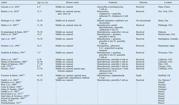
Our review of the worldwide literature revealed 26 cases of actinomycosis of the middle ear and mastoid since 1945. These are distributed broadly across the world, with a clustering in the USA (see Table I).Reference Gazzano, Chanteret, Duvillard, Folia and Romanet7–Reference Preobrazhenskii31 We speculate that this clustering is an artificial effect due a higher publication rate from this region. We present the first case in the UK in over 60 years.
• Actinomycosis is a bacterial disease caused by actinomyces species
• Middle-ear and mastoid actinomycosis is rare, presenting as recurrent otorrhoea with granulation tissue
• This paper presents the first case of middle-ear actinomycosis in the UK for 60 years
• Actinomyces form basophilic and eosinophilic colonies with club-shaped filaments radiating into rosette patterns (‘sulphur granules’)
• Histochemical profiling of colonies revealed silver avid, diastase-resistant periodic acid-Schiff negative staining
• Our recommended treatment is full surgical clearance of diseased tissue with six months amoxicillin
Cases of middle-ear actinomycosis*

* Cases presented in the worldwide literature since 1945. †Foreign language articles. Y = years; F = female; mth = months; po = per os (by mouth); IV = intravenous; M = male; wks = weeks; d = days
In the last documented case from the UK, Townrow and Barrie (1945) reported a case of a 64-year-old horseman with actinomycosis of the middle ear.Reference Townrow and Barrie20 The patient had presented with a 57-year history of ear discharge, which had worsened over the previous 8 days. He underwent mastoid exploration, but suffered a large venous bleed. He developed meningitis which was unsuccessfully treated with a sulphonamide antibiotic, ultimately resulting in his death. The authors reviewed the literature up to 1945 and noted that 16 out of 17 reported cases were fatal, with meningitis being the leading cause of death. Diagnosis in the pre-antibiotic age was based on post-mortem assessment. Following the introduction of penicillin, prognosis radically improved: the condition, previously associated with a near 100 per cent mortality rate, became a non-fatal and eminently treatable pathology.
It is our recommendation that complete surgical removal and histological examination of all infected tissue should be undertaken in similar cases. If actinomyces are found, we recommend six months oral amoxicillin as the first-line treatment. Other antibiotics including erythromycin, chloramphenicol and tetracycline have also been used successfully.Reference Wong, Turmezei and Weston1