INTRODUCTION
Helminth infections in livestock remain a global challenge despite decades of research efforts. In pigs, infections with the nematode Ascaris suum occur worldwide in both intensive and extensive production systems (e.g. Peng et al. Reference Peng, Zhou, Cui, Crompton, Whitehead, Xiong, Wu, Peng, Yang, Wu, Xu and Yan1996; Nissen et al. Reference Nissen, Poulsen, Nejsum, Olsen, Roepstorff, Rubaire-Akiiki and Thamsborg2011) and a recent survey showed that A. suum was found on 76% of Danish sow farms (Haugegaard, Reference Haugegaard2010). Ascaris suum infections may cause decreased weight gain and also give rise to liver condemnation at slaughter due to white spots caused by migrating larvae (reviewed by Thamsborg et al. Reference Thamsborg, Nejsum, Mejer and Holland2013). With the risk of emergence of anthelmintic resistance, alternatives to chemotherapy are highly warranted to ensure sustainable control in the future (reviewed by Hoste and Torres-Acosta, Reference Hoste and Torres-Acosta2011).
Since the early decades of the last century there has been a growing awareness that livestock breeds differ in resistance to gastrointestinal nematodes due to genetic variation (Ackert et al. Reference Ackert, Eisenbrandt, Wilmoth, Glading and Pratt1935; Johnson et al. Reference Johnson, Stewart and Hale1975; Mugambi et al. Reference Mugambi, Bain, Wanyangu, Ihiga, Duncan, Murray and Stear1997; Gauly et al. Reference Gauly, Bauer, Preisinger and Erhardt2002; Oliveira et al. Reference Oliveira, Alencar, Chagas, Giglioti and Oliveira2009; reviewed by Saddiqi et al. Reference Saddiqi, Jabbar, Sarwar, Iqbal, Muhammad, Nisa and Shahzad2011). Work in laboratory models has shown similar differences in resistance between host strains (Dow and Jarrett, Reference Dow and Jarrett1960; Mitchell et al. Reference Mitchell, Hogarth-Scott, Edwards, Lewers, Cousins and Moore1976; Lewis et al. Reference Lewis, Behnke, Stafford and Holland2006). We here define resistance as the ability to suppress establishment and/or subsequent development of infection due to development of immunity (Albers et al. Reference Albers, Gray, Piper, Barker, Le Jambre and Barger1987; Coustau et al. Reference Coustau, Chevillon and ffrench-Constant2000) and we define susceptibility as the inverse of resistance (Gray, Reference Gray, Gray, Woolaston and Eaton1995). Helminths typically induce a polarized TH2-type response which includes both innate and adaptive components (Dawson et al. Reference Dawson, Beshah, Nishi, Solano-Aguilar, Morimoto, Zhao, Madden, Ledbetter, Dubey, Shea-Donohue, Lunney and Urban2005; reviewed by Anthony et al. Reference Anthony, Rutitzky, Urban, Stadecker and Gause2007 and Pulendran and Artis, Reference Pulendran and Artis2012).
At the population level helminths are usually highly over-dispersed with a fraction of the individuals harbouring the majority of the parasites (e.g. Crofton, Reference Crofton1971; Croll et al. Reference Croll, Anderson, Gyorkos and Ghadirian1982) and there is now increasing evidence that this distribution to a large extent can be explained by genetic variation among hosts (reviewed by Quinnell, Reference Quinnell2003). Indeed, quantitative genetic studies of different nematode infection traits (worm burden, fecal egg counts (FEC), worm length) have found medium to high heritabilities in both human and domestic animal populations, including pigs (reviewed by Kloosterman et al. Reference Kloosterman, Parmentier and Ploeger1992; Stear et al. Reference Stear, Bairden, Duncan, Holmes, McKellar, Park, Strain, Murray, Bishop and Gettinby1997; Williams-Blangero et al. Reference Williams-Blangero, Subedi, Upadhayay, Manral, Rai, Jha, Robinson and Blangero1999; Zinsstag et al. Reference Zinsstag, Ankers, Njie, Smith, Pandey, Pfister and Tanner2000; Nejsum et al. Reference Nejsum, Roepstorff, Jorgensen, Fredholm, Goring, Anderson and Thamsborg2009a ). QTL (quantitative trait locus) studies have found several regions of the genome that apparently are involved in resistance to helminths (Iraqi et al. Reference Iraqi, Behnke, Menge, Lowe, Teale, Gibson, Baker and Wakelin2003; Dominik et al. Reference Dominik, Hunt, McNally, Murrell, Hall and Purvis2010; Silva et al. Reference Silva, Sonstegard, Hanotte, Mugambi, Garcia, Nagda, Gibson, Iraqi, McClintock, Kemp, Boettcher, Malek, Van Tassell and Baker2012). However, in many cases, these QTLs have not been validated in independent populations and the underlying genes explaining the differences in susceptibility have not yet been identified.
In pigs, a QTL on chromosome 4 associated with resistance to A. suum was recently detected (Skallerup et al. Reference Skallerup, Nejsum, Jorgensen, Goring, Karlskov-Mortensen, Archibald, Fredholm and Thamsborg2012). The authors found two single nucleotide polymorphisms (SNPs), 0_TXNIP_DS087128.1_2_2 (TXNIP) and 0_ARNT_DS076761.1_18 (ARNT), which were associated with total worm burden and/or adult worm burden in cross-bred Danish Duroc/Landrace/Yorkshire (DLY) pigs. Hence, pigs with the ‘resistant’ AA genotype at the TXNIP locus had a mean total burden of 16 worms, whereas pigs with the ‘susceptible’ AB genotype had a mean burden of 41 worms, i.e. the heterozygotes had a 2·5-fold higher total worm burden than the homozygotes (Skallerup et al. Reference Skallerup, Nejsum, Jorgensen, Goring, Karlskov-Mortensen, Archibald, Fredholm and Thamsborg2012). The present study was conducted in an independent group of cross-bred DLY pigs from a commercial farm which were selected based on their SNP TXNIP genotype (AA, AB). These pigs were then experimentally infected with A. suum to validate the hypothesis that pigs with the AA genotype would have lower worm burdens than pigs with the AB genotype.
MATERIALS AND METHODS
Experimental animals
Previous research has shown that individuals homozygous for the B allele at the SNP 0_TXNIP_DS087128.1_2_2 (TXNIP) locus have a low frequency in Danish mixed breed pigs and that this allele seems to be associated with A. suum susceptibility (Skallerup et al. Reference Skallerup, Nejsum, Jorgensen, Goring, Karlskov-Mortensen, Archibald, Fredholm and Thamsborg2012). Hence, it was only possible to include two genotypes (AA, AB) in the study. Power analysis showed that a group size of 24 pigs would be sufficient to demonstrate a significant difference in macroscopic A. suum burden between the two SNP TXNIP genotypes, assuming an effect size similar to our previous study (∼2·5-fold higher mean worm burden in heterozygous pigs compared with pigs homozygous for the allele increasing resistance). In order to identify pigs with the genotypes needed for the study, 112 cross-bred piglets (Danish Landrace/Yorkshire/Duroc) on a commercial specific pathogen-free farm were screened prior to inclusion. Mucosa cells from the oral cavity for DNA extraction were obtained using a gynobrush (Abena Ltd., Aabenraa, Denmark) and piglets were genotyped as described below. All genotypes were validated using DNA from blood samples taken on day 55 post first infection (PI).
Housing
The study was approved by the Animal Experiments Inspectorate, Ministry of Justice, Denmark (Ref. 2010/561-1914). Care and maintenance of all animals were in accordance with applicable Danish and European guidelines. Fifty-two pigs (N AA = 27; N AB = 25) from 10 different litters (two to eight piglets from each litter) were selected for the study. The farmer used mixed semen to produce the littermates which were thus full-sibs or half-sibs. The pigs were kept at the animal facility at the National Veterinary Institute, Denmark; upon arrival at 7 weeks of age they were allocated into three pens with concrete floors ensuring an equal distribution of genotype, litter of origin, sex and weight in each pen. The pens were littered with wood shavings on a daily basis, and water was provided ad libitum. In order to optimize worm establishment, the animals were fed a diet consisting of ground barley supplemented with proteins and minerals (Petkevicius et al. Reference Petkevicius, Bjorn, Roepstorff, Nansen, Bach Knudsen, Barnes and Jensen1995; Bjorn et al. Reference Bjorn, Roepstorff and Nansen1996). The piglets were treated prophylactically against Lawsonia spp. (tiamulin (Denagard Vet®) 10–25 mg kg−1 intramuscularly for 3 days) during the first week. Mean weight (±s.d.) of the AA and AB groups on day 0 PI was 13·5 kg (±2·5) and 13·4 kg (±2·4), respectively.
Phenotypic traits
The experimental protocol to generate parasite indicator traits involved trickle infections with A. suum. Hence, after 1 week of acclimatization, pigs of AA and AB genotypes (N AA = 27; N AB = 25) were experimentally infected with A. suum eggs (25 eggs kg−1 day−1) twice per week. One pig of each genotype was euthanized on day 13 PI as described below; in addition, two pigs of genotype AA died during the experiment and were excluded from the analysis. Based on previous experience (Nejsum et al. Reference Nejsum, Roepstorff, Jorgensen, Fredholm, Goring, Anderson and Thamsborg2009a , Reference Nejsum, Thamsborg, Petersen, Kringel, Fredholm and Roepstorff b ), the utilized inoculation dose was expected to give a high prevalence of macroscopic worms. Embryonated parasite eggs used for infections were prepared from female A. suum worms collected at a Danish slaughterhouse as described elsewhere (Oksanen et al. Reference Oksanen, Eriksen, Roepstorff, Ilsoe, Nansen and Lind1990). The first egg suspension was given orally via a plastic syringe at the base of the tongue; subsequently, the eggs were given in the feed on a pen basis.
The pigs were euthanized using a captive bolt pistol followed by exsanguination on days 55–59 PI. After removing the viscera, the small intestine was opened longitudinally, washed in lukewarm saline (0·9%) and gently pulled between two fingers to remove the mucus. Any macroscopic A. suum worms (large juveniles and adults) were removed and counted. Ascarid larvae were isolated by the agar-gel method (Slotved et al. Reference Slotved, Barnes, Eriksen, Roepstorff, Nansen and Bjorn1997) and total larval burden calculated by extrapolation from a 20% aliquot count. All worms were stored in 70% ethanol until they were counted. The liver was carefully examined, and white spots caused by migrating larvae were counted and classified as the diffuse granulation-tissue type or the lymphonodular type (Ronéus, Reference Ronéus1966; Copeman and Gaafar, Reference Copeman and Gaafar1972); all livers were examined by the same person who was blinded to pig genotype. The infectivity of the A. suum egg batch was tested in two pigs (one AA and one AB) which were euthanized on day 13 PI, and the number of larvae in the small intestine assessed as described above (larval burden was calculated by extrapolation from a 50% aliquot count). The recovery was 32% (AA pig) and 42% (AB pig) of the total dose given on days 0 and 3 PI.
Feces were sampled rectally at day 0 PI and weekly from week 5 PI until slaughter. Egg counts were determined using a modified McMaster method with an analytical sensitivity of 20 eggs per gram of feces (epg) (Roepstorff and Nansen, Reference Roepstorff and Nansen1998). Samples taken at week 8 PI were processed and counted in duplicate, and the mean of the two measurements was used in the statistical analysis.
Blood samples were collected on days −3 (two pens), 0 (one pen) and 55 PI (all pens) to test the antibody response to A. suum antigen preparations. Blood samples were refrigerated overnight and subsequently centrifuged (1763 g , 10 min). Serum was then frozen at −20 °C until analysis. Whole-blood samples (EDTA tubes) were taken on day 55 PI and kept at −20 °C until analysis.
For the preparation of adult A. suum (adult-As) antigen, adult worms were collected at a slaughterhouse in Japan, homogenized and incubated at 5 °C overnight. The extract was centrifuged (14 000 g , 10 min, 5 °C) and the supernatant was then isolated.
Ascaris suum lung L3 larvae (LL3) were collected from male Japanese white rabbits (Kyodo, Kumamoto, Japan) using the Baermann method as described elsewhere (Yoshida et al. Reference Yoshida, Nagayasu, Horii and Maruyama2012). LL3 larvae were cultured (37 °C) in 6-well culture plates (Nunc, Roskilde, Denmark) containing RPMI 1640 medium (Wako, Osaka, Japan) supplemented with 100 μg mL−1 of streptomycin, 100 U mL−1 of penicillin and 250 ng mL−1 of amphotericin B (all from Gibco, Rockville, MD, USA). Supernatants were collected 7, 14, 21 and 28 days after incubation, pooled, and subsequently concentrated using ultrafiltration (Amicon® Ultra-15 3K, Millipore, Billerica, MA, USA). LL3 excretory/secretory antigen (LL3-ES) concentration was determined by Quant-iT™ Protein Assay Kit (Invitrogen, Carlsbad, CA, USA).
Infective A. suum L3 larvae (iL3) were produced from eggs isolated from female worms collected at a slaughterhouse in Japan. The eggs were kept in sulphuric acid (0·1 N) and embryonated (8 weeks, 27 °C), then mechanically hatched in a beaker with 1·5–2·5 mm glass beads (30 min, 37 °C). The suspension was passed through a 425 μm sieve to remove glass beads, then larvae were isolated by the Baermann method and cultured as described above. iL3 excretory/secretory antigen (iL3-ES) was obtained from pooled culture supernatants collected 7, 14, 21 and 28 days after incubation, then concentrated and its protein concentration assessed as described for LL3-ES.
Flat-bottomed 96-well plates (MaxiSorp, Nunc, Roskilde, Denmark) were coated with adult-As, LL3-ES or iL3-ES A. suum antigen (1 μg mL−1; 50 μL well−1) in 0·05 m carbonate-bicarbonate coating buffer (pH 9·6) and incubated at 5 °C overnight. The plates were washed three times with PBST (0·01 m phosphate, 0·15 m sodium chloride, pH 7·2 and 0·05% Tween 20) in a microplate washer (BioTek, Winooski, VT, USA), blocked with 150 μL 10% caseine TBS per well and incubated for 2 h at room temperature (RT). Serum diluted 1:1000 (adult-As), 1:2000 (LL3-ES), or 1:500 (iL3-ES) in 10% caseine TBS was added to each well (50 μL well−1), incubated for 1·5 h at RT, and wells were then washed again three times. Fifty μL horseradish peroxidase-labelled goat anti-swine IgG (gamma chain) (KPL, Gaithersburg, MD, USA) diluted 1:2000 in 10% caseine TBS buffer was added to each well and incubated for 1·5 h at RT. The plates were washed three times and incubated 10 min at RT after addition of 50 μL 3,3′,5,5′-tetramethylbenzidine (KPL, Gaithersburg, MD, USA) to each well. The reaction was stopped with 25 μL sulphuric acid (1 m) per well and OD values were read at 450 nm (Multiskan EX, MTX Lab Systems Inc, VA, USA). As positive control we used a single anti-A. suum IgG positive serum sample from a pig trickle-infected with A. suum.
DNA extraction and SNP genotyping
DNA was extracted from oral mucosa cells using the Wizard® Genomic DNA Purification kit (Promega, Madison, WI, USA) according to the manufacturer's instructions. The quantity and quality of DNA was measured using a NanoDrop 1000 spectrophotometer (Thermo Fischer Scientific). Genotyping was performed using a custom-made TaqMan® SNP Genotyping Assay (Applied Biosystems, Carlsbad, CA, USA) on a Stratagene Mx3000P™ (Agilents, Santa Clara, CA, USA). All assays were run in a total volume of 15 μL with 20 ng of genomic DNA as template according to the manufacturer's instructions. The PCR programme was as follows: Initial denaturation (95 °C, 10 min) followed by 48 cycles (92 °C, 20 s; 60 °C, 1 min). The MxPro software (Stratagene) was used for allele discrimination. All animals included in the final analysis (N = 48) had their genotypes validated using DNA from whole-blood samples. DNA was extracted using a salting out procedure as described by Miller et al. (Reference Miller, Dykes and Polesky1988) with minor modifications. The quantity and quality of DNA was measured as described above.
Location of single nucleotide polymorphisms and quantitative trait locus associated with A. suum burden
In the recent assembly (sscrofa10.2) of the porcine genome (Groenen et al. Reference Groenen, Archibald, Uenishi, Tuggle, Takeuchi, Rothschild, Rogel-Gaillard, Park, Milan, Megens, Li, Larkin, Kim, Frantz, Caccamo, Ahn, Aken, Anselmo, Anthon, Auvil, Badaoui, Beattie, Bendixen, Berman, Blecha, Blomberg, Bolund, Bosse, Botti and Bujie2012) (http://www.ensembl.org), the QTL associated with resistance to A. suum (Skallerup et al. Reference Skallerup, Nejsum, Jorgensen, Goring, Karlskov-Mortensen, Archibald, Fredholm and Thamsborg2012) maps to SSC4: 106,139,259-109,133,240. This region is flanked by the genes TCHHL1 and POLR3C. In sscrofa10.2, SNP TXNIP maps to SSC4: 108,992,540; SNP ARNT maps to SSC4: 107,609,772.
Statistical analyses
Power analysis (80% power; 5% significance level) was performed in R (R Core Team, 2012) using power.t.test. Effect size (0·73) and variance estimates were calculated from log-transformed macroscopic worm burden data from a previous trickle-infection study using the same dose and which showed that the ‘susceptible’ genotype had 2·5-fold higher mean worm burden than the ‘resistant’ genotype (Nejsum et al. Reference Nejsum, Roepstorff, Jorgensen, Fredholm, Goring, Anderson and Thamsborg2009a ; Skallerup et al. Reference Skallerup, Nejsum, Jorgensen, Goring, Karlskov-Mortensen, Archibald, Fredholm and Thamsborg2012).
The association analyses between the two single nucleotide polymorphisms (SNPs) and traits related to A. suum infection were performed using a measured genotype approach (Boerwinkle et al. Reference Boerwinkle, Chakraborty and Sing1986) as implemented in the software package SOLAR version 4.2.7 (Texas Biomedical Research Institute, San Antonio, TX, USA) (Almasy and Blangero, Reference Almasy and Blangero1998). Sex and starting weight were fitted as fixed effects and litter and pen were fitted as random effects (to account for additive genetic effects and shared environmental effects, respectively). SNP genotypes were converted into measured covariates (fixed effects) by coding the number of copies of the rarer allele (0–2) thus assuming additivity of allelic effects. The measured genotype analysis tests whether there is a significant difference between the genotypic means (Havill et al. Reference Havill, Dyer, Richardson, Mahaney and Blangero2005).
In the analyses, each pen was treated as a separate household with shared environment. Since the farmer used mixed semen, we had no information about paternal origin of the piglets. Hence, we conservatively modelled that all piglets in each litter had the same father (i.e. assuming that piglets were full sibs and thus more related to each other than they probably were in reality), and that different litters were unrelated (i.e. we modelled that each sow was sired to one of nine designated model boars).
Data were normalized prior to analysis, if needed, using either the log10 transformation (total A. suum burden, FEC, lymphonodular type liver white spots) or the inverse Gaussian density distribution (macroscopic A. suum) to ensure that the phenotypes did not violate the assumptions underlying variance components analysis. Based on previous work (Skallerup et al. Reference Skallerup, Nejsum, Jorgensen, Goring, Karlskov-Mortensen, Archibald, Fredholm and Thamsborg2012) we expected a priori pigs in the AB group to have higher worm burdens than pigs in the AA group. Hence, in the analyses of macroscopic worm burden, total worm burden and FEC, we used a one-sided test whereas for the other traits we used a two-sided test. We chose a significance level of P = 0·05. The correlation between worm burdens and A. suum-specific antibody titres was determined from the Spearman correlation coefficient calculated in GraphPad Prism version 5.04 for Windows (GraphPad Software, San Diego, CA, USA).
RESULTS
Piglets were confirmed negative for A. suum infection as tested by FEC day 0 PI. In addition, all pigs were found seronegative to adult-As antigen before the start of the experiment. Two pigs of genotype AA died during the experiment for reasons not related to the study treatment and were excluded from the analysis.
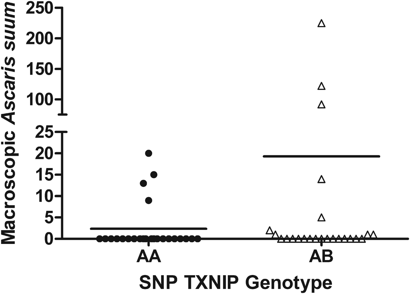
A total of 13 pigs (4 of AA genotype and 9 of AB genotype) had macroscopic A. suum at slaughter (Fig. 1; Table 1). Three pigs (all in the AB group) had more than 90 macroscopic A. suum. Mean A. suum intensity of AB pigs was 3·6 times higher than in AA pigs and the difference in abundance between the two genotypes approached significance (P = 0·06).
Number of macroscopic Ascaris suum in individual pigs homozygous (N = 24) or heterozygous (N = 24) at the single nucleotide polymorphism (SNP) 0_TXNIP_DS087128.1_2_2 (TXNIP) locus. Means are indicated with horizontal lines.

Statistics for association of two single nucleotide polymorphisms (SNPs) with macroscopic Ascaris suum burden, total A. suum burden and fecal egg counts. Sex and weight at start of experiment were included as fixed effects. Litter and pen were fitted as random effects

Previous work in our group has confirmed the association of another SNP, ARNT, with adult A. suum burden (Skallerup et al. Reference Skallerup, Nejsum, Jorgensen, Goring, Karlskov-Mortensen, Archibald, Fredholm and Thamsborg2012), and pigs were thus genotyped at the ARNT locus as well. The linkage disequilibrium (r 2) between the two SNPs (TXNIP, ARNT) was estimated to be 0·78. All three genotypes of the ARNT polymorphism were found and genotypic means for the different traits are presented in Tables 1 and 2. The AB genotype (SNP ARNT) had higher mean macroscopic A. suum burden than the AA genotype (18·4 vs 2·2; Table 1; P = 0·15), with the direction of effect being as expected. Only one of the three pigs with genotype BB harboured macroscopic worms (one worm; Table 1).
Statistics for association of two single nucleotide polymorphisms (SNPs) with number of liver white spots (total number of white spots and number of lymphonodular type white spots) and serum IgG antibody levels (OD450) specific for adult Ascaris suum antigen (adult-As), excretory-secretory (ES) antigen from A. suum L3 larvae (iL3-ES), and ES antigen from A. suum lung L3-larvae (LL3-ES). Sex and weight at start of experiment were included as fixed effects. Litter and pen were fitted as random effects

a One pig (genotype AB at both polymorphisms) did not have liver white spots counted; hence for this trait N AB = 23 (SNP TXNIP) and N AB = 25 (SNP ARNT).
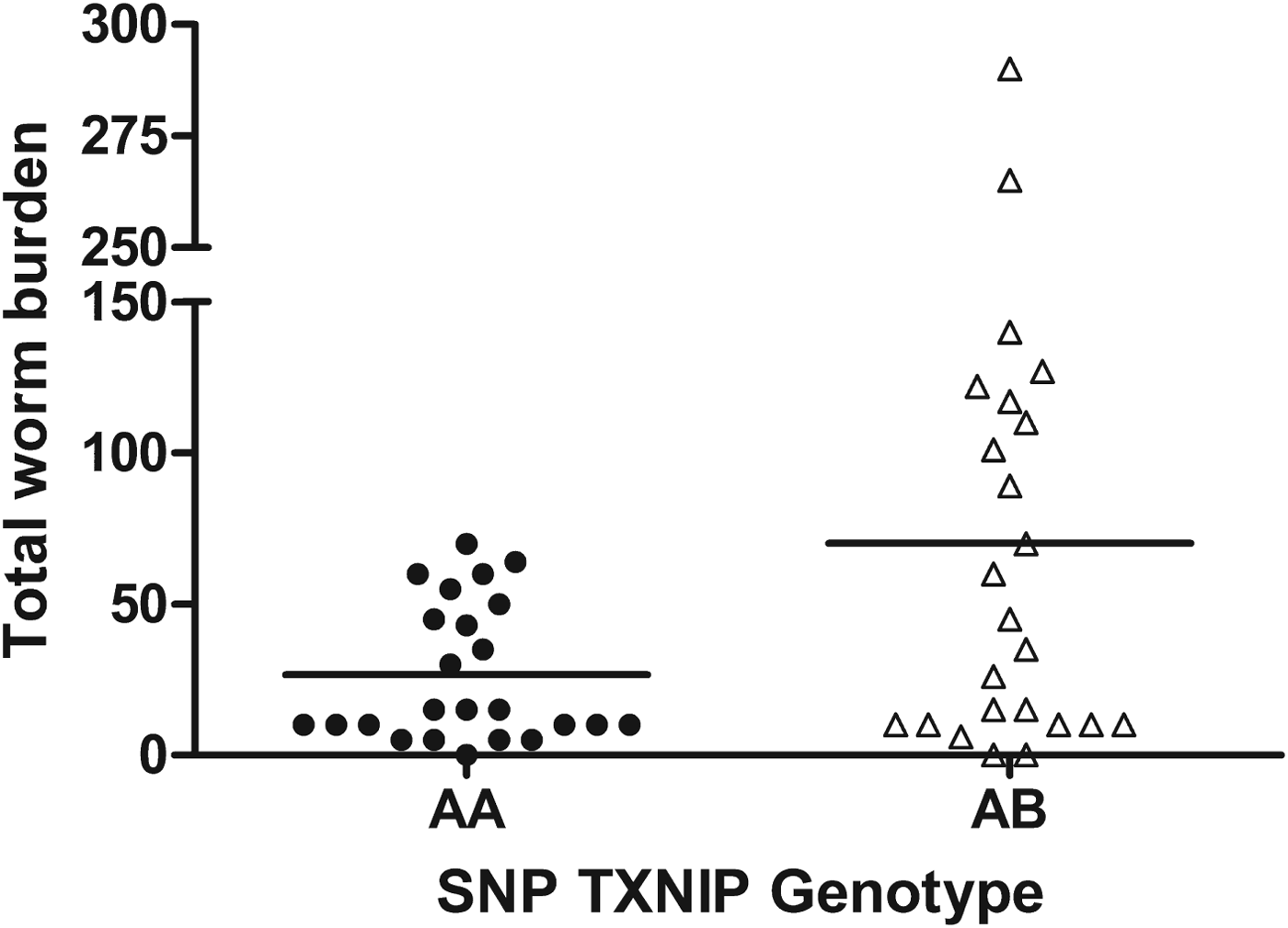
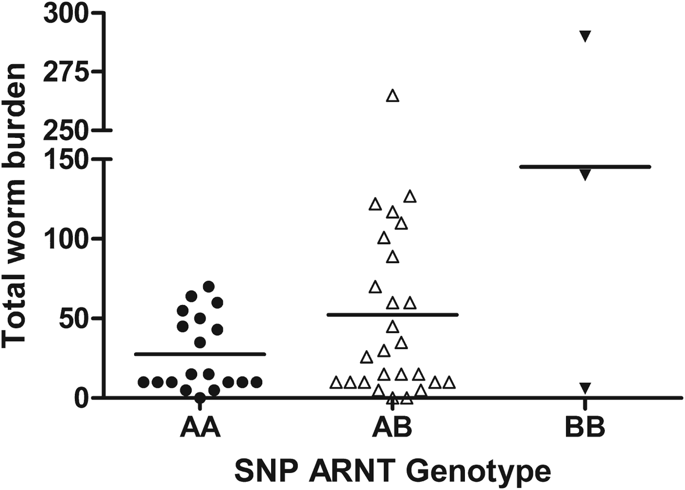
For total worm burden we found the same trend as for the macroscopic worms; pigs with the AB genotype (SNP TXNIP) had ∼2·6 times higher total worm burden (both measured as mean and median) than the AA genotype (Table 1, Fig. 2; P = 0·09). A similar tendency was seen for SNP ARNT; one copy of the susceptibility allele (B) doubled the total worm burden (both measured as mean and median) whereas carriers of two copies (BB) had a mean worm burden nearly three times higher than the heterozygotes (Fig. 3; Table 1; P = 0·06).
Total number of Ascaris suum in individual pigs homozygous (N = 24) or heterozygous (N = 24) at the single nucleotide polymorphism (SNP) 0_TXNIP_DS087128.1_2_2 (TXNIP) locus. Means are indicated with horizontal lines.

Total number of Ascaris suum in individual pigs homozygous (N AA = 19; N BB = 3) or heterozygous (N AB = 26) at the single nucleotide polymorphism (SNP) 0_ARNT_DS076761.1_18 (ARNT) locus. Means are indicated with horizontal lines.

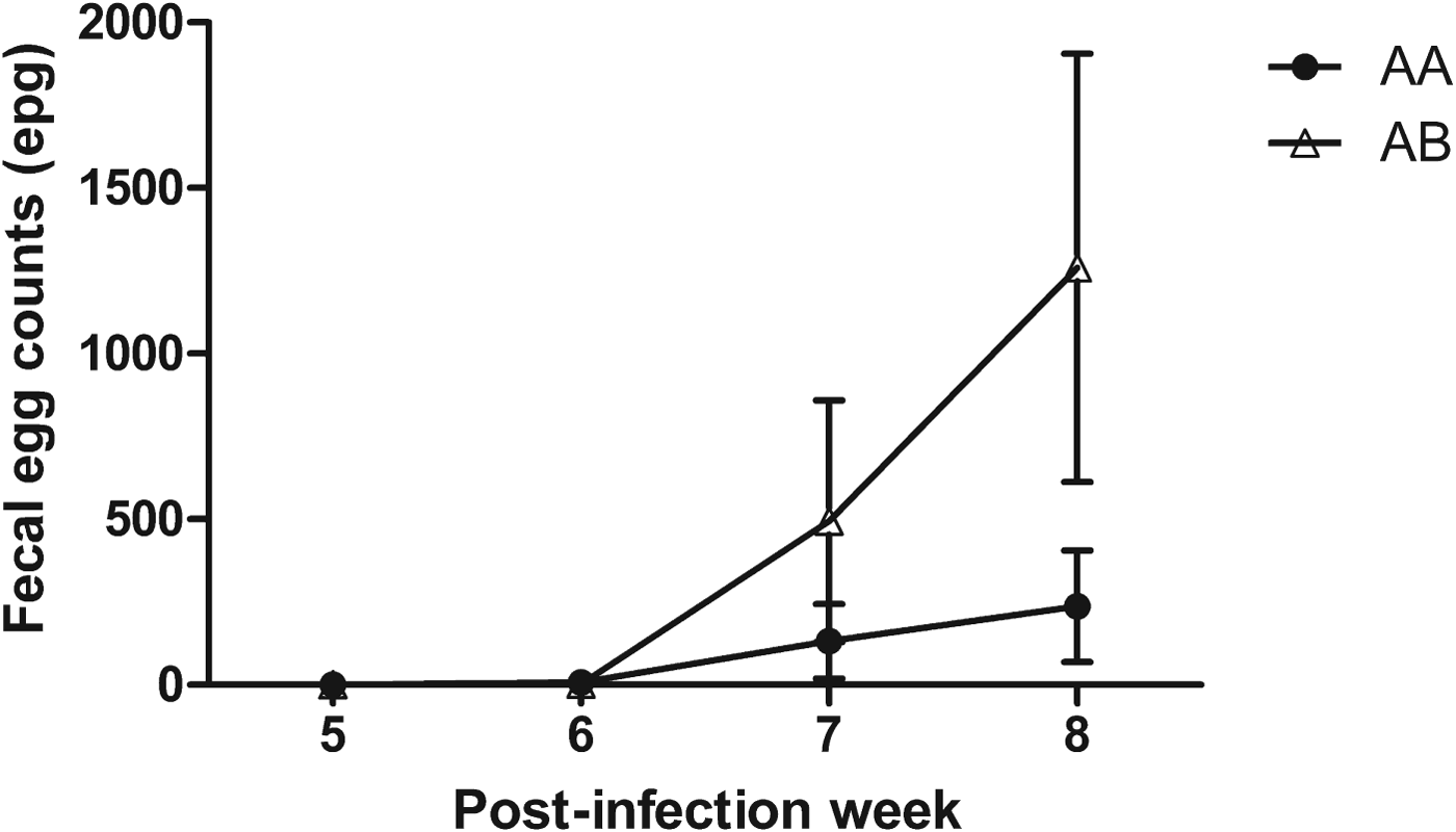
Ascaris suum eggs were detected from week 6 PI and onwards (Fig. 4). At week 8 PI, AB pigs (SNP TXNIP) excreted ∼5 times more eggs than AA pigs but the difference was not significant (Table 1). A similar picture was seen for SNP ARNT.
Mean Ascaris suum fecal egg count (epg) during infection in pigs homozygous (circles; N AA = 24) or heterozygous (triangles; N AB = 24) at the single nucleotide polymorphism 0_TXNIP_DS087128.1_2_2 (TXNIP) locus. Error bars represent s.e. of the mean.

All pigs had liver white spots at necropsy (Table 2). There was hardly any difference in total number of liver white spots between the AA and AB genotypes for both SNPs, although pigs of the BB genotype (SNP ARNT) tended to have higher numbers (Table 2). When considering only the pearl-like white spots (lymphonodular spots), the heterozygotes (SNP TXNIP) had a slightly higher mean number than homozygotes (10·6 vs 7·6; Table 2; P = 0·33). A similar effect was seen for the AA and AB genotypes at SNP ARNT while carriers of two copies of the susceptibility allele had twice as many lymphonodular spots as the heterozygotes (P<0·05).
OD values for serum IgG antibody response to the different antigens are presented in Table 2. For all three antigens tested (adult-As Ag, iL3-ES Ag and LL3-ES Ag), the AB group (TXNIP, ARNT) had higher antibody titres than the AA group. The effect of SNP TXNIP was strongest for the adult A. suum-specific antibody response but did not reach significance (P = 0·11). By merging the two genotypes, we found a significant positive correlation between total worm burden and adult A. suum-specific IgG antibody titre (P<0·05), and between total worm burden and LL3-ES specific IgG antibody titre (P<0·05). Macroscopic worm burden was not correlated with serum antibody response to any of the three antigens tested (data not shown).
DISCUSSION
Previous work by our group has demonstrated that A. suum burden is under genetic regulation (Nejsum et al. Reference Nejsum, Roepstorff, Jorgensen, Fredholm, Goring, Anderson and Thamsborg2009a ); in addition, we have detected a QTL encompassing one or more of these genes on the porcine chromosome 4 (Skallerup et al. Reference Skallerup, Nejsum, Jorgensen, Goring, Karlskov-Mortensen, Archibald, Fredholm and Thamsborg2012). The present study was designed to provide proof-of-principle that pigs resistant to A. suum infection can be identified based on their specific SNP genotype at a specific locus (SNP TXNIP) on chromosome 4. We selected piglets of two SNP TXNIP genotypes (AA, AB) from 10 different litters which were then A. suum trickle-infected for 8 weeks after which we compared the two groups. We used different indicators of resistance (worm burden, FEC, number of liver white spots and A. suum-specific serum IgG antibody levels) of which the first two traits were considered core traits and the last two traits were associated traits. Although the differences between the two genotypes were not significant, we found a consistent trend particularly across the core phenotypic traits examined; hence, the ‘resistant’ AA (TXNIP) genotype had fewer A. suum (both measured as macroscopic worm burden and total worm burden) and excreted fewer eggs throughout the study than the ‘susceptible’ AB genotype (Table 1). Hence, our data suggest that resistant pigs can be selected using the TXNIP marker and that the B allele is associated with susceptibility to A. suum infection.
The burden of macroscopic A. suum is a useful and convenient measure of resistance. In pigs that are continually exposed to A. suum, most larvae are prevented from reaching the liver (Urban et al. Reference Urban, Alizadeh and Romanowski1988; Stankiewicz and Jeska, Reference Stankiewicz and Jeska1990; Jungersen et al. Reference Jungersen, Eriksen, Roepstorff, Lind, Meeusen, Rasmussen and Nansen1999) and large, patent worms seem to originate from the early phase of exposure (Mejer and Roepstorff, Reference Mejer and Roepstorff2006; Nejsum et al. Reference Nejsum, Thamsborg, Petersen, Kringel, Fredholm and Roepstorff2009b ). In this early phase, the pre-hepatic barrier is not very effective and although some larvae are trapped in the liver parenchyma (Ronéus, Reference Ronéus1966; Pérez et al. Reference Pérez, García, Mozos, Bautista and Carrasco2001) and possibly the lung, nearly 50% may be recovered in the small intestine shortly after infection (Roepstorff et al. Reference Roepstorff, Eriksen, Slotved and Nansen1997; Helwigh and Nansen, Reference Helwigh and Nansen1999). The majority will normally be expelled between day 14 and 30 PI, depending on the infection regime, and this immune reaction (Miquel et al. Reference Miquel, Roepstorff, Bailey and Eriksen2005) leaves a small fraction of larvae which grow to maturity (Schwartz, Reference Schwartz1959; Jorgensen et al. Reference Jorgensen, Nansen, Nielsen, Eriksen and Andersen1975; Roepstorff et al. Reference Roepstorff, Eriksen, Slotved and Nansen1997; Nejsum et al. Reference Nejsum, Thamsborg, Petersen, Kringel, Fredholm and Roepstorff2009b ).
Unfortunately, the surprisingly low prevalence of macroscopic worms in our study made it difficult to determine to what extent the SNP TXNIP marker was associated with this trait; only 38% of AB (TXNIP) pigs were positive for this phenotype vs 17% of AA pigs which, however, is twice as high in ‘susceptible’ pigs compared with ‘resistant’ pigs. The prevalences of macroscopic A. suum in the present study were lower than in previous experimental trickle infections using the same infection protocol (Nejsum et al. Reference Nejsum, Roepstorff, Jorgensen, Fredholm, Goring, Anderson and Thamsborg2009a , Reference Nejsum, Thamsborg, Petersen, Kringel, Fredholm and Roepstorff b ). In two of the pens we found a lower prevalence (24 and 13%) than in the third (44%) even though the conditions were kept similar, i.e. same A. suum egg batch, feeding regime, stocking density and housing. Autoinfection is unlikely to have taken place because embryonation of A. suum eggs requires 10–14 days at 30 °C and more than 45 days at 17 °C (reviewed by O‘Lorcain and Holland, Reference O'Lorcain and Holland2000). In our study, egg excretion started week 6 PI and the study was terminated 2 weeks later. Thus, there was no possibility for uncontrolled infection. The two pigs examined on day 13 PI (one from a ‘high-prevalence’ pen and one from a ‘low-prevalence’ pen) showed recovery percentages similar to previous studies (Roepstorff et al. Reference Roepstorff, Eriksen, Slotved and Nansen1997). An analysis of a simulated dataset (assuming that pigs in all three pens had had same prevalence and intensities of macroscopic A. suum as the third pen) showed a significant association (P = 0·012) between macroscopic A. suum burden and SNP TXNIP.
As a second direct measure of the host's ability to suppress establishment and/or subsequent development of infection (Albers et al. Reference Albers, Gray, Piper, Barker, Le Jambre and Barger1987) we used total worm burden. While larvae recovered from the small intestine in the present study had completed migration, they were still vulnerable to expulsion, i.e. some of these larvae would probably have been expelled from the small intestine if the pig had been necropsied at a later time-point (Mejer and Roepstorff, Reference Mejer and Roepstorff2006; Nejsum et al. Reference Nejsum, Thamsborg, Petersen, Kringel, Fredholm and Roepstorff2009b ). We therefore expected the total worm burden trait to contain more noise (i.e. a smaller difference between ‘susceptible’ and ‘resistant’ pigs) than the macroscopic worm burden trait. Nevertheless, we found a clear trend with the AB group (TXNIP) having more than twice as many worms as the AA group (Table 1), most of which were larvae. These data suggest that part of the difference in worm burden among the two genotypes could be explained by differences in pre-hepatic barrier and/or losses during the hepato-tracheal migration. Comparing total worm burdens for the same SNP TXNIP genotypes, Skallerup et al. (Reference Skallerup, Nejsum, Jorgensen, Goring, Karlskov-Mortensen, Archibald, Fredholm and Thamsborg2012) reported effects of a similar magnitude in their discovery and validation studies.
In addition to the two worm burden traits discussed above, we looked at FEC as another core trait. Pigs of the AA genotype (TXNIP) excreted fewer A. suum eggs throughout the experiment but the difference was not significant. Again, the lack of significance could be explained by the limited number of pigs with large, patent worms of which one pig did not even excrete eggs, possibly due to single-sex infection (Jungersen et al. Reference Jungersen, Eriksen, Nansen and Fagerholm1997). Although several studies have shown a positive correlation between FEC and A. suum worm burden (Roepstorff and Murrell, Reference Roepstorff and Murrell1997; Roepstorff et al. Reference Roepstorff, Eriksen, Slotved and Nansen1997; Boes et al. Reference Boes, Medley, Eriksen, Roepstorff and Nansen1998), density-dependent reductions of fecundity may obscure the association between genetic markers and FEC (Croll et al. Reference Croll, Anderson, Gyorkos and Ghadirian1982; Anderson and Schad, Reference Anderson and Schad1985). In addition, egg counts have a large day-to-day variability e.g. due to variations in feed intake and consistency of feces and because of a non-homogeneous distribution of eggs in the feces (Brown, Reference Brown1927; Scott and Headlee, Reference Scott and Headlee1938; Croll et al. Reference Croll, Anderson, Gyorkos and Ghadirian1982; Anderson and Schad, Reference Anderson and Schad1985). Studies of trichostrongyles in sheep have demonstrated that genetic resistance mainly is due to control of worm length and fecundity rather than worm burden (Stear et al. Reference Stear, Bairden, Duncan, Holmes, McKellar, Park, Strain, Murray, Bishop and Gettinby1997, Reference Stear, Strain and Bishop1999). In pigs, this does not seem to be the case since worm fecundity and worm size were not heritable in contrast to high heritability estimates for A. suum worm burden (Nejsum et al. Reference Nejsum, Roepstorff, Jorgensen, Fredholm, Goring, Anderson and Thamsborg2009a ; Skallerup et al. Reference Skallerup, Nejsum, Jorgensen, Goring, Karlskov-Mortensen, Archibald, Fredholm and Thamsborg2012). This might indicate that immunity against A. suum is directed towards the migrating and newly established larvae while different mechanisms could have evolved in non-migrating ruminant trichostrongyles.
Liver white spots develop as an immune-pathological response to migrating ascarid larvae (Ronéus, Reference Ronéus1966; Urban et al. Reference Urban, Alizadeh and Romanowski1988; Pérez et al. Reference Pérez, García, Mozos, Bautista and Carrasco2001). The number of liver white spots is related to number of larvae that have recently (within 3–6 weeks) migrated through the liver (Eriksen et al. Reference Eriksen, Andersen, Nielsen, Pedersen and Nielsen1980, Reference Eriksen, Lind, Nansen, Roepstorff and Urban1992; Nejsum et al. Reference Nejsum, Thamsborg, Petersen, Kringel, Fredholm and Roepstorff2009b ) and in this study we used this parameter as an associated indicator trait for resistance. We expected that resistant pigs would have fewer liver white spots than susceptible pigs because they would more effectively establish a pre-hepatic barrier (Nejsum et al. Reference Nejsum, Thamsborg, Petersen, Kringel, Fredholm and Roepstorff2009b ). We did not find any differences in the number of liver white spots between the two variants of the TXNIP polymorphism and our results indicate that even in ‘resistant’ AA pigs, the pre-hepatic barrier was far from being complete at 8 weeks PI (Tables 1 and 2).
As a second associated trait, we looked at the A. suum-specific antibody production. For all antigens tested, the IgG antibody response was slightly higher in the AB group than in the AA group (SNP TXNIP), though not statistically significant. Previous work in experimentally and naturally infected pigs has shown a positive correlation between worm burden/FEC and IgG antibody titres (Roepstorff et al. Reference Roepstorff, Eriksen, Slotved and Nansen1997; Roepstorff, Reference Roepstorff1998; Vlaminck et al. Reference Vlaminck, Nejsum, Vangroenweghe, Thamsborg, Vercruysse and Geldhof2012). Since AB pigs (SNP TXNIP) had higher worm burdens than AA pigs, our results are in agreement with these findings. Indeed, we found a significant positive correlation between total worm burden and adult A. suum-specific IgG antibody titres and between total worm burden and LL3-ES specific IgG antibody titres. Other studies in pigs, cattle and sheep have shown conflicting results (e.g. Gasbarre et al. Reference Gasbarre, Leighton and Davies1993; Roepstorff and Murrell, Reference Roepstorff and Murrell1997; Nejsum et al. Reference Nejsum, Thamsborg, Petersen, Kringel, Fredholm and Roepstorff2009b ; Zaros et al. Reference Zaros, Bricarello, Amarante, Rocha, Kooyman, De Vries and Coutinho2010; Hassan et al. Reference Hassan, Good, Hanrahan, Campion, Sayers, Mulcahy and Sweeney2011). The relationship between worm burden/FEC and antibody response is apparently complex and there are probably different genes regulating the two traits (Gasbarre et al. Reference Gasbarre, Leighton and Davies1993).
In addition to the TXNIP locus, the pigs were also genotyped at another locus, SNP ARNT. In contrast to Skallerup et al. (Reference Skallerup, Nejsum, Jorgensen, Goring, Karlskov-Mortensen, Archibald, Fredholm and Thamsborg2012) who only found two of the genotypes of SNP ARNT in their validation studies, we found all three genotypes of this SNP present in this study. In agreement with the high linkage disequilibrium between the two SNPs, we observed a similar phenotypic trend to SNP TXNIP for the AA and AB genotypes; hence, the AA genotype (SNP ARNT) had fewer A. suum (both measured as macroscopic worms and total worm burden), excreted fewer eggs at week 8 PI, and had lower serum IgG antibody levels at week 8 PI than the AB genotype (Tables 1 and 2). Comparing pigs with genotype BB (SNP ARNT) with AA and AB pigs, there was no consistent trend and the low number of pigs (N = 3) carrying two copies of this susceptibility allele made it difficult to test the hypothesis that these pigs would have a ‘hyper-susceptible’ phenotype. Nevertheless, for two traits, i.e. mean total A. suum burden and mean number of lymphonodular spots, there was a clear effect of the BB genotype (Tables 1 and 2).
A number of linkage studies using microsatellites have dissected the genetics underlying parasite resistance (e.g. Iraqi et al. Reference Iraqi, Behnke, Menge, Lowe, Teale, Gibson, Baker and Wakelin2003; Davies et al. Reference Davies, Stear, Benothman, Abuagob, Kerr, Mitchell and Bishop2006; Coppieters et al. Reference Coppieters, Mes, Druet, Farnir, Tamma, Schrooten, Cornelissen, Georges and Ploeger2009) but SNP chip studies are becoming more common (Riggio et al. Reference Riggio, Matika, Pong-Wong, Stear and Bishop2013). To date, the large majority of genome-wide association studies have identified SNP variants that explain only a small proportion of the genetic variance for complex traits (Dermitzakis and Clark, Reference Dermitzakis and Clark2009; Manolio et al. Reference Manolio, Collins, Cox, Goldstein, Hindorff, Hunter, McCarthy, Ramos, Cardon, Chakravarti, Cho, Guttmacher, Kong, Kruglyak, Mardis, Rotimi, Slatkin, Valle, Whittemore, Boehnke, Clark, Eichler, Gibson, Haines, Mackay, McCarroll and Visscher2009), including parasite resistance in domestic animals (Kemper et al. Reference Kemper, Emery, Bishop, Oddy, Hayes, Dominik, Henshall and Goddard2011; Sallé et al. Reference Sallé, Jacquiet, Gruner, Cortet, Sauvé, Prévot, Grisez, Bergeaud, Schibler, Tircazes, Francois, Pery, Bouvier, Thouly, Brunel, Legarra, Elsen, Bouix, Rupp and Moreno2012). Nevertheless, our SNP marker (TXNIP) seems to have a rather strong effect, with AB pigs having 2·5-fold higher mean total worm burden than AA pigs both in the initial genome scan (Skallerup et al. Reference Skallerup, Nejsum, Jorgensen, Goring, Karlskov-Mortensen, Archibald, Fredholm and Thamsborg2012) and in the present functional study. A significant single-marker effect on worm burden has also been found in sheep. Hassan et al. (Reference Hassan, Good, Hanrahan, Campion, Sayers, Mulcahy and Sweeney2011) produced twin lambs with different genotypes at the MHC-DRB1 locus which were then experimentally infected with Teladorsagia circumcincta and subsequently necropsied at various time-points post infection. Despite small group sizes (four lambs per genotype per sampling day), the authors found that carriers of the DRB1*1101 allele had significantly lower worm counts than non-carrier lambs. Non-carriers had approximately 0·8–4-fold higher mean worm burdens than carriers of the allele increasing resistance. While this significance could be explained by a stronger marker effect of MHC-DRB1 than our TXNIP marker, it is also possible that the species difference in worm counts plays a role; hence, T. circumcincta occur in much higher numbers and with higher prevalences than A. suum.
Genetic markers can identify animals with desired traits for breeding programmes (Charlier et al. Reference Charlier, Coppieters, Rollin, Desmecht, Agerholm, Cambisano, Carta, Dardano, Dive, Fasquelle, Frennet, Hanset, Hubin, Jorgensen, Karim, Kent, Harvey, Pearce, Simon, Tama, Nie, Vandeputte, Lien, Longeri, Fredholm, Harvey and Georges2008; Nielsen et al. Reference Nielsen, Jorgensen, Vernersen and Fredholm2009; reviewed by Bishop, Reference Bishop2012). Care should be taken, though, to weigh the advantage against the loss of other beneficial alleles in linkage disequilibrium with the marker (Wolc et al. Reference Wolc, Arango, Settar, Fulton, O'Sullivan, Preisinger, Habier, Fernando, Garrick, Hill and Dekkers2012), and ideally marker information from the whole genome (i.e. genomic selection) should be included (reviewed by Goddard and Hayes, Reference Goddard and Hayes2007; Kemper et al. Reference Kemper, Emery, Bishop, Oddy, Hayes, Dominik, Henshall and Goddard2011). Our data provide suggestive evidence that A. suum resistant pigs can be identified using a genetic marker, TXNIP. We observed a consistent effect on two core traits (worm burden and FEC) and one associated trait (serum IgG antibody levels). Although we did not find a significant effect of genotype on worm burdens, the effects are in the exact same direction as expected and we believe it would be overly conservative to interpret data as if there were no effect of the marker. In the light of these considerations, we encourage further studies of the TXNIP marker. We believe the TXNIP marker which is located in intron 2 of the TXNIP gene is not the causative mutation (‘disease locus’) but most probably in linkage disequilibrium with one or more genes within the QTL on chromosome 4 that confer resistance to A. suum (Skallerup et al. Reference Skallerup, Nejsum, Jorgensen, Goring, Karlskov-Mortensen, Archibald, Fredholm and Thamsborg2012). The present study suggests that SNP ARNT is another diagnostic marker candidate for A. suum resistance and hence provides further support to the QTL detected previously (Skallerup et al. Reference Skallerup, Nejsum, Jorgensen, Goring, Karlskov-Mortensen, Archibald, Fredholm and Thamsborg2012) in agreement with the high linkage disequilibrium between these two SNPs. The total worm counts and liver white spot data presented in this paper indicate that protective immunity in the resistant group (TXNIP, ARNT) is expressed not only at the pre-hepatic barrier level (Urban et al. Reference Urban, Alizadeh and Romanowski1988; Masure et al. Reference Masure, Vlaminck, Wang, Chiers, Van den Broeck, Vercruysse and Geldhof2013) but at other sites as well. Further studies are required to elucidate the different immunological responses mounted by the two TXNIP genotypes and to investigate the effect of the four candidate genes within the QTL (TNFAIP8L2, PIAS3, RBM8A and RFX5) suggested by Skallerup et al. (Reference Skallerup, Nejsum, Jorgensen, Goring, Karlskov-Mortensen, Archibald, Fredholm and Thamsborg2012).
ACKNOWLEDGEMENTS
The authors wish to thank all those who contributed to the research project which forms the basis of this paper: Lise-Lotte Christiansen, Christel Ammitzböll Halberg, Minna Jakobsen and Anne Strandsby provided excellent technical assistance. Hans Skaaning and his team took good care of the pigs and assisted with the data collection.
FINANCIAL SUPPORT
This work was supported by the Danish Agency for Science, Technology and Innovation (grant awarded to P.N.). P.S. is supported by a University of Copenhagen (Denmark) PhD grant.