So-called “heterotaxy” is usually considered to represent the situation in which the thoracic and abdominal organs are not arranged in their expected lateralised pattern.Reference Jacobs, Anderson and Weinberg 1 As such, the entity is frequently ascertained during evaluation for symptoms due to complex cardiovascular malformations. Although the existence of isomeric arrangements within the heart is still doubted by some, it has long been recognised that the presence of isomeric, rather than lateralised, arrangements of the bronchis is an essential feature in patients with heterotaxy.Reference Van Praagh and Van Praagh 2 – Reference Partridge, Scott, Deverall and Macartney 6 In this context, isomerism refers to morphological mirror imagery in the same patient. It is much more accurate to use “isomerism” to describe this entity, rather than “heterotaxy”, as the latter word, when used literally, means any patient with characteristics other than those expected. Attention to mirror imagery of the two sides of the same patient, furthermore, permits recognition, within the overall syndrome, of the subsets of right bronchial as opposed to left bronchial isomerism. From the cardiac stance, patients known to have right bronchial isomerism often present with unbalanced atrioventricular septal defects and double-outlet right ventricle, typically in association with totally anomalous pulmonary venous connection and pulmonary atresia, thus necessitating single ventricle palliation. This subset of patients is also known to be associated, in most cases, with absence of the spleen. Patients with left bronchial isomerism, in contrast, are recognised as having less complex congenital cardiac malformations, but frequently present with interruption of the inferior caval vein. These patients usually have multiple spleens. When first recognised, it was conventional to describe heterotaxy in terms of the splenic syndromes.Reference Ivemark 7
Clinical recognition of heterotaxy is important because of its association with the aforementioned abnormalities. Equally important, however, is to differentiate its two subsets, as the risk of post-operative morbidity and mortality can vary, particularly for those with functionally univentricular hearts. It is also important to distinguish the subsets for purposes of genetic counselling and to establish the abnormal genetic cues that underscore the syndromes. In this regard, manipulation of developing mice has shown that it is possible to produce unequivocal right isomerism, including obvious isomerism of the right atrial appendages, by knocking out the Pitx1 or Cited-1 genes.Reference Bamforth, Braganca and Farthing 8 Knocking out the Lefty-1 or sonic hedgehog genes has subsequently been shown to produce left isomerism, including obvious isomerism of the left atrial appendages.Reference Meno, Ito and Saijoh 9 – Reference Hildreth, Webb and Chaudhry 11 The problem remains as how best to distinguish between the subsets in the clinical setting. From the stance of cardiac diagnosis, autopsy studies have shown that the morphology of the atrial appendages, as based on the extent of the pectinate muscles, provides the best discrimination. This feature shows good, but not absolute, correlation with bronchial morphology.Reference Uemura, Ho, Devine, Kilpatrick and Anderson 12 Distinction of the morphologically right from the left atrial appendages, nonetheless, remains difficult as judged by echocardiography, which is the standard modality currently used to interrogate cardiac anatomy during life. Advances in CT now allow for detailed imaging of the atrial appendages, but not all children with heterotaxy undergo tomographic scanning.Reference Wolla, Hlavacek, Schoepf, Bucher and Chowdhury 13 As already emphasised, however, bronchial morphology is known to correlate well with the anatomy of the atrial appendages. With this in mind, therefore, we have conducted a review of the findings from imaging of patients diagnosed clinically as having heterotaxy, assessing the utility of chest radiographs, cardiac angiography, CT, and MRI to demonstrate bronchial morphology. We have subsequently correlated our findings concerning right as opposed to left bronchial isomerism with the anticipated features of isomerism of the right as opposed to the left atrial appendages, the latter morphology being inferred from the findings in the remaining systems of organs, including the cardiac malformations. A previous study has already assessed these correlations using necroscopy specimens.Reference Calder 14 We have focussed on the evaluation of bronchial morphology in living patients.
Methods
Data collection
We reviewed medical records of all patients cared for since 1998 who were diagnosed with so-called “heterotaxy”. This was the year that electronic medical records were implemented at our institution. The data set included both patients born in or after 1998, as well as those born earlier who transitioned their care to our heart centre after 1998. Patients were identified through multiple strategies of searching so as to ensure the highest yield of patients. Medical records, medical billing data, and the cardiothoracic surgical database were queried for “heterotaxy”, “asplenia”, “multiple spleens”, and “polysplenia”. The echocardiography database was queried for “heterotaxy”, “interrupted inferior vena cava”, “double-outlet right ventricle”, “bilateral superior vena cava”, and “atrioventricular canal defect”. The cardiac catheterisation database was queried for “heterotaxy”.
The resulting lists of patients were then combined, and redundant entries were removed. The remaining patients were reviewed and included if they had both CHD – including interruption of the inferior caval vein – and recognised features of so-called “heterotaxy”.Reference Jacobs, Anderson and Weinberg 1 CHD was defined as any intra-cardiac lesion or an abnormality of venous returns to the heart. We considered “heterotaxy” to be present when there was a cardiovascular malformation in addition to evidence of abnormal arrangement of the abdominal organs, including the spleen, or pulmonary isomerism. Cardiovascular malformation included both intra-cardiac lesions as well as abnormalities of the systemic venous return. The diagnosis of heterotaxy was based on our subsequent review of the clinical data, and not simply by previous diagnosis, or lack thereof.
The details of cardiac anatomy were obtained primarily from the echocardiographic findings, although data from CT, MRI, and cardiac catheterisation studies were also available for some patients. The splenic status was based on findings from abdominal ultrasonography. We excluded patients if they did not have evidence of cardiovascular malformation. At our institution, genetic evaluation of heterotaxy patients is carried out on a case-by-case basis. Heterotaxy patients known to have chromosomal anomalies were not excluded.
The proposed methodology was approved by the Institutional Review Board at our institution.
Patient characteristics
To identify patients who differed from each other with regard to the arrangement of their organs, we compared the findings between patients as being likely to have isomerism of the right or left atrial appendages as opposed to usual or mirror-imaged atrial arrangement, using χ2 analysis for categorical data and independent t-tests or Mann–Whitney U-tests where appropriate. Patient characteristics were also compared between those identified as having isomerism of the right or left bronchi.
For our baseline analysis, we categorised the cardiac diagnoses into primary and secondary lists. The primary diagnosis was the lesion considered to be the most haemodynamically significant, whereas the secondary diagnosis included other associated lesions – for instance, if a patient had a complete atrioventricular septal defect, double-outlet right ventricle, and an interrupted inferior caval vein, then the atrioventricular septal defect was coded as the primary diagnosis, with double-outlet right ventricle and interrupted inferior caval vein listed as secondary diagnoses. If a patient simply had interruption of the inferior caval vein, then this was coded as the primary diagnosis. It was implicit that those with bilateral superior caval veins of necessity had a left-sided superior caval vein.
Inferences regarding the presence of isomeric right or left atrial appendages
The intra-cardiac features considered to indicate the presence of isomeric right as opposed to left atrial appendages were assessed independently by two authors (R.L. and R.H.A.), with cognizance taken of the findings in the other systems of organs as well. These included, but were not limited to, overall cardiac anatomy, venoatrial connections, and splenic anatomy. The results of the independent assessments were then compared, and any differences were discussed, with a consensus being reached by both authors regarding the likely presence of isomeric right as opposed to left atrial appendages. In some instances, the assessors agreed that there was probable usual arrangement of the atrial appendages, despite the presence of heterotaxy and bronchial isomerism.
An aggregate of several features was used to help determine whether isomerism of the right or left atrial appendages was present. The course of the systemic veins was utilised in this regard with interruption of the inferior caval vein being associated more so with isomerism of the left atrial appendage. The presence or absence of a coronary sinus was also utilised, with the presence of a coronary sinus being associated with isomerism of the left atrial appendage while its absence being associated with isomerism of the right atrial appendage. The presence of multiple spleens or the absence of a spleen was also used to help segregate isomerism of the atrial appendages with multiple spleens. Pulmonary arterial branching pattern by catheterisation was also used when available.
Assessment of bronchial angle
Bronchial angle was assessed using chest radiographs, cardiac angiography, and images obtained using CT or MRI. Bronchial angles (Fig 1) were measured using chest radiographs obtained in the posterior–anterior projection, cardiac angiography obtained in the posterior–anterior projection, and tomographic images obtained in a coronal plane. When both CT and MRI images were available, then the tomographic images were used for measurements. Averages of the bronchial angles, as measured by the various imaging modalities, were also calculated to assign bronchial morphology using this aggregate measure. Bronchial angles <135° were considered to indicate the presence of left isomerism, with bronchial angles >135° considered as indicative of right isomerism. Angles of the left- and right-sided bronchi were then compared within the groups of patients inferred to have overall right or left isomerism, using a paired t-test to determine variation within the specific groups. Angles of the left- and right-sided bronchi were then compared between those deemed to have the subsets of isomerism using an independent t-test. This permitted calculation of the sensitivity and specificity of bronchial morphology for specifying the presumed presence of isomeric right or left atrial appendages. The agreement in measurement of the bronchial angles by different imaging modalities was then analysed by the Bland–Altman analysis.
The Bland–Altman plot demonstrating the correlation between bronchial angle measurements made by chest radiograph and angiographic images. Labels on the plot simply convey what imaging modalities are represented. The x-axis represents the average of both tests, whereas the y-axis represents the difference between readings from the two tests.

Assessment of bronchial length
Right and left bronchial lengths were assessed using chest radiographs and images obtained using CT or MRI. The lengths of the right-sided and left-sided bronchi were measured from the site of bifurcation of the trachea to the point of the tangent produced by their first branches (Fig 1). A ratio of the right to left bronchial lengths between 0.67 and 1.5 was considered consistent with bronchial isomerism. The diagnoses of bronchial isomerism were then compared with the presumed diagnoses of isomeric right or left atrial appendages as assessed for the overall group of patients. The ratios of bronchial lengths between those presumed to have right as opposed to left isomeric appendages were further assessed using an independent t-test. The agreement in the ratios between the modalities used for imaging was assessed using the Bland–Altman analysis. The ratio of bronchial lengths was then normalised to tracheal width as described previously by Partridge et al,Reference Partridge, Scott, Deverall and Macartney 6 this also being compared between those with presumed right or left isomeric appendages.
Pulmonary arterial branching
The pattern of branching of the pulmonary arteries was assessed from images obtained during cardiac catheterisation, assessing the number of branches arising from the right-sided or left-sided pulmonary artery. Presence of three such branches was considered to be consistent with morphologically right pulmonary arterial morphology, whereas instances with two such branches were taken to indicate morphologically left pulmonary arteries.
Results
Patient characteristics
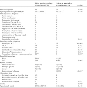
A total of 83 patients fulfilled the criteria for inclusion. Of these, we were able to include only 73 patients for the purpose of determining bronchial morphology; 10 patients were excluded due to large amount of missing clinical data or inadequate imaging data. In Table 1, we outline the baseline characteristics of the cohort, comparing the characteristics of the 53 patients considered likely to have isomerism of the right atrial appendages with those of the 20 patients considered to have features more indicative of the presence of isomerism of the left atrial appendages. There was no difference in the frequency of prenatal diagnosis between those with presumed isomerism of the right and left atrial appendages. In terms of the initial primary cardiac diagnosis, those subsequently presumed to have isomerism of the right atrial appendages were more likely to have a double-inlet left ventricle and double-outlet right ventricle (p<0.042). Those considered subsequently as having isomerism of the left atrial appendages were more likely to have coarctation of the aorta, hypoplastic left heart syndrome, the “ostium primum” variant of atrioventricular septal defect, tetralogy of Fallot, and ventricular septal defect (p<0.042).
Patient characteristics between those with right and left isomerism.

AV=atrioventricular; ICV=inferior caval vein; LSCV=left superior caval vein; VA=ventriculoarterial.
Secondary cardiac defects more commonly associated with presumed isomeric right atrial appendages included discordant ventriculoarterial connections (p=0.004) and totally anomalous pulmonary venous connections (p=0.009). Secondary cardiac defects more commonly associated with presumed isomerism of the left atrial appendages included interruption of the inferior caval vein (p<0.001). In those with presumed isomeric left atrial appendages, 25% had bilateral connections of the pulmonary veins. Of those deemed likely to have overall right isomerism based on the analysis of all the clinical findings, 81% had absence of a spleen, 6% had multiple spleens, and 13% had a solitary spleen, which were right-sided in all. Of those deemed to have overall left isomerism, 15% had absence of a spleen, 75% had multiple spleens, 5% had a solitary right-sided spleen, and 5% had indeterminate or unknown splenic anatomy (p<0.001). Intestinal malrotation was more frequently present in those considered to demonstrate right isomerism (p<0.001).
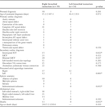
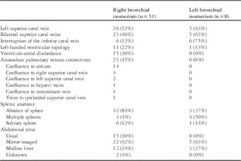
In Table 2, the baseline characteristics of the selected cohort are compared in terms of the 59 patients deemed to have right bronchial isomerism based on the measurements taken as opposed to the 24 patients considered to have left bronchial isomerism. No differences were found between these groups with respect to frequency of prenatal diagnosis, age of postnatal diagnosis, primary cardiac diagnosis, and arrangement of the abdominal organs. Secondary cardiac diagnoses did not differ significantly between the two groups, except for interruption of the inferior caval vein, which occurred with greater frequency in those with left bronchial isomerism (p=0.022). A significant difference was also noted with respect to splenic anatomy. A majority of those with right bronchial isomerism had absence of the spleen, whereas a majority of those with left bronchial isomerism had multiple spleens (p=0.043). Death was more frequently noted in those with right bronchial isomerism (p=0.043).
Patient characteristics between those with right and left bronchial isomerism.

AV=atrioventricular; ICV=inferior caval vein; LSCV=left superior caval vein; VA=ventriculoarterial.
It is known from autopsy studies that, in patients with isometric right atrial appendages, all will have totally anomalous pulmonary venous connections, even if the pulmonary veins all return to the same atrial chamber.Reference Hildreth, Webb and Chaudhry 11 This diagnosis, however, was initially made in less than half of our cohort subsequently considered likely to have isomeric right appendages. Similarly, it is known that the pulmonary veins frequently drain in a symmetrical and bilateral manner in those with isomeric left appendages. This diagnosis had not been made initially in any of our patients, although the feature was recognised in one-quarter of those with anticipated left isomerism on retrospective analysis of the echocardiographic findings.
Analysis of our findings, therefore, reveals discordances in a minority of patients between bronchial isomerism as determined from measurements of the bronchial tree and the presumed arrangement of the atrial appendages, along with multiple instances of discordant splenic morphology. In those with such discordances, most showed differences between the bronchial arrangement and the presumed isomeric arrangement of the atrial appendages. In four patients, our analysis suggested the presence of usual arrangement of the atrial appendages (atrial situs solitus), despite the presence of bronchial isomerism.
Bronchial angle
All 73 patients in whom we were able to analyse the bronchial morphology were confirmed to have bronchial isomerism. Based on the bronchial angles, we categorised 85% of the patients as having right isomerism based on their chest radiograph, 83% by angiography, 81% by CT and MRI, and 85% by the aggregate of all imaging modalities, the findings in the remaining patients being taken as indicating the presence of left bronchial isomerism for each imaging modality. We took the aggregate assessment as the definitive value when finally assigning individual patients to the groups with presumed right as opposed to left bronchial morphology.
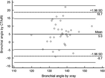
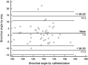
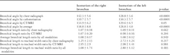
In those then deemed to have right bronchial isomerism, the average bronchial angle was 142° as assessed by chest radiograph, 140° by angiography, and 135° by the tomographic techniques, producing 139° as the aggregate value. In those with left bronchial isomerism, the average bronchial angle was 129° by chest radiograph, 130° by angiography, and 130° by the tomographic techniques, with 129° as the aggregate. These differences were all statistically significant (Table 3). When angles of the right-sided and left-sided bronchi were compared within individual patients, we found statistically significant, but clinically insignificant, differences, with mean differences between the two bronchi being ∼2°. When the Bland–Altman analysis was used to compare the variability in bronchial angle measurements, we found 95% confidence intervals of −11.4 and 14 for chest radiograph and angiography (Fig 1), 95% confidence intervals of −7.6 and 17.7 for catheterisation and tomographic images (Fig 2), and 95% confidence intervals of −8.7 and 18.7 when chest radiographs were compared with the tomographic images, respectively (Fig 3).
The Bland–Altman plot demonstrating the correlation between bronchial angle measurements made by CT/MRI and chest radiography. Labels on the plot simply convey what imaging modalities are represented. The x-axis represents the average of both tests, whereas the y-axis represents the difference between readings from the two tests.

The Bland–Altman plot demonstrating the correlation between CT/MRI and angiographic images. Labels on the plot simply convey what imaging modalities are represented. The x-axis represents the average of both tests, whereas the y-axis represents the difference between readings from the two tests.

Bronchial angles, bronchial length ratio, and bronchial to tracheal ratio by various imaging modalities in patients with isomerism of the right and left bronchus.

Bronchial length
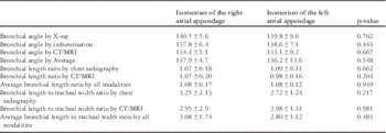
Bronchial length was measured for both the right- and left-sided bronchi. A ratio of bronchial lengths was then calculated as previously described by Partridge et al.Reference Partridge, Scott, Deverall and Macartney 6 The ratios for those with right and left bronchial isomerism was 1.08 and 1.07 as determined using chest radiography, 1.02 and 1.13 using angiography, and 1.07 and 1.11 when taking the aggregate of all modalities, respectively. None of the differences in ratios were statistically significant. In addition, all these values were <1.5, confirming the value of assessment of ratios as a means of identifying bronchial isomerism (Table 3). An average of the right- and left-sided bronchial lengths for each patient was then calculated and normalised to tracheal width, as also described by Partridge et al.Reference Partridge, Scott, Deverall and Macartney 6 The ratio of the bronchial length to tracheal width was 3.08 and 3.09 by chest radiograph, 2.38 and 5.78 by angiography, and 2.87 and 3.96 when using the aggregate of all imaging modalities, respectively (Table 2). We found no statistically significant differences in these ratios between those with right as opposed to left atrial appendage isomerism (Table 4).
Bronchial angles, bronchial length ratio, and bronchial to tracheal ratio by various imaging modalities in patients with overall right and left isomerism.

Discrepancies between bronchial and presumed atrial morphology
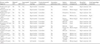
In 15 (21%) patients, we found discordances between the identified bronchial morphology and the presumed morphology of the atrial appendages, the latter being inferred on the basis of the known intra-cardiac morphology, along with the splenic anatomy. Of these patients with such discordances, in 12 (80%) patients, the measurements of the bronchial tree suggested the likely presence of isomerism of the right atrial appendages, although the findings in the remaining systems of organs, along with the intra-cardiac anatomy, were more suggestive of either left isomerism or usual arrangement of the atrial appendages. The most common primary intra-cardiac lesions in these patients were complete atrioventricular septal defect in six (40%) and double-outlet right ventricle in three (20%). Bilateral superior caval veins in seven (47%) and interruption of the inferior caval vein in 10 (67%) patients were also frequent findings. Left-handed ventricular topology was present in two (13%) patients, with two (13%) patients also having discordant ventriculo-arterial connections. The pulmonary venous connections were also known to be totally anomalous in two (13%) patients. In this group of patients, one (7%) had absence of the spleen, nine (60%) had multiple spleens, four (27%) had a normally located and solitary spleen, with one (6%) having unknown splenic anatomy. Table 5 demonstrates an overview of the findings in the patients with discordance between bronchial morphology, showing the findings we considered indicative of either usual atrial arrangement or discordant isomerism of the atrial appendages. Characteristics for those with concordance between the measured findings for bronchial morphology and the presumed atrial appendage isomerism are demonstrated in Table 6.
Patient characteristics of those with bronchial/atrial discordance.

AV=atrioventricular; ICV=inferior caval vein; LSCV=left superior caval vein; SCV=superior caval vein.
Patient characteristics of those with bronchial/atrial concordance.

Discussion
An isomeric arrangement of the bronchial tree in the setting of so-called heterotaxy has been recognised for several decades. It was Van Mierop et alReference Van Mierop, Eisen and Schiebler 3 who first pointed out this feature, which was confirmed by Landing et al.Reference Landing, Lawrence, Payne and Wells 5 Partridge and his colleagues then showed that the isomeric arrangements could be recognised based on routine clinical imaging. Using penetrated chest radiography, they demonstrated that bronchial isomerism was indicated by a ratio of <1.5 between the lengths of the right-sided and left-sided bronchi. Ratios between 1.5 and 2.0, in their opinion, were suggestive of isomerism, whereas those >2.0 were consistent with asymmetric bronchi. They also assessed the angulation of the bronchial bifurcation, showing that angles of >135° were consistent with right bronchial isomerism, whereas those <135° indicated left bronchial isomerism. They were also able to discriminate between right and left bronchial isomerism using a ratio of bronchial length to tracheal width,Reference Partridge, Scott, Deverall and Macartney 6 although we were unable to confirm this finding. Bronchial isomerism, therefore, is now an accepted finding in the setting of the syndrome currently described in terms of heterotaxy.Reference Jacobs, Anderson and Weinberg 1 This association was also demonstrated by Calder by assessing the findings in an archive of autopsied specimens.Reference Calder 14 These syndromes, of course, were initially differentiated on the basis of splenic morphology. Absence of the spleen, however, or the presence of multiple spleens, does not always correlate well with bronchial morphology.Reference Ivemark 7 , Reference Uemura, Ho, Devine and Anderson 15 The frequency of such discrepancies in the clinical setting, to the best of our knowledge, is currently unknown.
In this study, we demonstrated isomeric arrangements in all 73 patients in whom we were able to measure the bronchial lengths. The isomeric arrangements could be determined using chest radiographs, from angiography in which the trachea and bronchi were visible, as well as in images generated using CT or MRI. Correlation between the various methods was strong. We were then able to use the angulation of the bronchial bifurcation at 135°, as suggested by Partridge et al,Reference Partridge, Scott, Deverall and Macartney 6 to discriminate between those with right as opposed to left isomerism. There was a moderate to strong correlation between the differences shown by the various imaging modalities, with the variation likely secondary to properties such as rotation, penetration, and phases of respiration. We then discovered that a significant proportion of our patients exhibited discordance between their known bronchial morphology and the pattern of isomerism anticipated on the basis of the findings in the remaining systems of organs, including the potential presence of usual atrial arrangement. Such discordance was present in over one-fifth of our cohort, with no specific sub-group being identified in which such discordance was more frequent. As already emphasised, such discordance between bronchial isomerism and intra-cardiac morphology, either based on splenic morphology or on the anatomy of the atrial appendages, is well recognised. This has been established by investigations based on both clinical and necroscopy findings. The frequency of such discordances has tended to be higher in the investigations based on clinical evaluation, as these have typically depended on splenic morphology, rather than precise determination of the anatomy of the atrial appendages. Although not always in concordance with presumed isomerism within the heart, nonetheless, bronchial morphology, which is an easily obtainable clinical datum, can provide initial discrimination of patients with so-called heterotaxy into the sub-groups of right as opposed to left isomerism. Comparison with other features, such as intra-cardiac lesions, caval venous anatomy, splenic anatomy, and the arrangement of the liver, stomach, and intestines, then permits inferences concerning the likely presence of isomerism of the right or left atrial appendages. Based on this, we determined that a small number of patients were likely to have usual arrangement of the atrial appendages, despite the presence of bronchial isomerism.
Such distinctions were exemplified by two of the patients in our cohort known to have bronchial isomerism. They had isolated interruption of the inferior caval vein, with continuation through the azygos venous system, but in the absence of intra-cardiac lesions. The interruption of the inferior caval vein, of course, was also known to be associated with left isomerism. The first patient, known to have left bronchial isomerism, presented with Group B Streptococcal bacteraemia and meningitis in the 1st year of life, with a history significant for several respiratory infections and ear infections. Evaluation including abdominal ultrasonography had demonstrated the presence of multiple spleens. The detection of Howell–Jolly bodies, however, was indicative of functional asplenia. A single right-sided superior caval vein was found, which returned normally to the right-sided atrium, with all the pulmonary veins draining in normally to the left-sided atrium. The abdominal organs were mirror-imaged, with the stomach being right-sided and the liver located on the left. There was left bronchial isomerism. Genetic testing revealed a mutation in the DNAH11 gene, generally considered causative of laterality defects.
The second patient also had isolated interruption of the inferior caval vein with azygos continuation and no intra-cardiac lesions. There was a single right-sided superior caval vein, and all pulmonary veins returned normally to the left-sided atrium. In this patient, abdominal ultrasonography demonstrated absence of any splenic tissue and there was right bronchial isomerism. Although the findings in the second patient, therefore, were consistent with overall right isomerism, the cardiac morphology was indicative of usual arrangement of the atrial appendages. The findings in these patients show that, although discordances can be found between the systems of organs, even in the setting of normal cardiac anatomy, the separate description of each system of organs makes it possible to remove any suggestion of ambiguity in the overall bodily arrangement.
All of this is of importance when considering the controversy that still exists regarding the existence of isomerism within the heart.Reference Van Praagh and Van Praagh 2 Part of this controversy devolves on the parts of the heart that are considered to be isomeric. More than 50 years ago, Van Mierop and GessnerReference Van Mierop and Gessner 16 showed that the sinus nodes were isomeric in patients with asplenia. The subsequent review of the “splenic syndromes” by Van Mierop et alReference Van Mierop, Gessner and Schiebler 17 then illustrated the isomeric morphology of the atrial appendages. It was unfortunate, therefore, that Macartney et al,Reference Macartney, Zuberbuhler and Anderson 18 when claiming to demonstrate isomerism within the heart, described their findings in terms of “atrial isomerism”. As has now been shown unequivocally by genetic manipulation of mice, it is only the atrial appendages that are truly isomeric.Reference Meno, Ito and Saijoh 9 , Reference Hildreth, Webb and Chaudhry 11 , Reference Ho, Seo, Brown, Cook, Fagg and Anderson 19 , Reference Seo, Brown, Ho and Anderson 20 Moreover, as was shown by Uemura et al,Reference Uemura, Ho, Devine, Kilpatrick and Anderson 12 the isomeric nature of the appendages can readily be distinguished by determining the extent of their pectinate muscles relative to the atrioventricular junctions. As suggested by our present review, nonetheless, not all patients with bronchial isomerism will have isomeric atrial appendages, and the cardiac malformations are not always as expected based on the known bronchial arrangement. Thus, even in those presumed based on the intra-cardiac anatomy, venoatrial connections, and splenic morphology to have isomeric appendages, there can be discordances when comparisons are made with bronchial morphology as defined by measurements of bronchial angles. It remains difficult, therefore, to specify overall isomeric arrangements within the group of patients usually currently specified as having heterotaxy. In those with potential discordances between the arrangements of the thoracic and abdominal organs and the intra-cardiac anatomy, nonetheless, any suggestion of ambiguity, as might be inferred by the alternative classification of “situs ambiguous”, is removed by describing each system of organs independently and by providing a full account of cardiac anatomy, including the venoatrial connections.Reference Van Praagh and Van Praagh 2
Our review has shown, therefore, that bronchial isomerism can now readily be diagnosed using current clinical techniques. This allows for anticipation and evaluation of other associated findings and can raise suspicion for functional abnormalities such as functional asplenia or ciliary dyskinesia.Reference Nakhleh, Francis and Giese 21 Both these features have important clinical implications, particularly in post-operative patients in a hospital setting. Although previous studies have shown morbidity and mortality to be higher in those with presumed right as opposed to left isomerism, it is unclear whether right or left bronchial isomerism, in and of themselves, are responsible for the noted increases.Reference Bartz, Driscoll and Dearani 22 , Reference Peoples, Moller and Edwards 23 Of particular interest are respiratory outcomes as well as whether or not there is an association between right or left bronchial isomerism and ciliary dyskinesia, which has been found to be increasingly common in those with so-called heterotaxy. The abnormal ciliary function has been demonstrated to be associated with increased morbidity.Reference Harden, Tian and Giese 24 Within the heart itself, it is important for the surgeon to recognise the presence of isomeric right or left atrial appendages because of the implications these features carry with regard to the disposition of the conduction tissues.Reference Ho, Seo, Brown, Cook, Fagg and Anderson 19 , Reference Kurosawa and Kawada 25 – Reference Ho, Fagg, Anderson, Cook and Allan 27
Within our own cohort, we determined that four (5%) patients were likely to have usual arrangement of the atrial appendages, despite the evidence of isomerism in these patients based on the extra-cardiac features. Such cases have previously been described using terms such as “heterotaxy with isolated levocardia”. The patients had normally structured hearts, without any caval venous abnormalities.Reference Ghawi, Zghouzi, Emahbes and Awad 28 – Reference Imamura, Momoi, Go, Ogasawara, Sato and Hosoya 33 The possibility that some might have had isomeric left appendages, however, cannot be discounted, as others have noted conduction abnormalities in these settings, such as sick sinus syndrome, or disordered atrioventricular nodal conduction.Reference Fukuzawa, Haneda and Ishii 34 , Reference Kakura, Miyahara and Sohara 35
Our study has its obvious limitations. First and foremost, its retrospective nature introduces the bias inherent in all studies having a retrospective design. As our study was retrospective, we were unable specifically to assess the patients to determine the morphology of the atrial appendages. Our assessment of presumed isomerism of the atrial appendage, therefore, is based on inferences made from the clinical findings. It is also possible that our acceptance of the “cut-off” of 135° suggested by Partridge and colleagues did not accurately differentiate all the patients with right as opposed to left bronchial morphology. Misclassification in this manner could have increased the number of patients classified as having discordance between the known bronchial morphology and the presumed arrangement of the atrial appendages.Reference Landing, Lawrence, Payne and Wells 5 , Reference Partridge, Scott, Deverall and Macartney 6 Unfortunately, imaging of the atrial appendages was not carried out in these patients by any modality. This, however, likely does represent the usual clinical scenario in which the morphology of the appendages may not be certain.
Conclusion
Our study has confirmed that there are typical constellations of findings associated with patients anticipated to have isomerism of the right and left atrial appendages, although highlighting that discordant findings are possible between the arrangement of the different systems or organs. This emphasises the need to explicitly describe each system in a clear and concise manner. The simple description of right as opposed to left isomerism does not account for any discordance between the systems. We conclude that terms such as “situs ambiguous” are best avoided, as they can be replaced with more specific descriptions such as “midline liver with right- or left-sided stomach”. This, coupled with accurate description of splenic morphology, bronchial arrangement, and all cardiac features, removes any suggestion of ambiguity. Such descriptions have obvious implications in forming conclusions from genetic studies, as well as conveying information critical to clinical management. Proper analysis of ongoing genetic studies will require comparison of like patients. This can only be achieved when the findings in all the systems are fully described.
Acknowledgements
None.
Financial Support
This research received no specific grant from any funding agency, commercial, or not-for-profit sectors.
Conflicts of Interest
None.
Ethical Standards
This research was based on already-available patient data collected via chart review. Institutional review board permission was received to conduct this study.