Introduction
Surgery is the primary treatment for breast cancer. For selected patients, adjuvant treatments including chemotherapy, hormonal therapy and radiotherapy will be given. Post-operation adjuvant radiotherapy has proved to reduce cancer recurrence from 35 to 19·3% and cancer death from 25·2 to 21·4%.Reference Darby, McGale and Correa1 However, breast cancer radiotherapy may cause normal tissue complications such as skin erythema, radiation pneumonitis and increased risk of long-term cardiac mortality especially for breast cancer on the left side of the body.Reference Darby, Ewertz and McGale2
Many previous studies have evaluated the risk to the heart from radiotherapy to the breast. The anterior part of the heart and the left anterior descending (LAD) coronary artery are common sites of atherosclerosis causing myocardial infarction as they receive the greatest dose for most radiotherapy techniques. Irradiation of these structures might have contributed to the excess risk of death from heart disease as seen from some past breast cancer radiotherapy regimens.Reference Taylor, Nisbet, McGale and Darby3 In a study, a 4% increased risk of heart disease was estimated for each 1 Gy increase in mean heart dose.Reference Sardaro, Petruzzelli, D’Errico, Grimaldi, Pili and Portaluri4 In another study, the rate of major coronary events increased linearly with the mean dose to the heart by 7·4% per Gy, with no apparent threshold.Reference Darby, Ewertz and McGale2 As a result, it is important to reduce radiation dose to the heart during radiotherapy without compromising the dose to the target.
In order to reduce the irradiated volume and dose to the heart and the LAD of left breast cancer patients, respiratory management tools such as respiratory gating, surface contour monitoring and active breath holding are widely adopted in radiotherapy. DIBH has been found to significantly reduce the irradiated volume of the heart and the LAD coronary artery.Reference Bergom, Currey, Desai, Tai and Strauss5–Reference Remouchamps, Letts and Vicini11 Deep inspiration breath hold (DIBH) is a reproducible and stable technique for breast irradiation, with significant promise in reducing the late cardiac toxicities associated with radiation therapy.Reference Berson, Emery and Rodriguez12–Reference Taylor and Kirby15
The Self-Held Respiration Monitoring Device
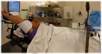
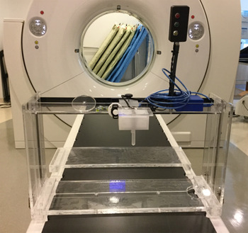
Our team designed a monitoring device to trace the patient’s breathing motions during radiotherapy. This self-held respiration monitoring device (SHRMD) consists of two parts: (1) a mechanical sensor connected with a controller (Figure 1) and (2) a computer software, which records, processes and monitors the patient’s respirations. The mechanical sensor is in contact with the patient during respiration or breath holding. The sensor reflects patient’s chest wall motions and provides mechanical motion data to the computer software. In addition, the controller gives a green light signal together with an alarm sound to guide the patient during respiration monitoring and to indicate the optimum level of breathing threshold (Figure 2). The computer software records the patients breathing pattern by transferring the sensor mechanical movement into digital readings. After setting the optimal breathing thresholds by staff, it monitors patient’s respiratory motion and gives light signal with alarm sound when the patient’s breathing threshold is within the optimal range. Using this device, patient’s actual breathing motion in the anterior–posterior dimension can be detected and monitored.
Setup of SHRMD.

SHRMD.

Objectives
The purpose of this study is to evaluate the use of SHRMD in DIBH radiotherapy for patients with left breast cancer. The study consists of three objectives:
to evaluate the inter-fractional reproducibility of the breath-holding threshold in using the SHRMD;
to evaluate the intra-fraction regularity of patient’s breath-holding ability in using the SHRMD and
to evaluate the potential dosimetric benefits in using SHRMD for DIBH compared with free breathing (FB) for Left breast cancer.
Methods and Materials
Patient selection
Sixteen patients with left breast cancer, who underwent breast conservative surgery and planned for tangential beam chest wall irradiation only, were recruited within 14 months of research period. The research protocol was approved by the hospital ethics committee. Details of study were explained to all the patients and consent forms were signed. Before planning computed tomography (CT), a session of breath-holding training was provided. After training, suitable patients were expected to stably hold their breath at moderate to deep inspiration level for at least 15 seconds.
Planning CTs
Two sets of planning CTs were acquired with FB and DIBH, respectively.
1. Treatment plans: A field of 6 MV in field tangential opposed plans for FB and DIBH were generated for each of the patients. The planning system was Eclipse 13.6 (Varian Medical Systems, Palo Alto, CA, USA). The prescribed doses of the treatment plans were 40–43·2 Gy in 15–16 fractions using 6 MV photons. All the plans were generated with at least 95% isodose, encompassing the whole breast and there was no hotspot over 110% isodose.
The plan with DIBH was used for treatment. The FB plan was the backup plan in case the patient could no longer hold the breath as planned. The FB plan was also used for dose comparison to investigate the benefits of DIBH.
Dose parameters for comparison between FB and DIBH include:
i. Maximum dose (Dmax), mean dose (Dmean) and median volume receiving more than 50% of the prescribed dose (V median > 50%) of the heart.
ii. Dmax, Dmean and median volume receiving more than 50% of the prescribed dose (V median > 50%) of the LAD.
iii. Dmean and relative median volume receiving more than 50% of the prescribed dose (V median > 50%) of the left lung.
Treatment delivery
All 16 patients underwent uneventful radiotherapy as per DIBH plans using SHRMD. The treatment was delivered with 6 MV photon to 40–43·2 Gy at 100% isodose level in 15–16 daily fractions by tangential opposed beams. Megavoltage cine images were acquired during treatment for each beam as quality assurance for intra-fractional position verification.
Data analysis
Statistical Package for the Social Sciences (SPSS) (SPSS Inc. Released 2007. SPSS for Windows, Version 16.0. Chicago, SPSS Inc.) was used for data analysis. Non-parametric signed rank test was used for the dosimetric comparison between BH and FB. (H0: No difference between BH and FB, H1: BH can reduce dose to organs at risk; p < 0·01: H0 can be rejected and H1 is true).
The chest wall excursions measured on the first MV Cine images of each treatment were used to evaluate the inter-fraction set-up reproducibility. Furthermore, the average chest wall excursions of all MV Cine images for each fraction were used to investigate the accuracy of the device by comparing with those on the planning CT images. The average fractional lung depths were used to evaluate the inter-fractional set-up reproducibility. The average lung depths variability between the medial and the lateral fields were used to evaluate the intra-fraction variability, and the range of lung depths within each beam was used to evaluate the intra-beam variability.
Results
Analysis of MV cine images
Non-significant intra-fractional overall mean error showed that the treatment positions were stable during treatments. Non-significant inter-fractional overall mean error showed that the treatment positions were reproducible (Table 1).
Results of reproducibility and stability

Dosimetric comparison of DIBH and FB plans
Heart dose
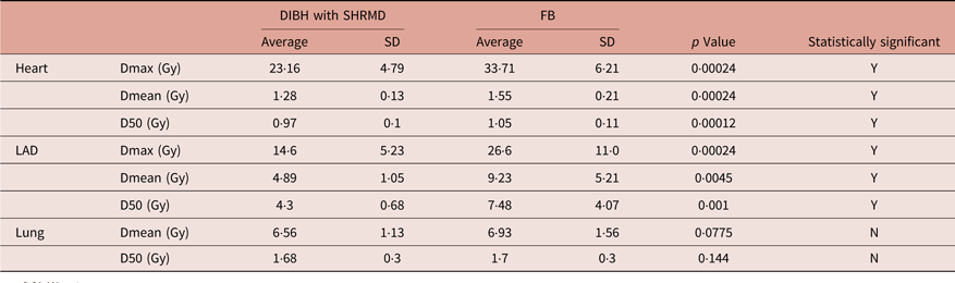
Using the DIBH plans, the results showed that the maximum dose of the heart was reduced by 29·9 ± 15·6%. The mean dose of heart was reduced by 16·6% ± 9·9%. The D50 of hearts was reduced by 7·3 ± 4·2% (Table 2).
Results of dosimetric comparison between DIBH and FB

p < 0·01 H1 = true.
Abbreviations: DIBH, deep inspiration breath hold; SHRMD, self-held respiration monitoring device; FB, free breathing; LAD, left anterior descending.
LAD dose
The results showed the maximum dose of the LAD in DIBH was reduced by 41·6 ± 18·3%. The mean dose of LAD was reduced by 31·9 ± 32·9%. The D50 of LAD was reduced by 27·5 ± 31·2%.
Lung dose with DIBH plan compared with FB plan: The results showed no difference in Dmean and D50 of lung between FB and BH plans.
Discussion
Comparison of heart dose
Our results showed that SHRMD provided significant dose reduction to the heart and the LAD artery. The maximum heart doses were significantly reduced by 29·9%, while the mean heart doses were reduced by 16·6%. The results of this study are consistent with other DIBH studies that were published previously. Many DIBH studies also showed that this technique can reduce the radiation dose to the heart and the assumed the risk of long-term side effects to the hearts could also be reduced.Reference Borst, Sonke and den Hollander6, Reference Jarvis, Maxim and Horst7, Reference McIntosh, Shoushtari, Benedict, Read and Wijesooriya9, Reference Pedersen, Korreman, Nystrom and Specht10 Using DIBH in left-sided intact breast radiation, the maximum heart dose is significantly reduced and the mean heart dose is reduced to a lesser degree.Reference Walston, Quick, Kuhn and Rong16
Comparison of lung dose
Although the benefits for reducing heart dose using DIBH seems clear, the results of previous DIBH studies were inconsistent regarding the reduction of lung dose.
Swanson et al.Reference Swanson, Grills and Ye17 showed there was a statistically significant reduction in all the heart and lung DVH parameters with moderate DIBH over FB plans. Conway et al.Reference Conway, Conroy and Harper18 also found that in DIBH the ipsilateral lung volume of V20Gy was reduced by more than 5% in the majority of patients. However, Hayden et al.Reference Hayden, Rains and Tiver19 found that there was no significant difference between DIBH and FB plans for lung V20 and mean lung dose. The DIBH plans demonstrated significantly larger total lung volume in their study. Differences in the lung dose between FB and DIBH plans were also not significant in the study by Al-Hammadi et al.Reference Al-Hammadi, Caparrotti and Naim20 Our study showed there was no significant difference in the Dmean and D50 of lungs between FB and BH plans.
Stability and reproducibility of treatment positions
Although the superiority of DIBH for the heart compared to FB is clear, the reproducibility and stability of treatments must be assured for the realisation of the dosimetric benefits. A systematic review of the cardiac dose-sparing benefits of DIBH in left breast irradiation by Smyth et al.Reference Smyth, Knight, Aarons and Wasiak14 concluded that the studies investigated the reproducibility and stability of DIBH showed small inter-fraction and intra-fraction variability in the set-up position for DIBH treatments.
Similar to our SHRMD, Jensen et al.Reference Jensen, Abramova, Frengen and Lund21 also introduced an in-house DIBH system. Portal images were acquired and the reproducibility and stability of DIBH treatment were compared to FB. Their in-house system gave a reproducible and stable DIBH treatment where the systematic error, ∑, and random error, r, were less than 2.2 mm in all directions but were not significantly better than that at FB.
The results of our study with SHRMD regarding the reproducibility and stability of DIBH treatments are consistent with other studies. Using this in-house established SHRMD, the treatment positions are stable and reproducible as other in-house devices or commercial devices in other studies.
Breath-hold coaching
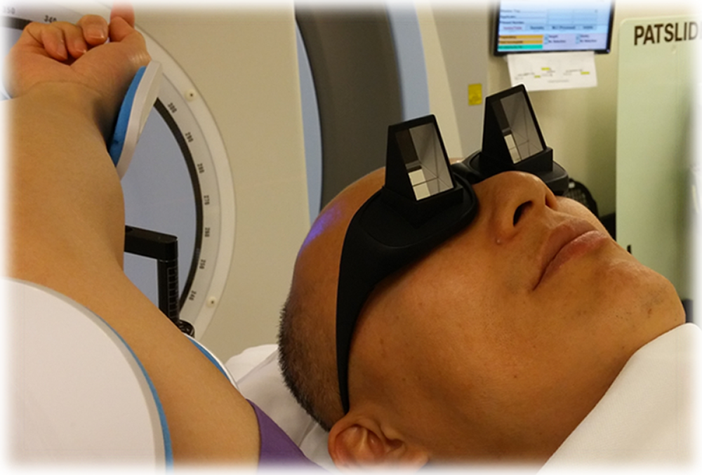
DIBH requires patient compliance.Reference Latty, Stuart, Wang and Ahern13 Patient education regarding the DIBH technique is important as it requires the patient to actively breathe in before every treatment field to ensure successful delivery. One of the reasons for stability and reproducibility of using SHRMD is good breath-hold coaching for the patients. Our SHRMD provides visual traffic light and audible alarm. The patients were asked to wear special glasses (Figure 3), so that they could see the traffic light system which allows them to control their breath hold to keep at green light level during treatments. The comments from our patients were positive about this traffic light system.
Special glasses.

Limitations
One of the limitations of our study is the small sample size of patients. Only 16 patients were recruited during this period because of the limitation of selection criteria and patients’ ability to hold their breath. The prescription doses (40–43·2 Gy) were different for plans for different patients. The numbers of fractions were also different (15 or 16 fractions), and the maximum dose of the LAD artery cannot be compared between different patients.
Conclusion
This study demonstrated the potential of using the SHRMD for DIBH to reduce the heart and LAD dose in the common technique of adjuvant irradiation of left breast cancer.
Results showed that patients undergoing radiotherapy to left breast cancer were able to perform a stable and reproducible DIBH using the SHRMD. The system was well tolerated and all patients completed their treatment sessions with DIBH.
Acknowledgements
The authors would like to thank Professor Maria Yuen Yee Law (School of Medical and Health Sciences, Tung Wah College, Hong Kong) for her valuable and constructive suggestions. The authors would also like to thank the great effort of staff of Department of Radiotherapy and the Medical Physics and Research Department of Hong Kong Sanatorium & Hospital, 2 Village Road, Happy Valley, Hong Kong.
Financial Support
This research received no specific grant from any funding agency, commercial or not-for-profit sectors.
Conflicts of Interest
None.
Ethical Standards
The authors assert that all procedures contributing to this work comply with the ethical standards of Good Clinical Practice; Code of Federal Regulations and Code of Professional Conduct and with the Helsinki Declaration of 1975, as revised in 2008, and has been approved by Hong Kong Sanatorium & Hospital Research Committee (RC-2016-10).