Introduction
Multiple symmetrical lipomatosis, also known as Madelung disease, is a rare disease caused by abnormal lipid metabolism. Most reports describe its occurrence in middle-age men with a history of alcohol abuse.
The aetiology of multiple symmetrical lipomatosis is unknown. However, mitochondrial DNA mutations have been found on muscle biopsy, suggesting one possible pathophysiological mechanism.Reference Klopstock, Naumann, Seibel, Shalke, Reiners and Reichmann1
Histologically, the lipomatous mass is seen to consist of benign, hyperplastic adipocytes.
The disease is characterised by insidious, progressive growth of the lipomatous mass, which has a high propensity for recurrence after removal. Advanced cases of neck lipomatosis with mediastinal extension can develop airway and vascular compression.
We report a patient with multiple symmetrical lipomatosis and deep neck infiltration. The proposed pathogenesis and treatment options are discussed.
Case report
A 45-year-old man was referred to us with a massive, diffuse neck mass. The mass had been noticed for the previous 10 years, and had gradually increased in size. There was no symptom of upper aerodigestive compression. The patient's head and neck movements were unrestricted. Similar subcutaneous masses were found on both arms and the upper back. The patient was a chronic smoker and social drinker. He was otherwise well, with no neurological symptoms.
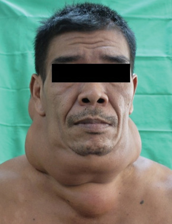
On examination, the mass was seen to involve the entire circumference of the neck. It extended from the lower border of the mandible to the supraclavicular region (Figure 1).
Clinical photograph showing symmetrical fat distribution mainly localised in the neck region.

There were no significant findings on nasal or laryngeal endoscopy.
Fine needle aspiration cytology showed benign adipocytes, consistent with a diagnosis of lipoma.
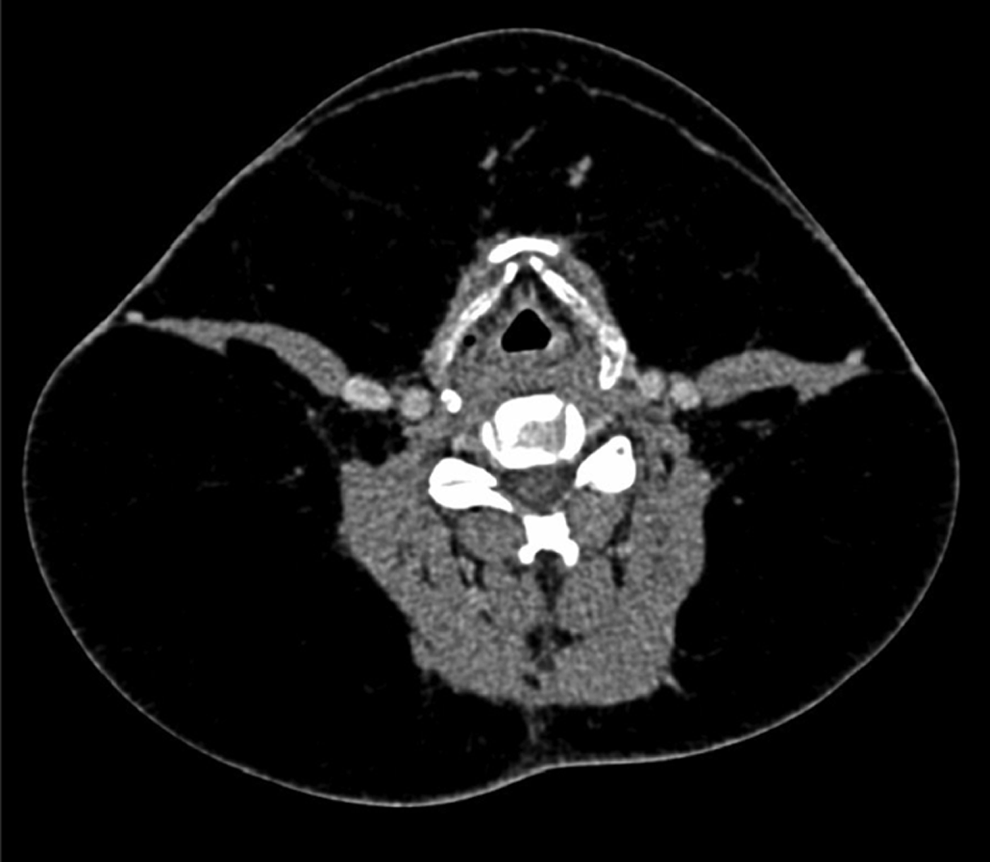
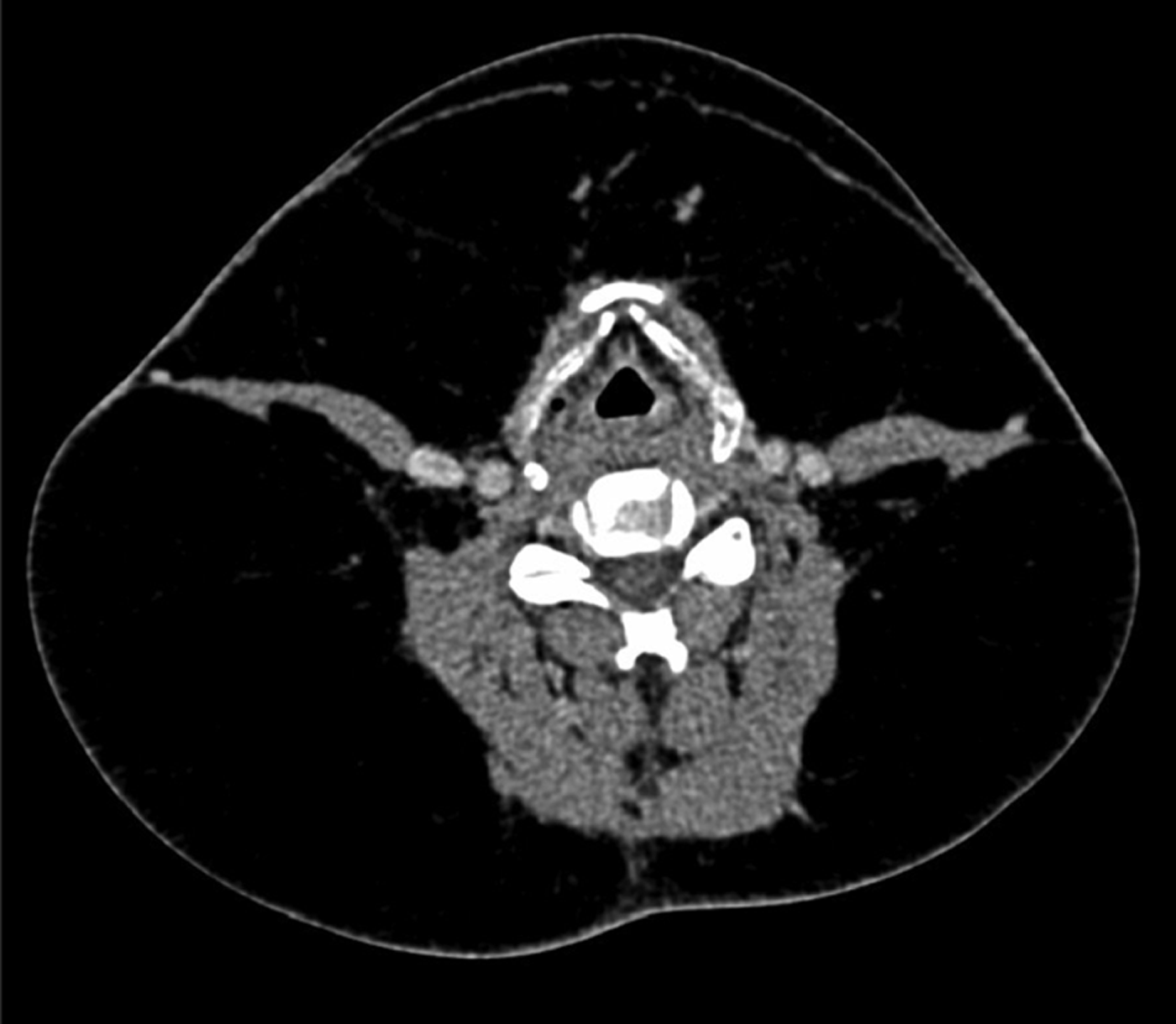
Computed tomography of the neck showed diffuse neck lipomatosis distributed between the neck muscles (i.e. the semispinalis, splenius capitis and trapezius) and abutting both carotid sheaths (Figure 2). There was no mediastinal extension or significant cervical or mediastinal lymphadenopathy.
Axial computed tomography scan of the neck showing circumferential lipomatosis. The fatty tissue has infiltrated into the muscle plane, abutting the carotid sheaths bilaterally.
The patient was scheduled for surgical excision of the lipomatous mass. Intra-operatively, extensive, unencapsulated fat tissue was found. This involved the parapharyngeal space and was in contact with the carotid sheaths bilaterally. The surgical dissection was difficult due to the ill-defined dissecting plane. Debulking surgery was performed, with removal of 2 kg of fat tissue.
Post-operatively, there was seroma formation, which resolved after repeated needle aspiration.
Histopathological examination of the surgical specimen showed benign lipoma with reactive lymph nodes.
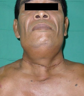
The neck wound healed well, with a good aesthetic outcome seen at follow up (Figure 3).
Clinical photograph showing a good cosmetic outcome, one week after surgical excision of the lipoma.

Discussion
Multiple symmetrical lipomatosis is characterised by extensive fat tissue deposition, especially on the neck, trunk and upper extremities. Its most common presentation is an unsightly neck mass. Advanced cases may have mediastinal extension of fat tissue which leads to aerodigestive compression symptoms.
Although the aetiology of this rare disease remains obscure, mitochondrial dysfunction is believed to play a role in the pathogenesis. Several studies had reported the identification of a deletion mutation of mitochondrial DNA on muscle biopsy in some patients.Reference Klopstock, Naumann, Seibel, Shalke, Reiners and Reichmann1, Reference Silvestri, Ciafaloni, Santorelli, Shanske, Servidei and Graf2 The identified mutations cause a decrease in oxidative phosphorylation which would result in lipoma formation. Klopstok T et al. have also identified a mitochondrial DNA mutation within a sural nerve biopsy.Reference Klopstock, Naumann, Seibel, Shalke, Reiners and Reichmann1 Furthermore, these authors also performed electrophysiological tests which demonstrated axonal neuropathy in 60 per cent of subjects with multiple symmetrical lipomatosis.
Head and neck malignancies have been reported in patients with multiple symmetrical lipomatosis; however, this association remains uncertain.Reference Ujpál, Németh, Reichwein and Szabó3–Reference Ruzicka, Vieluf, Landthaler and Braun-Falco5 It is essential to perform a complete clinical assessment of the head and neck region in patients with a history of alcohol abuse, as the latter is an aetiopathogenetic factor for both head and neck malignancy and multiple symmetrical lipomatosis. Aerodigestive compression symptoms should not be attributed to lipoma compression until regional malignancy has been excluded by endoscopic and imaging investigations; malignant tumour and cervical lymphadenopathy may lurk unnoticed underneath a massive lipoma.
Extremely rare malignant transformation of multiple symmetrical lipomatosis has also been reported.Reference Guastella, Borsi, Gibelli and Berta6 Tizian et al. have described the appearance of intramyxoid sarcoma in a case of benign lipomatosis after six years’ follow up, while Durand et al. reported a case of liposarcoma transformation.Reference Tizian, Berger and Vykoupil7, Reference Durand, Thomine, Tayot, Foucault and Deshayes8
The treatment for multiple symmetrical lipomatosis ranges from surgery to various modes of medical therapy. Surgical excision and liposuction are still the most effective surgical options.Reference Alameda, Torres, Perez-Mitchell and Riera9, Reference Hasegawa, Matsukura and Ikeda10 Liposuction has gained popularity recently due to its minimal scar. Liposuction is considered less invasive and technically easier, compared with surgical excision. It is also more suitable for patients who represent a higher surgical and/or anaesthetic risk.Reference Faga, Valdatta, Thione and Buoro11 Based on the high propensity of lipoma for recurrence, some authors suggest that the goal of treatment should be a palliative result with restoration of function. It is unnecessary to subject patients to the risks of radical surgery for this benign condition. Surgical excision should be limited to cases with airway compression or cosmetic unacceptability.Reference Verhelle, Nizet, Van den Hof, Guelinckx and Heymans12 Liposuction is safer and less aggressive, and suffices for debulking purposes.
On the other hand, liposuction in the neck region performed by an inexperienced surgeon always carries the risk of injury to vital neck structures, which could be life-threatening. Faga et al. have described ultrasound-assisted liposuction in a case with neck movement restriction.Reference Faga, Valdatta, Thione and Buoro11 This technique allows gentler and more precise suction without major damage to subcutaneous tissue and vascular structures. Nevertheless, in the case in question the procedure was performed for the purpose of functional relief, and the cosmetic outcome was unsatisfactory.Reference Faga, Valdatta, Thione and Buoro11 The limitations of liposuction include inadequate aspiration of lipoma, especially in cases with a dense, fibrous constitution. Furthermore, liposuction is often restricted in the submental region, and in areas where previous surgery has been performed with subsequent scarring.Reference Verhelle, Nizet, Van den Hof, Guelinckx and Heymans12 Thus, patients should be made aware of the possibility of residual lipoma due to inadequate liposuction. Despite the above risks, at present there has been no reported case of major complications from liposuction.
The main disadvantage of surgical excision is the long, unsightly scar.
In our patient's case, we opted for surgical excision because we believed this was safer as the lipoma abutted both carotid sheaths. Direct visualisation during surgery may reduce the risk of injury. We anticipated a difficult dissection as the lipomatous mass was unencapsulated and had deep tissue infiltration.Reference Sreekrishna, James and Rosemary13 At operation, the pathological hyperplastic lipoma was hard to distinguish from normal subcutaneous fat and the lymphofatty tissue along the carotid sheaths. After the lipoma debulking surgery, redundant skin was apparent. Excess skin was trimmed to restore the normal neck contour, enabling optimal cosmesis.
• The pathophysiology of multiple symmetrical lipomatosis (Madelung disease) is unclear
• Cosmetic deformity is the commonest problem
• Airway and vascular compression are rare
• Pre-operative computed tomography is crucial to assess extent and proximity to vital structures
• Open excision is best for massive, deeply infiltrating lipomatosis
Lifestyle modification, with alcohol abstinence and control of blood sugar and lipids, may curb fat growth but will not shrink a pre-existing lipoma.Reference González-García, Rodríguez-Campo, Sastre-Pérez and Muñoz-Guerra14 Toshio et al. have reported the use of phosphatidylcholine as mesotherapy (via intralesional injection), with satisfactory results.Reference Hasegawa, Matsukura and Ikeda10 Other reported methods have included the use of an oral β2 agonist (i.e. salbutamol) and a peroxisome proliferator-activated α receptor agonist (fibrate). However, at present the consistent efficacy of these treatments has not been demonstrated.Reference Zeitler, Ulrich-Merzenich, Richter, Vetter and Walger15
Conclusion
Complete clinical assessment of patients with multiple symmetrical lipomatosis is imperative in order to exclude any associated head and neck malignancy. Surgical excision and liposuction are complementary treatment modalities in the management of this condition. Patients should be aware of the risks and benefits of these surgical options, and of the risk of lipoma recurrence, before surgery. The decision on the mode of surgery depends upon the disease extent, the patient's expectations and the surgeon's experience.