Introduction
In the course of a scanning electron microscopy study using energy-dispersive spectroscopy of an old collection of platinum-group minerals from the Norilsk ore district, the senior author of the present paper encountered various phases containing Pd and Sn as major elements. However, their unambiguous assignment to a particular mineral species on the basis of chemical composition alone turned out to be quite difficult. The most problematic issue was the correct identification of stannopalladinite, as the available literature data on this mineral turned out to be very contradictory. In order to better understand the problem and try to solve the above issue, a detailed investigation of the latter was undertaken.
The official International Mineralogical Association (IMA) List of Minerals (Pasero, Reference Pasero2023) includes stannopalladinite as a valid mineral species with ‘grandfathered’ status which applies to minerals discovered before the establishment of the IMA in 1958. Indeed, stannopalladinite dates back to 1947 when this name first appears in the paper of Maslenitskiy et al. (Reference Maslenitskiy, Faleev and Iskyul1947). However to date, the mineral remains poorly studied. Its official chemical formula is reported as Pd3Sn2 with a question mark. Various literature sources contain contradictory information regarding its physical properties, chemical composition and X-ray data. The structure of stannopalladinite is unknown.
The aim of this paper is to provide new data obtained on what we believe is the exact analogue of the original material studied by Maslenitskiy et al. (Reference Maslenitskiy, Faleev and Iskyul1947). New chemical and X-ray analyses conducted in this work combined with the critical perception of the data published in the literature led us to propose a new chemical formula and unit-cell parameters for stannopalladinite and unambiguously demonstrate its difference from other minerals of the Pd–Sn and Pd–Cu–Sn systems.
Historical background
Maslenitskiy et al. (Reference Maslenitskiy, Faleev and Iskyul1947) reported on a mineral with the formula Pd3Sn2 called “stannopalladinite” from “the placer and concentrates of the processing plant”. The new mineral was described as very small but abundant inclusions in well-formed cubic skeletal, platy and prismatic elongated crystals of ferroplatinum. Apart from very limited physical (Table 1) and chemical properties (weak reaction with concentrated ΗΝΟ3 and fast reaction with aqua regia), the authors provided a bulk chemical composition of grains where stannopalladinite was “maximally concentrated”. Because of the substantial copper content, the mineral was also named “cuprian stannopalladinite”.
Literature data on stannopalladinite.

* Probably a typo, correct range is likely to be 220–328.
n.d. = no data available
One year later, Maslenitskiy (Reference Maslenitskiy1948) added more information on the above findings. In particular, he provided the detailed description and photos of ferroplatinum crystals and stannopalladinite inclusions in them and reported eight more chemical analyses related to this mineral.
Twenty four years later, Berry and Thompson (Reference Berry and Thompson1962) published under the name “stannopalladinite” the powder X-ray data of a synthetic material with the formula Pd3Sn2 and assigned to it a P63/mmc space group and hexagonal unit cell with a = 4.399 and c = 5.666 Å, referring to the paper of Nowotny et al. (Reference Nowotny, Schubert and Dettinger1946). The latter authors were first to provide the powder X-ray data of synthetic Pd3Sn2, its hexagonal unit cell similar to the one mentioned above and suggested the structure of this compound to be a filled NiAs type.
Genkin (Reference Genkin1968) disclosed the type locality of stannopalladinite described by Maslenitskiy et al. (Reference Maslenitskiy, Faleev and Iskyul1947) as the platinum placer of Ugol'nyi Ruchei (‘Coal stream’) Mine in the Norilsk Cu–Ni deposit. Obviously, this locality could not be mentioned in 1947 given that in 1935–1956 the Norilsk deposits were managed by Noril'lag (a soviet concentration camp that was part of the infamous GULAG system), mined by prisoners and all the relevant mining information was under secrecy. Apart from the type locality, A.D. Genkin also reported on stannopalladinite from the 203 m level of the Taimyr Mine in the Oktyabrskoe Cu–Ni deposit as small rounded, oval or elongated grains up to 0.3 mm rimmed by “tetragonal ferroplatinum” in chalcopyrite veins. A lot of other important information was added by the author to further characterise this mineral such as optical properties, including a reflectance dataset, hardness and powder X-ray diffraction data (Table 1). It was suggested that the latter are similar to synthetic Pd3Sn2. The direct comparison of the data revealed that several d spacings of the synthetic compound are absent in natural stannopalladinite, however, the author explained it by the chemical features of the mineral. The microspectral analysis showed Cu to be an essential part of the mineral along with Pd and Sn, so after Maslenitskiy et al. (Reference Maslenitskiy, Faleev and Iskyul1947) the mineral was called “cuprian stannopalladinite”.
Chernyaev and Yushko-Zakharova (Reference Chernyaev and Yushko-Zakharova1968) reported on stannopalladinite from the Monchegorsk Cu–Ni deposit (Murmansk Oblast) as grains up to 0.1 mm in size intergrown with niggliite (PtSn). Some of their data, however, differed from the stannopalladinite from Norilsk. In particular, the reported hardness was substantially larger and the chemical composition, measured for the first time by the electron microprobe method, showed the absence of Cu and the empirical formula to be closer to Pd5Sn3 rather than Pd3Sn2. No X-ray data were provided.
Vyalsov (Reference Vyalsov1973) provided a new reflectance dataset and some other optical data for stannopalladinite “from Norilsk” with no more details on the locality. The chemical formula was given with admixed Pb (Table 1).
L.J. Cabri in his handbooks on PGM minerals (Cabri, Reference Cabri1972, Reference Cabri and Cabri1981) compiled the data on stannopalladinite given by predecessors and assigned to natural material the hexagonal unit-cell parameters (e.g. a = 4.399 and c = 5.666 Å) similar to those reported by Berry and Thompson (Reference Berry and Thompson1962). For powder X-ray data, he referred to the PDF card #4-801 of the synthetic Pd3Sn2.
Genkin et al. (Reference Genkin, Distler, Gladyshev, Filimonova, Evstigneeva, Kovalenker, Laputina, Smirnov and Grokhovskaya1981) noted the difficulties in calculating the formula and characterisation of stannopalladinite and stressed the necessity to conduct a detailed check of all chemical compositions mentioned in the literature along with structural studies in order to establish the real position of copper atoms in the unit cell of the mineral. Nevertheless, the authors reported on stannopalladinite from another locality – the Talnakh deposit in the Norilsk ore district where the mineral forms a central part of zoned grains included in massive chalcopyrite and talnakhite ores. Stannopalladinite is often intergrown with plumbopalladinite, polarite and silver and is rimmed by isoferroplatinum. Its intergrowths with arsenides of Pd and Ni are rarer. Optical and chemical data of this material were given. The authors noted the constant presence of copper (~6 wt.%) as the main feature of the chemical composition of the mineral but, at the same time, stressed the significant variations in the content of other elements (Pd, Pt, Sn, Pb, Bi, Sb and As). Their chemical analyses were calculated in three possible ways: (1) Pd and Cu in one position; (2) Cu and Sn in one position; and (3) Pd, Cu and Sn all in independent positions. The last option gave the most stoichiometric formula close to (Pd,Pt)6Cu1(Sn,Pb,Bi,Sb,As)3, or Pd6CuSn3. The reported d spacings of the debyegram (Table 1) were suggested to be close to the synthetic Pd3Sn2.
Evstigneeva and Nekrasov (Reference Evstigneeva and Nekrasov1984) studied in detail natural and synthetic compounds belonging to the ternary Pd–Cu–Sn system. They provided chemical and powder X-ray data for stannopalladinite but, unfortunately, with no information on its provenance. These authors came to the conclusion that stannopalladinite is the natural analogue of the synthetic Pd5Sn2Cu for which they suggested an orthorhombic symmetry with a ≈ 7.8 and b ≈ c ≈ 3.9 Å and Pmmm space group. Optical data were given for the synthetic material.
Cabri (Reference Cabri and Cabri2002) tried to summarise all the previous information on stannopalladinite from various publications. He assigned to stannopalladinite the formula Pd5Sn2Cu (with a question mark) and the orthorhombic cell proposed by Evstigneeva and Nekrasov (Reference Evstigneeva and Nekrasov1984) but clearly noted that the rarity of this phase, the existence of other Pd–(Pt) minerals with Cu and Sn (atokite, paolovite, cabriite and taimyrite), the very small grain size and frequent occurrence as intergrowths with other minerals have caused confusion in the literature. He stated that there is no certainty that the mineral studied by Maslenitskiy et al. (Reference Maslenitskiy, Faleev and Iskyul1947) is the same as that studied by subsequent authors, including Genkin et al. (Reference Genkin, Distler, Gladyshev, Filimonova, Evstigneeva, Kovalenker, Laputina, Smirnov and Grokhovskaya1981) and Evstigneeva and Nekrasov (Reference Evstigneeva and Nekrasov1984). The key to this problem is X-ray diffraction, but most publications contain no X-ray data and all conclusions are made solely on the basis of electron probe microanalyses. The author suggested that a formal redefinition of stannopalladinite is needed, including the re-examination of type material, determination of ideal formula/range of compositions and re-investigation of powder X-ray diffraction/symmetry.
A summary of the literature data on stannopalladinite, as compiled in Table 1, clearly demonstrates a lack of unified approach to what this species should be considered. It is obvious that some of the data assigned to stannopalladinite probably belong to other phases and, as was correctly suggested by Cabri (Reference Cabri and Cabri2002), more work is required to properly define this mineral.
Samples and experimental methods
Unfortunately, no information is available on the location of the original material of stannopalladinite from Ugol'nyi Ruchei studied by Maslenitskiy et al. (Reference Maslenitskiy, Faleev and Iskyul1947), Maslenitskiy (Reference Maslenitskiy1948) and Genkin (Reference Genkin1968). The nomenclature designations for the type mineral specimens and the encouragement to authors to deposit such specimens in museums were approved by the Commission on New Minerals and Mineral Names (CNMNC) of the IMA much later (Dunn and Mandarino, Reference Dunn and Mandarino1987).
However, the collections of the Fersman Mineralogical Museum comprise three samples with catalogue numbers 41647, 45863 and 46887 that originate from Ugol'nyi Ruchei and whose general appearance look very similar to the material studied by the above-mentioned predecessors. These samples were registered to the Museum's collection in 1938 and 1949 and were probably mined during panning out in the placer of Ugol'nyi Ruchei in this period of time. They represent cubic, prismatic, flattened elongated crystals up to 10 mm with features of skeletal growths in many of them. A more detailed description of these three samples and their photographs are given by Generalov and Pautov (Reference Generalov and Pautov2005).
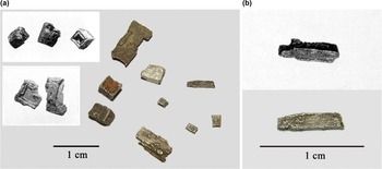
The visual comparison of the samples from the Fersman museum with those studied by Maslenitskiy et al. (Reference Maslenitskiy, Faleev and Iskyul1947) and Maslenitskiy (Reference Maslenitskiy1948) show their very close resemblance (Fig. 1a). Also, similarly to the original material, considerable parts of crystals from the Fersman museum show strong ferromagnetism. Finally, the chemical analysis of one of the cubic crystals from sample 41647 done by Generalov and Pautov (Reference Generalov and Pautov2005) revealed its inhomogeneous composition with most of the grain matching Pt–Fe-bearing minerals (tetraferroplatinum, ferronickelplatinum and isoferroplatinum) with inclusions of Pd–Cu–Sn–Pb-phases, some of which were interpreted as stannopalladinite. This is also in line with the information provided by Maslenitskiy et al. (Reference Maslenitskiy, Faleev and Iskyul1947) and Maslenitskiy (Reference Maslenitskiy1948).
Original material from Ugol'nyi Ruchei studied by Maslenitskiy et al. (Reference Maslenitskiy, Faleev and Iskyul1947) and Maslenitskiy (Reference Maslenitskiy1948) in comparison with the material from the Fersman museum's collections: (a) cube-shaped (top left) and platy skeletal (mid left) crystals of Maslenitskiy (Reference Maslenitskiy1948, their figures 1 and 4) and crystals of different habit from sample 41647; (b) prismatic crystal pictured by Maslenitskiy (Reference Maslenitskiy1948, their figure 3) (top) and a similar-looking crystal from sample 41647 (bottom), from which a fragment was separated for further research.

All the above information led us to undertake a more detailed investigation of the material from the Fersman museum's collections. As the cube-shaped crystal was already chemically studied by Generalov and Pautov (Reference Generalov and Pautov2005) we decided to investigate one of the flattened, prismatic crystals, 6 mm long, from sample 41647 (Fig. 1b), especially as Maslenitskiy (Reference Maslenitskiy1948) suggested these to be richer in stannopalladinite than cubic crystals. A small fragment of the crystal was separated, put in the thin section and examined using a variety of analytical techniques.
Micro-indentation hardness was measured with a PMT-3 instrument calibrated with respect to NaCl.
The optical properties in reflected light were studied using POLAR-3 and POLAR-215 polarising light microscopes. Reflectance values have been measured in air using a MSF-R (LOMO, St. Petersburg, Russia) microspectrophotometer with silicon used as a standard. The photometric aperture diameter was 0.3 mm, monochromator exit slit size – 0.1 mm, spectral interval – 6 nm.
The chemical composition was analysed with a Cameca SX-100 electron microprobe (Masaryk University, Brno) using the wavelength dispersive spectroscopy mode mode, 25 kV, 10 nA, 2 μm beam diameter. Analysed elements, analytical X-ray lines, analysing crystals (in parentheses) and standards were as follows: FeKα (LLIF) – FeS2; NiKα (LLIF) – pararammelsbergite; CuKα (LLIF) – Cu; PdLα (LPET) – Pd; SnLβ (PET) – Sn; PtLα (LLIF) – Pt; and PbMα (LPET) – PbSe. Other elements heavier than F were sought for but their contents were below the detection limit. The raw intensities were converted to concentrations using X-PHI matrix-correction software (Merlet, Reference Merlet1994).
In order to get single-crystal X-ray diffraction (SCXRD) data, grains of minerals studied optically and by electron microprobe were extracted from the thin section and analysed using a Rigaku Oxford Diffraction SuperNova diffractometer (University of Padova) equipped with a Pilatus 200K Dectris detector and an X-ray micro-source (MoKα radiation) with accelerating voltage of 50 kV, beam current of 0.8 mA and beam size on the sample ~0.12 mm. The detector-to-sample standard distance was 68 mm. The powder X-ray diffraction (PXRD) data were collected on the same instrument using microdiffraction mode with 0–360° rotation scan and exposure time varying from 15 minutes (tetraferroplatinum) to 12 hours (for stannopalladinite).
In an attempt to improve the quality of the SCXRD data, an additional experiment with the same grain of stannopalladinite was carried out at a low temperature (100 K) using a Rigaku Oxford Diffraction XtaLAB Synergy-S diffractometer (Research Centre for X-ray Diffraction Studies, St. Petersburg State University) equipped with a HyPix-6000HE detector and an X-ray micro-source (MoKα radiation) with accelerating voltage of 50 kV, and beam current of 1.0 mA. The detector-to-sample distance was 34 mm, and the exposure time per each frame was 60 seconds.
Results and discussion
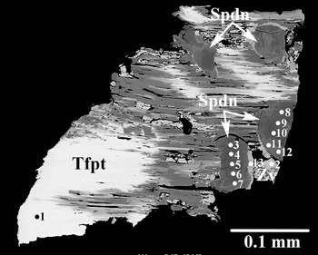
Optically and chemically, the studied fragment represents an intimate intergrowth of mainly two phases: tetraferroplatinum and a Pd–Cu–Sn-bearing mineral that we determined as stannopalladinite on the basis of the data below (Fig. 2). Tetraferroplatinum is white in reflected light and isotropic. The mineral is strongly magnetic. Its composition corresponds to a nickeloan–cuprian variety (Table 2, an. 1) and its tetragonal unit-cell parameters calculated from the powder X-ray diffraction data are: a = 2.7003(3), c = 3.6833(6) Å and V = 26.86(6) Å3. Very minor Sn-bearing zvyagintsevite (Table 2, an. 2) is also present in the association studied.
Intimate intergrowth of stannopalladinite (Spdn, various shades of grey) and tetraferroplatinum (Tfpt, white) in association with zvyagintsevite (Zv). Analytical spots have the same numbers as in Table 2. Thin section, back-scattered electrons image.

Chemical composition (wt.%) of minerals from Ugol'nyi Ruchei.

1 – tetraferroplatinum; 2 – zvyagintsevite; 3–14 – stannopalladinite (sample 41647, our data).
Analysis 14 (bold) is the mean of analyses 3–13.
Analyses 15, 16 and 17 correspond to analyses 8, 10 and 11, respectively, from Generalov and Pautov (Reference Generalov and Pautov2005).
Analysis 18 is the mean data calculated by us from the values reported in Table 2 by Maslenitskiy et al. (Reference Maslenitskiy, Faleev and Iskyul1947).
Analysis 19 is the mean of eight analyses calculated by us from the values reported in by Maslenitskiy (Reference Maslenitskiy1948) their table 3; total includes 0.45 wt.% of Au.
Empirical formulas are calculated on the basis of atoms sum of 2 (an. 1) and 4 (an. 2–19).
The Pd–Cu–Sn mineral forms spherical and ovoid-shaped grains up to 0.08 × 0.05 mm. It is dark grey, opaque and has a metallic lustre. The values of micro-indentation hardness (VHN, 30 g load) are between 233 and 266 kg/mm2, and the average of 4 measurements is 244 kg/mm2, corresponding to a Mohs hardness of 4.
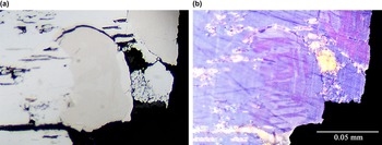
The mineral has very distinct optical properties. It is pale pink in reflected light (Fig. 3a). The bireflectance is noticeable only in oil immersion. In crossed nicols, the mineral exhibits strong anisotropy with colour effects changing from reddish purple to greyish blue and polysynthetic twinning of grains (Fig. 3b). Its reflectivity is high but lower than that of neighbouring tetraferroplatinum. Measured reflectance values are given in Table 3 and plotted in Fig. 4 in comparison with the literature data for stannopalladinite. Note a very distinct anomalous dispersion of reflectance curves.
Enlarged fragment of Fig. 2 (right lower part) in reflected light: (a) at one nicol; (b) in crossed nicols.

Reflectance values (%) for stannopalladinite.

Reflectance curves of stannopalladinite.

As we can see, the optical data of our mineral and its hardness are a very close match to those reported by Maslenitskiy et al. (Reference Maslenitskiy, Faleev and Iskyul1947) and Genkin (Reference Genkin1968) for the original stannopalladinite from Ugol'nyi Ruchei (Table 1).
Chemically, the Pd–Cu–Sn mineral is heterogeneous, as shown in the back-scattered electron image (Fig. 2), and contains various admixtures of Pb and Pt. Dark grey areas are almost Pb-free (Table 2, an. 3–10) whereas in light grey zones the content of Pb (Table 2, an. 11–12) is up to 19.15 wt.% in the most Pb-rich parts (Table 2, an. 13). We do not exclude that Pb admixture is due to an intimate intergrowth of our Pd–Cu–Sn-mineral with zvyagintsevite. Generalov and Pautov (Reference Generalov and Pautov2005) reported on the compositions of apparently the same mineral with 15.13 wt.% Pt (Table 2, an. 15) and up to 17.72 wt.% Pb (Table 2, an. 16 and 17). Such chemical variety, however, does not affect the general stoichiometry which remains always the same for all analyses obtained by us and Generalov and Pautov (Reference Generalov and Pautov2005), i.e. (Pd,Cu,Pt) : (Sn,Pb) ≈ 3 : 1. The empirical formula obtained on the sample studied by us as a mean of 11 analyses and calculated on the basis of 4 atoms pfu is (Pd2.42Cu0.56Pt0.02)Σ3.00(Sn0.92Pb0.08)Σ1.00.
To compare the chemical composition of our mineral with the original stannopalladinite, we calculated the empirical formulae for the latter using the average values reported by Maslenitskiy et al. (Reference Maslenitskiy, Faleev and Iskyul1947) and eight chemical analyses given by Maslenitskiy (Reference Maslenitskiy1948) – see Table 2, an. 18 and 19, respectively. Surprisingly, we found out that, even bearing in mind the imperfectness of the chemical data of that time and probable admixture of other minerals in them, the calculated stoichiometry (Pd,Cu,Pt,Fe,Ni) : Sn is much closer to 3:1 obtained by us rather than 3:2 suggested by Maslenitskiy et al. (Reference Maslenitskiy, Faleev and Iskyul1947) and following authors.
The combination of factors, such as the similar general appearance of the material and its origin, similar mineral association, physical properties (hardness, unique optical properties etc.) and close chemical composition, led us to the conclusion that the Pd–Cu–Sn-bearing mineral discovered by us in sample 41647 from the Fersman museum collections seems to be the same as the original stannopalladinite described by Maslenitskiy et al. (Reference Maslenitskiy, Faleev and Iskyul1947), Maslenitskiy (Reference Maslenitskiy1948) and Genkin (Reference Genkin1968).
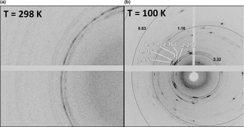
In order to perform X-ray studies, we extracted from the thin section the most homogenous, almost Pb-free fragment. Unfortunately, it was impossible to obtain reliable single-crystal structure data at ambient temperature as the mineral turned out to be of a completely ‘powdery’ nature (Fig. 5a). The SCXRD data collection of the same stannopalladinite sample at low temperature (100 K) slightly improved the quality of the diffraction patterns, which is seen from the appearance of a few discrete Bragg reflections (Fig. 5b) that were totally absent on the patterns collected at room temperature. However, it was still impossible to process these data as the main portion of the diffraction results in diffraction arcs (sections of Debye-Scherrer rings), while discrete reflections belong to different domains. So, the final unit cell parameters were determined from the PXRD data collected earlier at room temperature. However, we note that the diffraction arcs from the SCXRD experiment at low temperature fully correspond to the PXRD data collected at ambient temperature (Fig. 5b, Table 4).

Powder X-ray data for stannopalladinite and synthetic Pd3Sn2.

The obtained PXRD data (Table 4) contain all the d spacings reported by Genkin (Reference Genkin1968) and, to our opinion, are yet another proof of the identity of our studied mineral to the original stannopalladinite from Ugol'nyi Ruchei. The comparison with the powder X-ray data of Evstigneeva and Nekrasov (Reference Evstigneeva and Nekrasov1984) gives an even better match (Table 4) and their chemical formula Pd5Sn2Cu has the same ratio (Pd,Cu):Sn = 3:1 as our mineral. Regrettably, these authors provided no information on the locality of their stannopalladinite.
In comparison, the PXRD data of synthetic Pd3Sn2 (Table 4) – based on the data of Nowotny et al. (Reference Nowotny, Schubert and Dettinger1946), as well as data of Schubert et al. (Reference Schubert, Lukas, Bhan and Meissner1959) who distinguished two modifications (α and β) of Pd3Sn2, and data of Sarah et al. (Reference Sarah, Alasafi and Schubert1981) for the phase Pd20Sn13 with a Ni13Ga3Ge6 type structure – only poorly match with our mineral. The structural correspondence of stannopalladinite to the synthetic analogue of α-Pd3Sn2 given by Genkin (Reference Genkin1968) and Genkin et al. (Reference Genkin, Distler, Gladyshev, Filimonova, Evstigneeva, Kovalenker, Laputina, Smirnov and Grokhovskaya1981) seem to be improbable.
Using the DicVol indexing method in HighScore Plus software (Panalytical), assuming a unit-cell volume V < 6000 Å3, the only solution that used the 100% of the reflections in Table 4 and that was characterized by best Figure of Merit (FOM) provided an orthorhombic cell with the following unit-cell parameters: a = 14.634(2), b = 8.5253(6), c = 4.5946(3) Å and V = 573.24(7) Å3 (Z = 8).
The calculated density of stannopalladinite using the above unit cell data and empirical formula (Table 2, an. 14) is 9.781 g/cm3.
Our data clearly show that the hexagonal unit cell with a ≈ 4.4 and c ≈ 5.7 Å attributed to stannopalladinite in some literature sources (Table 1) and databases cannot refer to this mineral.
As the structure of stannopalladinite cannot be refined due to the poor quality of the material studied, we can only speculate it if Cu is a species-defining element in stannopalladinite or if it partly substitutes Pd as an admixture. Various synthetic compounds of the Pd–Cu–Sn ternary system were produced by Evstigneeva and Nekrasov (Reference Evstigneeva1980, Reference Evstigneeva and Nekrasov1984) and later by Kareva et al. (Reference Kareva, Kabanova, Kalmykov, Zhmurko and Kuznetsov2014) but none of these were good enough to refine their structures. Moreover, the authors themselves admitted that the quality of the collected powder X-ray patterns was usually poor and most of the lines were broad and diffuse. We infer, therefore, that the ‘revised’ chemical formula Pd5Sn2Cu as proposed by Evstigneeva and Nekrasov (Reference Evstigneeva and Nekrasov1984) and used in subsequent publications (i.e. Spiridonov et al., Reference Spiridonov, Korotayeva, Kulikova, Mashkina and Zhukov2011; Krivolutskaya et al., Reference Krivolutskaya, Tolstykh, Kedrovskaya, Naumov, Kubrakova, Tyutyunnik, Gongalsky, Kovalchuk, Magazina, Bychkova and Yakushev2018; Tolstykh et al., Reference Tolstykh, Krivolutskaya, Safonova, Shapovalova, Zhitova and Abersteiner2020 etc.) cannot be applied to stannopalladinite as long as there is no proof of Cu ordering in its structure.
The Cu-free phase from the Monchegorsk deposit described by Chernyaev and Yushko-Zakharova (Reference Chernyaev and Yushko-Zakharova1968) differs from our data by composition, stoichiometry and hardness and, thus, is unlikely to be stannopalladinite.
In any case, as we are not yet aware of any chemical analyses of Cu-free stannopalladinite and until the position of Cu in the structure of the mineral is resolved, we propose writing the chemical formula of stannopalladinite as (Pd,Cu)3Sn instead of the official formula Pd3Sn2 used by the IMA and various databases. At the same time, the lack of structural data, unfortunately, precludes the official procedure of the redefinition of this mineral through the CNMNC of the IMA.
Currently, there are five valid mineral species having Pd and Sn or Pd, Cu and Sn as essential elements in their chemical compositions. Considering their ideal formulae, they are assigned to the binary Pd–Sn (atokite and paolovite) or the ternary Pd–Cu–Sn (cabriite, taimyrite and stannopalladinite) systems (Evstigneeva, Reference Evstigneeva1980; Evstigneeva and Nekrasov, Reference Evstigneeva and Nekrasov1984). The comparative data for all of these are summarised in Table 5.
Comparative data for the minerals belonging to the Pd–Sn and Pd–Cu–Sn systems.

a Calculated by us as an average of 3 analyses from Genkin et al. (Reference Genkin, Evstigneeva, Vyalsov, Laputina and Troneva1974)
b Calculated by us as an average of 5 analyses from Begizov et al. (Reference Begizov, Zavyalov and Pavlov1982)
c Calculated by us as an average of 8 analyses from Evstigneeva and Genkin (Reference Evstigneeva and Genkin1983)
d For sample 1 from Genkin et al. (Reference Genkin, Evstigneeva, Vyalsov, Laputina and Troneva1974)
e At 480, 546, 589 and 656 nm, respectively
f For sample 1 from Evstigneeva and Genkin (Reference Evstigneeva and Genkin1983)
The chemically closest phase to stannopalladinite is taimyrite (Begizov et al., Reference Begizov, Zavyalov and Pavlov1982) for which the same formula (Pd,Cu)3Sn is given. Cabriite (Evstigneeva, Genkin, Reference Evstigneeva and Genkin1983) is richer in Cu than the other two but still keeps the same stoichiometry (Pd,Cu):Sn = 3:1. Atokite, another mineral with the ratio Pd:Sn = 3:1, was reported as being Cu-free (Mihálik et al., Reference Mihálik, Hiemstra and de Villiers1975), however, Evstigneeva (Reference Evstigneeva and Nekrasov1980) noted a possible admixture of Cu in it leading to the same formula (Pd,Cu)3Sn. We do not exclude that some of these minerals are polymorphs but cannot prove it until their structures are determined. Unfortunately, to date there is no single case of a successfully refined structure of natural nor a synthetic Pd–Cu–Sn compound and proof of Cu ordering in it.
Despite having the same or similar chemical composition, all the above minerals possess different physical properties (optical data including reflectance values, density and hardness) and powder X-ray patterns. We suggest therefore that for the sake of a correct identification of these minerals a combination of analytical methods is necessary, not chemical data alone. As an example, we can refer to the paper of Krivolutskaya et al. (Reference Krivolutskaya, Tolstykh, Kedrovskaya, Naumov, Kubrakova, Tyutyunnik, Gongalsky, Kovalchuk, Magazina, Bychkova and Yakushev2018) where a number of chemical analyses from the Talnakh deposit are reported as “stannopalladinite Pd5Sn2Cu”. The authors noted, however, the excess of Cu in most of the studied grains and added it to Pd, writing the formula as (Pd,Cu)5Sn2Cu. As all their analyses can be recalculated to (Pd,Cu)3Sn, their assignment to stannopalladinite (and not to taimyrite, for example) cannot be considered unambiguous without additional analytical data.
Acknowledgements
The authors are grateful to Iain McDonald, two anonymous reviewers and Associate Editor Ian Graham for a careful review of the manuscript that improved its quality. Principal Editor Stuart Mills is acknowledged for his valuable comments. Maria D. Milshina is deeply thanked for help with photography. This research was supported by the Grant Agency of the Czech Republic (project No. 22-26485S to A.V.
Competing interest
The authors declare none.