Introduction
Gibberellin-induced cysteine-rich proteins (GIP), which have N-terminal signal peptides of varying lengths and contain 12 conserved cysteine residues at the C-terminal domain, play a key role in growth and developmental processes in plants, including the regulation of plant hormones (Herzog et al., Reference Herzog, Dorne and Grellet1995), defence against plant pathogens (Segura et al., Reference Segura, Moreno, Madueno, Molina and Garcia-Olmedo1999; Berrocal-Lobo et al., Reference Berrocal-Lobo, Segura, Moreno, Lopez, Garcia-Olmedo and Molina2002), root growth (Taylor and Scheuring, Reference Taylor and Scheuring1994), seed germination (Roxrud et al., Reference Roxrud, Lid, Fletcher, Schmidt and Opsahl-Sorteberg2007), fruit maturation (Medina-Escobar et al., Reference Medina-Escobar, Cardenas, Valpuesta, Munoz-Blanco and Caballero1997) and cell division and cell elongation (Schunmann et al., Reference Schunmann, Smith, Lang, Matthews and Chandler1997; de la Fuente et al., Reference de la Fuente, Amaya and Castillejo2006). Wigoda et al. (Reference Wigoda, Ben-Nissan, Granot, Schwartz and Weiss2006) reported that the protein could carry out its antioxidative activity in vivo, but the mechanism was not clear. It is therefore necessary to conduct experiments in vitro to unravel this antioxidative mechanism. In vitro studies can also provide useful structural information, such as the numbers of disulphide bonds, the connectivity modes, and the secondary as well as tertiary structures. This information is currently not available for these proteins.
In earlier work, we screened this type of gcgasa gene (Genbank accession no. EF222208) from the cDNA library of Gymanadenia conopsea R.Br. In order to investigate the structure and function of its encoding protein, we carried out prokaryotic expression using pET-32(a) as the recombinant plasmid, which has the advantages of simple operation and high efficiency. A comparison was made between the presence and absence of the signal peptide and, finally, soluble gcgasa protein was successfully expressed and purified.
Materials and methods
Materials
Gymanadenia conopsea R.Br. was kindly provided by GanZi ZangYao Institute in Sichuan Province, China. Escherichia coli JM109 and BL21(DE3) were used as cloning host and expression host, respectively, and were preserved in our laboratory. pET-32(a) (Novagen, Darmstadt, Germany) was used as the expression plasmid. The restriction endonuclease EcoR I and Hind III, as well as T4 DNA-ligase were purchased from TaKaRa, Dalian, Japan. For the Western blotting experiment, mouse anti-His-tag monoclonal antibody, horseradish peroxidase (HRP) conjugated goat anti-mouse IgG, and 3,3′-diaminobenzidine (DAB) were obtained from Invitrogen (Carlsbad, USA). A Ni2+-NTA column (Pierce, Rockford, USA) was used in the purification process. Other reagents were of analytical grade. Polymerase chain reaction (PCR) primers were synthesized by Shanghai Shenneng Bocai Biological Engineering Company (Shangai, China).
Methods
Construction of the expression vector
According to the oligonucleotide sequence of the gcgasa gene, which was screened from the G. conopsea R.Br. cDNA library in our previous work (Zhou et al., Reference Zhou, Yang and Bai2006), we designed two forward primers F1 and F2 at the first ATG position in the open reading frame (ORF) and at the second ATG position following the signal peptide, respectively. The reverse primer R was designed based on the sequence adjacent to the ending codon TGA. Two pairs of primers F1/R and F2/R were used to amplify the complete cDNA (gcgasa) and the signal peptide-truncated fragment (Δgcgasa), respectively:
Forward primer
F1: 5′-CACGAATTCATGACCCTCTTCTCAG-3′
EcoR I
F2: 5′-CACGAATTCATGGCCTCGTCACAAT-3′
EcoR I
Reverse primer
R: 5′-GAGAAGCTTTGGCAGATCAG-3′
Hind III
In the PCR experiments, the plasmid containing full-length cDNA was used as a template. The 25 μl reaction mixture comprised 2.5 μl 10× buffer, 1.5 μl MgCl2 (25 mM), 1 μl dNTP (10 mM), 1 μl forward primer, 1 μl reverse primer, 2 μl cDNA template, 0.5 μl Taq DNA polymerase (10 000 U/ml), and 15.5 μl distilled water. After pre-denaturation at 94°C for 4 min, the products were amplified for 35 cycles under the following conditions: initial denaturation for 1 min at 94°C, annealing for 1 min at 58°C, extension for 2 min at 72°C, and a final extension of 10 min at 72°C. The amplified product was electrophoresed against 1% agarose gel. After the fragment was recovered from the gel, it was digested with EcoR I and Hind III, and then ligated into EcoR I/Hind III-digested pET-32(a) plasmid. The resultant recombinant plasmid was transformed into E. coli JM109 cells. The positive clones, named pET-32(a)/gcgasa and pET-32(a)/Δgcgasa, were sequenced commercially.
Expression of the recombinant plasmid
The positive colony was grown overnight, and the recombinant plasmid extracted using a Plasmid Miniprep Kit (Shanghai Shenneng Bocai Biological Engineering Company), and then transformed into E. coli BL21(DE3) cells. A single selected colony was pre-cultured in Luria Bertani (LB) medium supplemented with 100 μg/ml ampicillin (AMP), in an incubator set at 37°C, 180 rpm, and then 500 ml of fresh LB medium was inoculated with 1 ml of the pre-culture. Once the optical density at 600 nm (OD600) reached 0.6–0.8, protein expression was induced by adding IPTG to the culture to a final concentration of 1 mmol/l by using empty vector pET-32(a) as control. After shaking at 37°C for 6 h, the cells were harvested by centrifugation at 4860g for 30 min at 4°C, washed three times with phosphate buffered saline (PBS), and re-suspended in lysis buffer (10 mM Tris-HCl, 300 mM NaCl, 10 mM imidazole). The cells were then sonicated on ice at 300 W for 90 cycles of 5 s sonication followed by 5 s no sonication. The supernatant was collected by centrifugation at 13 000 rpm for 20 min at 4°C. The whole cell extract and supernatant were checked using 15% SDS-PAGE for solubility analysis.
Western blotting
The supernatant obtained from the procedure described above was separated by SDS-PAGE, and then transferred to nitrocellulose membranes. Mouse anti-His-tag and goat anti-mouse IgG conjugated with HRP were used as the primary and secondary antibodies, respectively. DAB was used as the colorimetric substrate in Western blotting.
Purification of the soluble protein
The supernatant was applied to a Ni2+-NTA affinity column, which was first equilibrated with the binding buffer (20 mM Tris-HCl, 300 mM NaCl, 10 mM imidazole, 10% glycerol, 0.1% Triton X-100). After washing with the washing buffer (20 mM Tris-HCl, 300 mM NaCl, 20 mM imidazole, 10% glycerol, 0.1% Triton X-100), the target protein was eluted from the column with an elution buffer (20 mM Tris-HCl, 500 mM NaCl, 200 mM imidazole). Following SDS-PAGE, the fraction was further purified using a Sephadex G-75 column with 20 mM Tris-HCl (pH 7.4) containing 150 mM NaCl as an elution buffer.
Transmission electron microscopy of the cell suspension
In accordance with the procedure of Betton and Hofnung (Reference Betton and Hofnung1996), the cell suspension was harvested, fixed with 1% osmium tetroxide, and then stained with 0.5% uranyl acetate. After dehydration with methanol, the sample was embedded in paraffin, and ultra-thin sections, 50–60 nm thick, were examined using a Hitachi H-600 transmission electron microscope.
Results
Sequence analysis
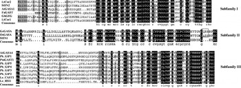
Amino-acid sequence alignment showed that (1) gcgasa protein (GcGASA) belongs to the subfamily II members of GIP and shares the highest identity (~60.8%) with the Oryza sativa homologue OsGASA and (2) different from the other proteins, as demonstrated in Fig. 1, GcGASA and OsGASA have three more amino acids at the C-terminal section, including the positively charged Arg.
The alignment of amino acid sequence among GIP proteins. Le_GAST1(P27057) and Le_RSI1(L22189), Lycopersicon esculentum; StSN1(AJ320185) and StSN2(AJ312904), Solanum tuberosum; AtGASA1(U11766) and AtGASA4(U15683), Arabidopsis thaliana; Ph_GIP1(X87225), Ph_GIP2(AJ417389), Ph_GIP3(AJ417390), Ph_GIP4(AJ417391) and Ph_GIP5 (AJ417392), Petunia hydrida; GhGEG(AJ005206), Gerbera hybrida; FaGAST(AF039183), Fragraria×ananassa; PmGAST1 (A051227), Picea marina; Ltcor11(A007784) and Ltcor12(AF060569), Lavatera thuringiaca; OsGASA(XP_469855), Oryza sativa. Black and grey shading indicates identical or similar amino acid residues, respectively.

Construction and identification of the expression plasmid
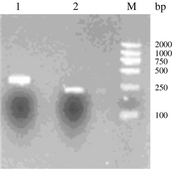
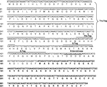
PCR amplification was carried out using cloning plasmid as a template, and F1/R and F2/R as primers. As shown in Fig. 2, two fragments having lengths 319 and 238 bp were detected by agarose gel electrophoresis. They were recovered, digested, ligated into pET-32(a), and then transformed into E. coli JM109. The resultant recombinant plasmid was confirmed by DNA sequencing. Taking pET-32(a)/Δgcgasa as an example, as well as the target protein, ATG, Trx-tag, His6-tag, S-tag and enterokinase were also distinguished in the amino-acid sequence (Fig. 3). The expression vectors were named pET-32(a)/gcgasa and pET-32(a)/Δgcgasa, and encoded 264 and 237 amino acids with predicted molecular weights 28.7 and 25.7 kDa, respectively.
Agarose gel electrophoresis of PCR products of gcgasa gene. Lanes: 1, complete coding region gcgasa; 2, signal peptide-truncated fragment Δgcgasa; M, DL2000 DNA marker.

Partial nucleotide sequences and deduced amino-acid sequences of recombinant vector pET-32(a)/Δgcgasa. gcgasa amino-acid sequences are indicated by bold letters; Trx.Tag, His.Tag, S.Tag and enterokinase are representative of thioredoxin tag, histidine tag, S fragment tag of RnaseA enzyme and enzyme site of enterokinase, respectively.

Expression, SDS-PAGE and Western blot
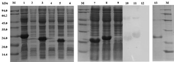
After successful construction of the recombinant plasmids pET-32(a)/gcgasa and pET-32(a)/Δgcgasa, they were transformed into E. coli BL21(DE3) cells. Once an OD600 of 0.6–0.8 at 37°C had been reached, 1 mM IPTG was introduced to the cells to induce expression. SDS-PAGE analysis revealed that there was no yield of heterogeneous proteins in the absence of IPTG, but the addition of IPTG gave rise to production of the heterogeneous protein with an apparent molecular weight of 26.0 kDa for pET-32(a)/gcgasa (lane 1, Fig. 4) and 25.2 kDa for pET-32(a)/Δgcgasa (lane 3, Fig. 4). The former was 2.7 kDa smaller than the calculated molecular mass, and the latter was almost consistent with the predicted value. No significant protein band was observed at ~26 kDa in the supernatant obtained from pET-32(a)/gcgasa (lane 9, Fig. 4), indicating that gcgasa was expressed mainly in the form of an inclusion body. This was not the case for pET-32(a)/Δgcgasa, however, in which a large amount of expressed protein was present in the supernatant (lane 8, Fig. 4). Western blotting results showed that the soluble protein could react with mouse anti-His-tag antibody, displaying a specific reaction band (lane 11, Fig. 4). Further purification was achieved using a Ni2+-NTA affinity column and Sephadex G-75 gel filtration chromatography (lane 13, Fig. 4). Induction of IPTG on the empty vector pET-32(a) gave one soluble protein band at 23.6 kDa, the size of which was larger than the calculated molecular weight (seen in lane 5 and lane 7 of Fig. 4). It could also be hybridized with antibody, mainly due to the occurrence of the His6 tag sequence (lane 10, Fig. 4).
SDS-PAGE and Western blotting result of gene engineering bacteria without induction and after induction with IPTG. Lanes: 1 and 2, 6-h induction and no induction with IPTG on pET-32(a)/gcgasa, respectively; 3 and 4, 6-h induction and no induction with IPTG on pET-32(a)/Δgcgasa, respectively; 5 and 6, 6-h induction and no induction with IPTG on empty vector pET-32(a), respectively; 7 and 10, SDS-PAGE and Western-blotting results of the supernatant obtained from pET-32(a); 8 and 11, SDS-PAGE and Western-blotting results of the supernatant obtained from pET-32(a)/Δgcgasa; 9 and 12, SDS-PAGE and Western-blotting results of the supernatant obtained from pET-32(a)/gcgasa; 13, purified protein; M, protein marker.

Location of the inclusion body
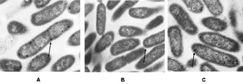
Figure 5 presents TEM images of ultra-thin sections of recombinant plasmids of pET-32(a)/gcgasa, pET-32(a)/Δgcgasa and empty vector pET-32(a). As shown in Fig. 5A, the presence of signal peptide led to the formation of an inclusion body predominantly in the periplasmic space. In contrast, the number of inclusion bodies in the periplasmic space dramatically diminished in the absence of the signal peptide (seen in Fig. 5B). For the empty vector pET-32(a), the majority of inclusion bodies were detected in the periplasmic space, rather than in the cytoplasmic space (Fig. 5C).
Electron micrograph of ultra-thin sections of recombinant plasmid pET-32(a)/gcgasa (A), pET-32(a)/Δgcgasa (B) and empty vector pET-32(a) (C). Arrows indicate the sites of inclusion bodies in cells.

Discussion
At present, studies pay much attention to regulation of the GIP gene, temporal and spatial regulation patterns and subcellular location (Aubert et al., Reference Aubert, Chevillard, Dorne, Arlaud and Herzog1998; Kotilainen et al., Reference Kotilainen, Helariutta and Mehto1999; Ben-Nissan et al., Reference Ben-Nissan, Lee, Borohov and Weiss2004; Furukawa et al., Reference Furukawa, Sakaguchi and Shimada2006). At least eight GAST1 homologues are characterized in Arabidopsis thaliana, and they show different expression patterns in various organs and in different developmental stages. For example, expression of both GASA1 and GASA4 are dependent on exogenous gibberellin (GA), and their mRNA is abundant in flower buds. However, GA has no effect on the closely related GASA2 and GASA3, which have been shown to have high mRNA levels in dry siliques and seeds (Roxrud et al., Reference Roxrud, Lid, Fletcher, Schmidt and Opsahl-Sorteberg2007). The Gerbera hybrida GASA homologue, GEG, is induced by GA and is strongly expressed in developing petals (Kotilainen et al., Reference Kotilainen, Helariutta and Mehto1999). Other homologues were identified, including SNAKIN1 and SNAKIN2 from the potato (Segura et al., Reference Segura, Moreno, Madueno, Molina and Garcia-Olmedo1999; Berrocal-Lobo et al., Reference Berrocal-Lobo, Segura, Moreno, Lopez, Garcia-Olmedo and Molina2002) and RSI1 from the tomato, which is expressed in the root and is induced by auxin (Taylor and Scheuring, Reference Taylor and Scheuring1994). The aforementioned results suggest that although various GA-induced proteins share highly conservative cysteine residues, they are involved in diverse developmental processes and exert different biological functions. This phenomenon raises the question of how function and structure are related to one another. Although nearly 50 GIP sequences have been reported in the NCBI databank, no useful information is currently available regarding their secondary and tertiary structures. In addition, it has been found that the protein is able to eliminate peroxide in vivo. We wonder whether it can still perform this antioxidative function in vitro. These questions need to be resolved in further work.
Based on our previous work, in which we succeeded in screening the gcgasa gene, in this paper we have investigated the effect of signal peptide on the expression of the heterogeneous gene using pET-32(a) as the expression vector. The results of SDS-PAGE, Western blotting and TEM revealed that gcgasa having signal peptide can be highly efficiently expressed in the form of an inclusion body in the periplasmic space of E. coli BL21(DE3). This can be explained as follows: (1) the protein synthetic rate is too fast to ensure formation of the appropriate disulphide bonds and (2) hydrophobic amino-acid residues in the signal peptide should be responsible for the aggregation or insolubility of the heterogeneous protein. As for the phenomenon whereby the target protein has a relative molecular weight ~2.7 kDa smaller than the calculated value, we have presumed that this might result from those basic amino acids and proteins rich in disulphide bonds. Also, we cannot exclude the possibility that this deviation might be within the margin of error. In contrast, the signal peptide-truncated fragment Δgcgasa was mainly expressed in the soluble form with high efficiency. Taking into account the fact that Δgcgasa has a high content of cysteine residues (~17%), we predicted that it is less likely to be produced in reductive cytoplasmic environments, where the formation of disulphide bonds is not favoured (Messens and Collet, Reference Messens and Collet2006). Further studies are needed to explore the cytoplasmic or periplasmic location of the protein.
In this paper, we compared the expression of pET-32(a)/gcgasa and pET-32(a)/Δgcgasa, and found that the absence of signal peptide could greatly enhance the solubility of the target protein, which provides a useful method to obtain the soluble protein. Additionally, this is the first time that soluble GIP has been expressed and purified using a Ni2+-NTA affinity column, laying the foundation for further studies on the structure and function of GIP.
Acknowledgements
This work was supported by the National Natural Science Foundation of China (grant no. 30730065).