INTRODUCTION
The Agassiz's parrotfish, Sparisoma frondosum (Actinopterygii: Labridae), occurs along the Brazilian coast from the Manoel Luiz reef (00°50′S 44°15′W) to Santa Catarina State (27°30′S 48°31′W), including the oceanic islands of Fernando de Noronha (3°54′S 32°25′W), Rocas Atol (3°52′S 33°49′W), the Archipelagos Saint Peter and Saint Paul (00°56′N 29°22′W) and Trindade (20°31′S 29°19′W). Moreover, the species has been recorded in Venezuela, in the Dutch Antilles, Grenada, Trinidad and Tobago (Moura et al., Reference Moura, Figueiredo and Sazima2001; Nelson, Reference Nelson2006; Padovani-Ferreira et al., Reference Padovani-Ferreira, Rocha, Ferreira, Francini-Filho, Moura, Gaspar, Feitosa, Choat, Myers and Russell2012) and at the Cape Verde Archipelago (00°56′N 29°22′W) (Freitas et al., Reference Freitas, Osmar, Silva, Floeter, Bernardi and Ferreira2014). Overall, along the Brazilian coast it is the second most abundant parrotfish species (Padovani-Ferreira et al., Reference Padovani-Ferreira, Rocha, Ferreira, Francini-Filho, Moura, Gaspar, Feitosa, Choat, Myers and Russell2012), locally called budião. The reclassification in 2001 of this parrotfish as a separate species from that which occurs in the Caribbean, suggests that the records of occurrence of this species in the Caribbean islands may be misidentifications – as this species is in fact endemic to Brazil (Freitas et al., Reference Freitas, Osmar, Silva, Floeter, Bernardi and Ferreira2014).
Sparisoma frondosum occupies reef areas located in depths from 1 to 54 m where it reaches sizes of 38 cm of length (total length – TL) (Moura et al., Reference Moura, Figueiredo and Sazima2001; Ribeiro, Reference Ribeiro2004). Males and females show different patterns of colours, displaying in the upper caudal peduncle a dark spot, which is a distinctive feature from other western Atlantic species from the same family. In north-eastern Brazil, this is the most frequent species of the Sparisoma genus (sensu Westneat & Alfaro, Reference Westneat and Alfaro2005) in catches of fish-traps, which also include other reef species such as the parrotfish S. axillare and the spotted goatfish Pseudupeneus maculatus (Ribeiro, Reference Ribeiro2004).
Analysing official catch data annual average landings of Sparisoma species were estimated around 200 t in Pernambuco for the period 1996 to 2001 (Lessa & Araújo, Reference Lessa, Araújo, Lessa, Bezerra and Nóbrega2009) as well as for 2002 and 2005 (136.5 and 280.5 t, respectively) (Véras, Reference Véras2008). The number of gear annually employed for obtaining those captures ranged from 150 traps day−1 in April to 4500 traps day−1 in September/October and January (Ribeiro, Reference Ribeiro2004).
There are indications of overfishing for this species in south-eastern Brazil, which is worrying since fishing pressures have been steadily increasing throughout the distribution range of the Sparisoma species (Padovani-Ferreira et al., Reference Padovani-Ferreira, Rocha, Ferreira, Francini-Filho, Moura, Gaspar, Feitosa, Choat, Myers and Russell2012). Because of this, the Agassiz's parrotfish was included in the IUCN red list as Data Deficient (DD) (Padovani-Ferreira et al., Reference Padovani-Ferreira, Rocha, Ferreira, Francini-Filho, Moura, Gaspar, Feitosa, Choat, Myers and Russell2012), and was classified in the Vulnerable (VU) category in recent regional assessments in Brazil (Brazil, Ordinance 445 of 17.12.2015) in recognition of the threats that include fisheries.
In addition to the fishing pressure, the continuing degradation and loss of coral reef habitat on which this species is dependent for shelter and food (Padovani-Ferreira et al., Reference Padovani-Ferreira, Rocha, Ferreira, Francini-Filho, Moura, Gaspar, Feitosa, Choat, Myers and Russell2012) is a matter of concern. Furthermore, based on life history, ecological characteristics and extinction vulnerability of marine fishes, Cheung et al. (Reference Cheung, Pitcher and Pauly2005) developed an index of intrinsic vulnerability for 835 species, among them some species from the Sparisoma genus, which obtained the index 48 (10 being the least vulnerable and 88 being the most vulnerable). Therefore, it is clear that the assessment of fisheries that allows the population age structure as well as the sizes most heavily impacted by fisheries to be known, are important in order to contribute meaningfully to fishing management (Longhurst & Pauly, Reference Longhurst and Pauly1987).
Aside from the general lack of knowledge about age distribution, reproductive aspects of this species are also unknown. Sparisoma frondosum is assumed to be a functional hermaphrodite, presenting a diandric protogynous pattern (Robertson & Warner, Reference Robertson and Warner1978), similar to other species of the same genus studied in the Caribbean. This hermaphrodite strategy encompasses a change in gender from female to male that may occur either from mature females or directly from the juvenile phase (Shapiro, Reference Shapiro1987; Sadovy de Mitcheson & Liu, Reference Sadovy de Mitcheson and Liu2008). However, in this species the pattern of protogyny has not yet been ascertained.
The impact of fishing in hermaphroditic species is complex and dependent on mating systems. Selective pressures on adult individuals can significantly alter sex change dynamics (Taylor, Reference Taylor2014). One concern is that sexual inversion in a long-lived and large-body species may be hindered by fisheries, which reduce the average length of the fish (Sparre & Venema, Reference Sparre and Venema1997), preventing individuals from reaching the minimum size for sex reversal (Warner, Reference Warner1984; Francis, Reference Francis1992). On the other hand, in contexts with less intensive fishing pressure, protogynous hermaphroditism may increase sufficient resiliency to prevent stock depletion (Petersen & Warner, Reference Petersen, Warner and Sale2002) – and it may be a better strategy than that of gonochorism (Bannerot, Reference Bannerot1984).
The knowledge that the inversion of sex in hermaphrodites can be triggered by social stimuli, has led to an increasing number of studies on behavioural, ecological and evolutionary aspects of hermaphroditism (Sadovy & Shapiro, Reference Sadovy and Shapiro1987; Alonso-Fernandez et al., Reference Alonso-Fernández, Alós, Gran, Domingues-Petit and Saborido-Rey2011; Collins & McBride, Reference Collins and McBride2011). However, overall information on this species is scarce and restricted to secondary literature.
Our aim in the current study was to estimate age through otolith analysis, fitting a first growth curve for S. frondosum. With this, we intend to provide information on size and sex composition of samples through microscopic analyses, so that size at first maturity can be estimated – which is essential for fisheries management.
MATERIALS AND METHODS
Monthly samples of S. frondosum were randomly collected in landings of vessels at Itamaracá Island (Pernambuco) from May 2009 to April 2010 (Figure 1). Daily fishing trips were conducted using hexagonal unbaited fish traps, 1 cm mesh size, measuring 90 cm in length and 72 cm in height (the largest diameter of aperture being 24 cm and the smallest 9 cm). Small motorized vessels of 8.5 to 12 m in length operating in depths of up to 60 m were used (IBAMA, 2005).
Location of the the area (shaded in dark grey) of collection of Agassiz's parrotfish S. frondosum in north-eastern Brazil from 2009 to 2010.

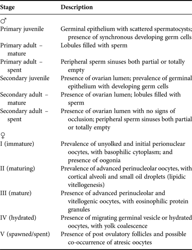
The total length (TL, cm) and gutted weight (GW, g) of each specimen caught were recorded and the gonads were removed and stored in 4% formaldehyde solution. Sections of the anterior, medium and posterior areas of gonads were taken for ascertaining the sexes. They were dehydrated in alcohol series of different concentrations, diaphanized in xylene, embedded in paraffin at 60°C, sectioned at 6 µm and finally stained with haematoxylin-eosin. After this, sections were examined under a light microscope and gender was assigned following the descriptions of Sadovy & Shapiro (Reference Sadovy and Shapiro1987) for males, Kume et al. (Reference Kume, Kubo, Yoshimura, Kiriyama and Yamaguchi2010) for females, and Hastings (Reference Hastings1981) for transitional individuals. Female gonads were also inspected for classification into maturity phases based on the relative proportions of the various development stages of germ cells in the ovary, following Shapiro et al. (Reference Shapiro, Sadovy and McGehee1993) and Sadovy & Colin (Reference Sadovy and Colin1995) (Table 1).
General histological description of the maturity stages of the functional males and females of Sparisoma frondosum from north-eastern Brazil.

The overall ratio of males to females in the catches and the monthly frequencies of length classes by sex were tested using two-tail Chi-square tests (χ2; gl = 1; α = 0.05) (Zar, Reference Zar1999). The relationship between gonad weight (GW) and TL of males and females was estimated, linearized and tested to show differences between sexes (ANCOVA, α = 0.05). Comparisons of mean length by sex were performed using the Mann–Whitney U-test (Zar, Reference Zar1999) through the two-tail Chi-square test (Zar, Reference Zar1999).
An estimate of the length at 50% of maturity for males and females (TL, cm), divided in class intervals of 2 cm, were obtained from a logistic equation. Parameters of the model were derived by linearization of the curve through the use of the least-squares method (King, Reference King1995):
 $$P = \displaystyle{1 \over {1 + {{\rm e}^{\left[ { - r \times \left( {L - {L_m}} \right)} \right]}}}}$$
where: P is the proportion of mature fish in length class; L is the midpoint of the length class, r is the slope of the curve and L m is the size at 50% maturity derived from |a/b.
The pair of the sagittae was removed, soaked in a solution of bleach (sodium hypochlorite) and rinsed in distilled water and ethanol. The left otolith of the pair was embedded in transparent polyester resin and transversally cut using a metallographic saw, preserving the core (Panfili et al., Reference Panfili, de Pontual, Troadec and Wright2002). The sections were affixed with crystalbond on glass slides, sanded and polished in alumina powder of different granulations (0.33 to 3.0 µm). Two independent readings along the major axis of the otolith were made by the same reader under 25 × magnifications using a binocular dissecting microscope and transmitted light. On each occasion the reader was unaware of the length of the individual or the result of the previous reading.
To estimate the accuracy of the readings, the average percentage error (APE) (Beamish & Fournier, Reference Beamish and Fournier1981) for comparing the reproducibility of age determination between the readings was used:
 $${\rm APE} = 100\% \times \displaystyle{1 \over R} \times \sum\limits_{i = 1}^R {\displaystyle{{\left\vert {{X_{ij}} - {{\overline X }_j}} \right\vert} \over {{{\overline X }_j}}}} $$
where N is the number of samples; R is the number of age determinations for the sample; X ij is the estimated age; and X j is the average of estimations.
The periodicity of ring deposition was inferred through Cadvallader's (Reference Cadvallader1978) equation. Statistical differences in monthly marginal increment ratios (MIR) were tested using the non-parametric Kruskal–Wallis test, and the post-hoc Dunn's test was employed to examine the differences among individual months (α = 0.05) (Zar, Reference Zar1999):
 $${\rm MIR} = \displaystyle{{RO - {R_n}} \over {{R_n} - {R_{n - 1}}}}$$
where RO is the distance from the core to the otolith edge; R n is the distance from the core to the last ring; and R n–1 is the distance from the core to the penultimate ring.
The relationship between the length of the otolith and individual fish length was examined through linear regression and ANCOVA (α = 0.05) was used for examining statistical differences between regression lines by sex. Distances from the otolith core to each ring were back-calculated through the Monastyrsky formula:
 $${L_i} = {\left( {\displaystyle{{{S_i}} \over {{S_c}}}} \right)^b} \times {L_c}$$
where L i is the back-calculated length at the moment of ring deposition; L c is the length at capture; S i is the distance between the core to each ring; S c is the otolith radius; and b is the slope of regression between otolith radius and L c .
The VBGF (von Bertalanffy, Reference von Bertalanffy1938) and Gompertz (Reference Gompertz1825) models were adjusted to total length vs age data. Parameters were estimated through the SOLVER routine of the Excel program, using the least squares method: VBGF: L t = L ∞ × [1 − e−k × (t−t 0)] and Gompertz: L t = L ∞ × e[−a×e(−k × t)], where L t is length at age t; L ∞ is the asymptotic length; k is the growth coefficient; t 0 is the theoretical age for L t zero; and a is a parameter of the equation.
Then, the Akaike's information criterion AIC = 2log(θ) − 2k (Akaike, Reference Akaike1974), differences in AIC values (Δ i ) and the weight of evidence (w i ) (Burnham & Anderson, Reference Burnham and Anderson2002) in favour of each model were computed to select the most suitable: Log(θ) being the maximal likelihood; and k being the number of parameters of the model.
The maximum longevity achieved was estimated from the size-at-age distribution (Choat et al., Reference Choat, Robertson, Ackerman and Posada2003). Moreover, the age at first maturity was estimated through the inverted VBGF:
 $${t_m} = {t_0} - \left[ {\left( {\displaystyle{1 \over K}} \right) \times \left( {1 - \displaystyle{{{L_m}} \over {{L_\infty}}}} \right)} \right]$$
Using the inverted von Bertalanffy growth function on individual total length, the catch curve (King, Reference King1995) was used for estimating the total mortality rate (Z). The Hewitt & Hoenig (Reference Hewitt and Hoenig2005) equation was used for estimating natural mortalities (M) for male, female and both sexes.
 $$M = \displaystyle{{4.22} \over {{t_{\max }}}}$$
Throughout the text all statistical inferences were made at a significance level of 0.05.
RESULTS
Population structure: size, sex and size at first maturity
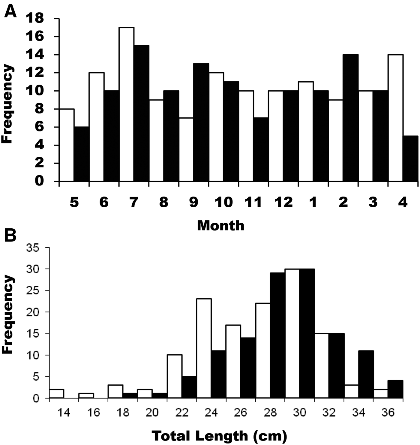
From the 251 parrotfish analysed, 121 were males and 130 were females, resulting in an overall male:female sex ratio of 1:1.07, not significantly different from 1:1 (χ 2 = 0.256; g.l. = 1; P > 0.05). Conversely, when monthly proportions were considered, significant differences were found in April (χ 2 = 4.263; g.l. = 1; P < 0.05), with significantly more females being captured (Figure 2A).
Monthly frequency of males and females (A) and length frequency distribution (B) of males (black columns) and females (white columns) of the parrotfish S. frondosum caught in north-eastern Brazil (Pernambuco) from 2009 to 2010.

Males of 17.5 to 36.6 cm (mode 27–31 cm) and females of 13.1 to 36.8 cm (mode 25–31 cm) were captured (Figure 2B). Mean length of males in the sample was 27.7 ± 3.1 cm TL, whereas mean length of females was slightly smaller (25.8 cm ± 2.3 cm TL), thus there was no significant difference between sexes (Mann–Whitney U-test = 0.067, g.l. = 248, P > 0.05).
However, the ratio between males and females did differ significantly among length classes. In length class 23–25 cm TL females were more numerous than males (67.6% females) (χ 2 = 4.000, g.l. = 1, P < 0.05) (Figure 2B), whereas length class 33–35 cm TL was dominated by males (78.6% of examined individuals). The stages of development of germ cells in ovaries were assigned as shown in Table 1.
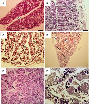
Secondary males were observed; both mature and spent (Figure 3A, B). The gonadal morphology was similar, presenting the ovarian lumen and a lamellar organization with peripheral sperm sinuses, but in the spent secondary males the peripheral region presented residual sperm (Figure 3).
Photomicrographs of male and female gonads of S. frondosum, in various reproductive stages stained with haematoxylin and eosin. (A) Transverse section of a secondary mature testis showing central lumen (white arrow); (B) Longitudinal section of a secondary spent testis showing central lumen (white arrow) and empty areas with residual spermatozoids (black arrows) in peripheral region. (C) Transverse section of functional ovary in initial development/immature stage showing initial perinucleolar oocytes (arrows); (D) Mature stage of functional ovary, showing cells in lipid vitellogenic phase (black arrows), with prevalence of vitellogenic oocytes (white arrows); € Hydrated, with residuals of hydrated oocytes (arrows); (F) Spent ovary showing atresic oocytes (arrows). Scale bars: 200 µm (A, B, C, D), 400 µm (E, F).

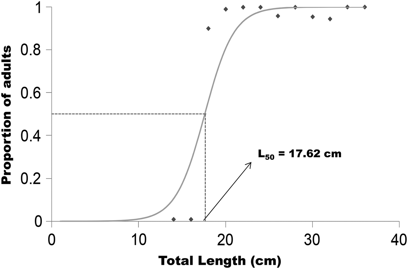
The estimated size at first maturity (L m ) was 17.6 cm (SE = 3.6 cm) for females (Figure 4). For males it was not possible to determine the size at first maturity due to the absence of immature individuals in the overall sample. However, the smallest mature males were found in size class 17.0–18.0 cm (TL).
Relationship between the proportion of adults and total length of females of S. frondosum caught in north-eastern Brazil (Pernambuco) from 2009 to 2010.

Age, growth and age composition of samples
A total of 251 otoliths had the ventral region inspected along an axis drawn from the nucleus to the edge of the structure. Alternate opaque and translucent zones considered as growth rings were counted and measured (Figure 5). In the whole sample 1 to 9 rings were recorded in individuals from 13.1 to 36.8 cm TL. The percentage of error among readings ranged from 0 to 3.3%, with an overall sample average of 1.1%.
Transverse section of an otolith of a 26 cm TL female of Sparisoma frondosum, caught in north-eastern Brazil from 2009 to 2010 under transmitted light showing four rings (black dots). D: dorsal edge, N: nucleus, V: ventral edge.

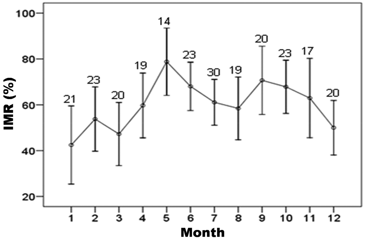
The mean relative marginal increment was lowest in January (Figure 6), suggesting that the otolith growth ring is laid down during this month. The Kruskal–Wallis test showed significant differences (H = 10.512; g.l. = 11; P < 0.05) among monthly median values that were caused by January and May.
Monthly relative marginal increment (IMR) of otoliths of S. frondosum caught in Pernambuco, Brasil. Vertical bars are mean values with standard deviations and numbers are the sample size per month.

The relationship between otolith radius and individual fish length (TL) was not significantly different between the sexes (ANCOVA; P > 0.05); it was represented by the potential regression TL = 8.390 RO 1.0801 (N = 178, r 2 = 0.7293, SE = 0.16) for both sexes combined.
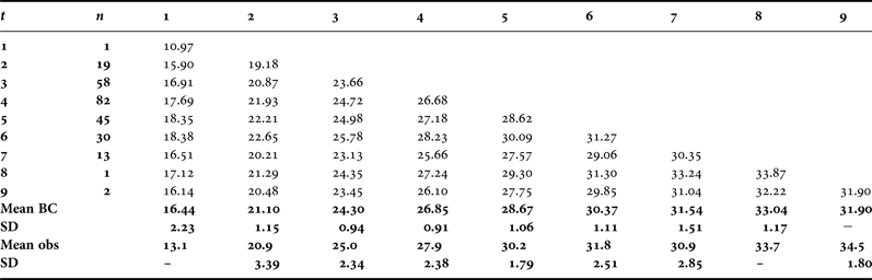
Mean length at observed and back-calculated ages were similar (Table 2). However, the lowest value for the sum of squares of residuals was obtained for mean observed lengths (OL), therefore, whenever age is mentioned hereafter, we refer to OL.
Mean observed (obs) and back-calculated (BC) lengths for age class, for sexes combined of S. frondosum, caught in north-eastern Brazil (Pernambuco) from 2009 to 2010.

SD = standard deviation, n = number of individuals.
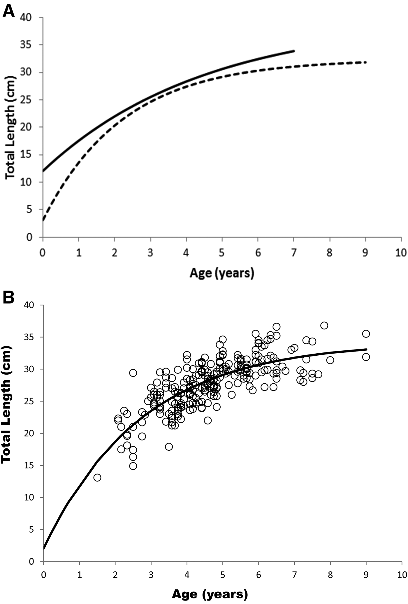
Growth parameters were estimated adjusting the von Bertalanffy and Gompertz models on the basis of relative observed ages (Table 3; Figure 7). Slightly lower AIC and higher w i values were obtained for the von Bertalanffy model, that was chosen for describing growth for this species (Table 3).
Growth curve estimated using the von Bertalanffy model (VBGF) for males (solid line) and females (dashed line), separate (A) and combined sexes (B) for S. frondosum caught in north-eastern Brazil (Pernambuco) from 2009 to 2010.

Growth parameters estimated for separate and combined sexes adjusting the von Bertalanffy (VB) and Gompertz (G) models to observed lengths for S. frondosum from north-eastern Brazil (Pernambuco) caught from 2009 to 2010.

Growth curves for separate sexes of S. frondosum were different according to the likelihood test (χ2 = 73.46, P > 0.05), so the parameters of VBGF for each sex separately indicated that both males (L ∞ = 39.7 cm TL, K = 0.22, t 0 = −1.63 years) and females (L ∞ = 32.3 TL cm, K = 0.44; t 0 = −0.23 years) display rapid growth in the early years of life, reaching about 50% of their maximum length (L ∞ ) at 2 years of age (Figure 7A). At 5 years of age, 75% of the maximum length was attained after which growth slows considerably.
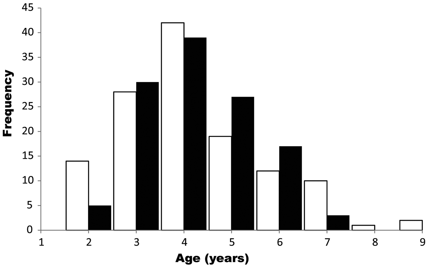
Parameters of VBGF estimated for combined sexes were: L ∞ = 33.6 cm TL, K = 0.41 and t 0 = −0.27 years (SE = 0.15) (Figure 7B). Age composition for the overall sample (N = 251) (Figure 8) showed that 55% of individuals were 3 and 4 years of age, the maximal age being 9 years. Females were present in all age classes from 1 to 9 years whereas males occurred in age classes from 2 to 7 years only. Mature females represented 45% of catches, since the age at first maturity was 1.6 years (17.6 cm TL), estimated on the basis of the inverted VBGF. A female from the age class 8–9 was the oldest specimen in the whole sample whereas the oldest male was estimated to be 7–8 years of age (Figure 8).
Age distribution of males (black columns) and females (white columns) of S. frondosum, caught in north-eastern Brazil (Pernambuco) from 2009 to 2010.

The natural mortality estimated using the Hewitt & Hoenig's equation for S. frondosum resulted in M = 0.469, whereas total mortality was Z = 0.739, estimated through the catch curve.
DISCUSSION
The Sparisoma species follow two patterns of sexuality, gonochorists or diandric protogynous hermaphrodites (Robertson et al., Reference Robertson, Karg, Moura, De Victor and Bernardi2006; Sadovy de Mitcheson & Liu, Reference Sadovy de Mitcheson and Liu2008), and this information is important to properly investigate how sexes are allocated throughout the range of sizes. An example of this is the current study, where sexes were microscopically assigned.
Corroborating with Robertson & Warner (Reference Robertson and Warner1978), who only found secondary males in samples, in the current study PM weren't detected in the whole sample (although smaller females starting at 13 cm TL were collected), suggesting that males were derived from females (Reinboth, Reference Reinboth1967; Alonso-Fernández et al., Reference Alonso-Fernández, Alós, Gran, Domingues-Petit and Saborido-Rey2011).
Furthermore, features observed in S. radians and S. chrysopterum (Robertson & Warner, Reference Robertson and Warner1978; Robertson et al., Reference Robertson, Karg, Moura, De Victor and Bernardi2006) were also found in S. frondosum, such as similarity of sex-ratios (with females prevailing by only 3.5%, not significantly different), females as large as males and females barely attaining identical maximal sizes. Therefore, considering that S. frondosum follows the same diandric protogynous hermaphroditism as the species above, the smallest individuals in the sample were a 13.1 cm TL female and a male of 17.5 cm TL. The latter also exhibited characteristics of a secondary mature male (SM), supposedly having changed sex at an undetermined smaller class, since only females were smaller than 17 cm. Results by Sadovy de Mitcheson & Liu (Reference Sadovy de Mitcheson and Liu2008) on S. aurofrenatum, suggest that the lack of gonads undergoing sexual transition in S. frondosum – a feature considered the strongest evidence of sex change in fishes – requires further investigation.
With respect to size at maturity, females in the overall sample were either clearly immature (under 17 cm TL) or fully mature in classes larger than 17 cm TL. Hence, the expected gradual increase of the percentage of mature individuals, with the increase of body size, was not perceived. The allocation of mature females throughout the range of sizes may have caused biases in the estimate. Still, in females of the largest fully mature sizes, the sex inversion is likely to result from a socially determined process driven by a decrease in the number of males, for instance, due to fisheries (Pauly et al., Reference Pauly, Christensen, Dalsgaard, Froese and Torres1998). The absence of males smaller than 17 cm TL (although females were recorded starting at 13 cm TL), hampered the estimation of the size of maturity for males, which in another study using a different approach was suggested at 17 cm (SL) off the coast of Pernambuco (Véras, Reference Véras2008). Thus, 17 cm TL was assumed as a speculative first maturity size for males.
In the present study sagittal otoliths of S. frondosum were sectioned with the objective of obtaining more accurate estimates, a requirement especially important for older fish (Crabtree et al., Reference Crabtree, Harnden, Snodgrass and Stevens1996) – which are more prone to the stacking of rings on the otolith edge (Lou, Reference Lou1992). It is essential to know the periodicity of increments for the accuracy of age estimates. Growth rings were clearly observed along the reading axis of otoliths with annual periodicity inferred through marginal increment analyses, as was also shown for S. viride in the Caribbean (Choat et al., Reference Choat, Robertson, Ackerman and Posada2003). In addition, the average percentage error (APE) indicated that errors of readings were within the recommended limits (Campana, Reference Campana2001), meaning that otoliths are suitable for age estimations and readings can be repeated.
The lowest value of the marginal increment in January (summer) was due to the deposition of the translucent ring with the new band forming from that month onwards. It may be related to the reproductive period, as suggested for species of this family which have peaks of spawning in summer (Jonna, Reference Jonna2003; Kume et al. Reference Kume, Kubo, Yoshimura, Kiriyama and Yamaguchi2010), when the greatest reproductive activity occurs and most of the energy of the body is assigned to reproduction (spawning period), not to somatic growth (Takada & Tachihara, Reference Takada and Tachihara2009).
Overall, ages ranged from 2 to 7 years in males and from 1 to 9 years in females, with maturity of females being 1.6 years of age. The maximal age for S. frondosum corroborates with Choat et al. (Reference Choat, Robertson, Ackerman and Posada2003) for S. viride in four populations of the Caribbean. Such a result may be an attempt by the fish to maximize reproductive success, where some females do not change sex, aiming to achieve higher fertility in larger classes when compared with other females of her social group. Thus, the postponement of sex reversal could be deemed as an advantage for the population (Muñoz & Warner, Reference Muñoz and Warner2003; Hixon et al., Reference Hixon, Johnson and Sogard2014).
The Akaike information criterion (AIC) has parsimony as its tenet, which implies that models with a smaller number of parameters are the most appropriate, expressing a satisfactory balance between bias and variance (Burnham & Anderson, Reference Burnham and Anderson2002). Thus, even if the lowest AIC value and the highest w i were estimated for the VBGF, the Gompertz model also had a performance that adequately described the growth of this parrotfish. However, the VBGF was chosen because it has described the growth of similar species, having a stronger biological basis and allowing parameters to be used in stock assessments (Campana, Reference Campana2001).
Considering the reliability of the parameters of VBGF regarding the L ∞ , observed lengths provided a result that was closer to the maximal recorded length for the species (L max = 36.8 cm TL), thus it was chosen as the most accurate for the growth curves of S. frondosum. The L ∞ for both sexes combined conforms to this upper limit of maximal size in the sample, whereas the lower limit agrees with the maximal sizes recorded in other places of the species’ distribution range (Vaske et al., Reference Vaske, Lessa, Ribeiro, de Nóbrega, Pereira and de. Andrade2006). The overall rapid growth in the early years of life, reaching about 50% of their maximum length (L ∞ ) at 2 years of age, complies with findings for other Sparisoma species of the Caribbean (Choat & Robertson, Reference Choat, Robertson and Sale2002; Choat et al., Reference Choat, Robertson, Ackerman and Posada2003).
The S. frondosum species is referred to as a sister species of S. chrysopterum, from the Caribbean (Moura et al., Reference Moura, Figueiredo and Sazima2001), which may suggest similar growth parameters (Choat & Robertson, Reference Choat, Robertson and Sale2002). However, higher longevity (t max) and faster growth (K) were estimated for S. frondosum, revealing different growth patterns. Moreover, the largest specimen of S. frondosum ever recorded was a 34.5 cm SL (Moura et al., Reference Moura, Figueiredo and Sazima2001) (~38 cm TL), which corresponds with the largest size in the current study. Also, the L ∞ estimated for females and both sexes combined are compatible with the largest sizes for the species. Finally, the L ∞ estimated for males exceeded the largest size in the sample by about 5% being quite acceptable according to Campana's criterion (Reference Campana2001). Overall, the VBGF for combined sexes (Figure 7B) provided a K parameter compatible with the values estimated for the Sparisoma species from the Caribbean as shown by Choat et al. (Reference Choat, Robertson, Ackerman and Posada2003).
In the current study natural mortality >0.45 was estimated, which together with the index of intrinsic vulnerability to fisheries (estimated for S. cretensis – 48 in a rank ranging from 10 to 88) (Cheung et al. Reference Cheung, Pitcher and Pauly2005) suggests that S. frondosum deserves attention to keep the population stable. However, according to Petersen & Warner (Reference Petersen, Warner and Sale2002) the protogynous hermaphroditism, which has not yet been completely ascertained in S. frondosum, may be a strategy that increases resiliency to fishing depletion when stocks are submitted to moderate fishing pressure, as seems to be the case in the study area.
In spite of that, in Brazil several species of the Sparisoma genus (including S. frondosum) were classified as vulnerable (VU) using the IUCN criteria (Brazil, Ordinance 445 of 17.12.2014), which demands the adoption of conservation measures, such as reduction of fishing efforts, among others (not necessarily related to fisheries). In this context, information on gonad maturation, growth rates, age structure and longevity is important for the assessment of the state of exploitation of this parrotfish in years to come.
ACKNOWLEDGEMENTS
The authors are indebted to local fishermen and to two anonymous reviewers for comments and suggestions that highly improved the manuscript.
FINANCIAL SUPPORT
Conselho Nacional de Desenvolvimento Científico e Tecnológico- CNPq supplied a Productivity Research Grant to the first author R.L. (PQ 303604/2007-7) and a Master's of Science Scholarship to C.R. da S. (580112/2008-0).