Introduction
Bronchoscopic removal of a foreign body from the upper airways is a common emergency procedure in paediatric otolaryngology. While many foreign bodies can be quickly and safely removed by an experienced surgeon, there are situations where the patient's state of respiratory compromise or the nature of the foreign body makes expeditious foreign body removal difficult or even impossible. Large foreign bodies that are difficult to grasp and manipulate can cause complete obstruction of the airway during attempts at removal, leading to hypoxic cardiac arrest.
Extracorporeal membrane oxygenation is used as a temporary form of respiratory support for potentially reversible conditions. Although extracorporeal membrane oxygenation is usually used for prolonged support in an intensive care setting, it can also be used for shorter periods to provide safe operating conditions in the absence of spontaneous or mechanical ventilation.
We present a case of a child who aspirated a foreign body; the object could not be extracted conventionally, but was successfully extracted via rigid bronchoscopy using veno-venous extracorporeal membrane oxygenation respiratory support.
Case report
A 12-year-old (40 kg) boy was playing with his friends when he picked up what he thought was a shiny blue sapphire. As his friends wanted to grab it from him, he put the stone in his mouth and ran, causing him to aspirate it and collapse. He was admitted to the local hospital, from which he was transferred to the tertiary care emergency service.
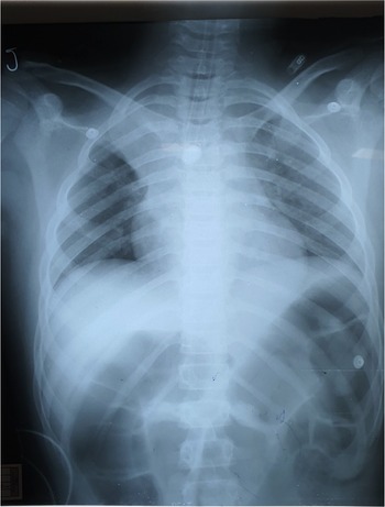
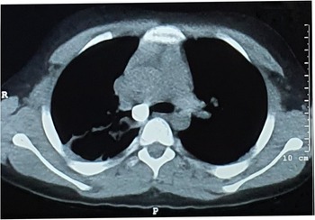
Multiple attempts were made by the otolaryngology team to remove the foreign body using rigid bronchoscopy under general anaesthesia. The foreign body proved difficult to remove because of its large size, smooth surface and oval shape, which caused it to lodge transversely across the carina and obstruct both major bronchi during manipulation (Figures 1 and 2). As a temporary measure, the foreign body was pushed into the right bronchus, and the patient was stabilised on single lung ventilation and transferred to the paediatric intensive care unit.
Chest X-ray showing the foreign body.

Axial computed tomography showing the foreign body. R = right; P = posterior

The patient was referred to the cardiothoracic unit for surgical removal of the foreign body from the right bronchus. An urgent multidisciplinary meeting was called to consider the best available options. As a result, it was decided to reattempt rigid bronchoscopy with the options to proceed to extracorporeal membrane oxygenation support and/or surgery, and parental consent was taken accordingly.
Another attempt was made at bronchoscopic removal of the foreign body with additional operators and instruments being available. During each attempt, there was hypoxia and progressive hypercarbia, making the continuation of instrumentation dangerous. Therefore, it was decided to use veno-venous extracorporeal membrane oxygenation support to stabilise respiratory functions so that there could be a prolonged, uninterrupted period for upper airway instrumentation.
After partial heparinisation (75 IU/kg), the child was percutaneously cannulated for veno-venous extracorporeal membrane oxygenation using size 19F drainage and 18F return cannulae (Bio-Medicus cannula; Medtronic, Minneapolis, Minnesota, USA) inserted by the Seldinger technique into the right femoral and right internal jugular vein, respectively. The cannulae were connected to an extracorporeal membrane oxygenation circuit incorporating a Levitronix blood pump (Abbott Laboratories, Pleasanton, California, USA) and a polypropylene hollow fibre adult oxygenator (Sorin, Milan, Italy).
Extracorporeal membrane oxygenation flows were started at 50–60 ml/kg/minute flow, and the child rapidly became 100 per cent saturated. As the child was hypercarbic at the onset, the oxygenator gas flow was commenced at 5 litres per minute for 10 minutes to reduce the partial pressure of carbon dioxide (CO2) to normal levels, after which it was reduced to 3 litres per minute. The initial activated clotting time was 145 seconds, and further heparin was given, bringing the activated clotting time to 191 seconds. (The normal activated clotting time target range for extracorporeal membrane oxygenation with this system is 180–200 seconds.)
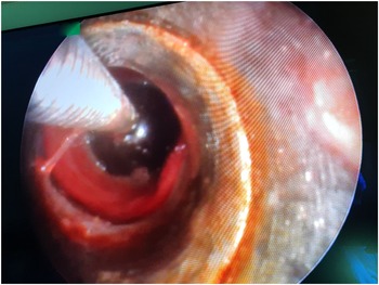
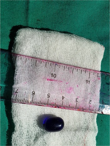
Having stabilised the blood gases (pH 7.4; partial pressure of CO2 = 40 mmHg; partial pressure of oxygen (O2) 240 mmHg), a 7.5 mm rigid ventilating bronchoscope was inserted. Secretions were removed to expose the foreign body, which was now situated within the carina and was partially wedged into the right bronchus. Using a combination of Fogarty catheters, forceps and a Dormia® basket, the foreign body was eased into the trachea below the vocal folds and then extracted through them intact within the Dormia basket (Figure 3). The sapphire-blue oval glass bead measured 10 × 6 mm (Figure 4). Despite the total period of uninterrupted manipulation being 25 minutes, there was no bleeding. During the procedure, the partial pressure of CO2 and pH remained as before, although the O2 saturation levels dropped to 84 per cent at one point. This was compensated for by increasing the extracorporeal membrane oxygenation flows to 80 ml/kg/minute.
Foreign body in right bronchus within Dormia basket.

Blue glass bead after removal.

Having further cleared secretions from the trachea, a 6.5 mm cuffed endotracheal tube was inserted. Mechanical ventilation was started at conventional tidal volumes. The extracorporeal membrane oxygenation flows were reduced and then stopped as the partial pressure of O2 and partial pressure of CO2 remained at normal levels. The duration of extracorporeal membrane oxygenation support was 57 minutes. The extracorporeal membrane oxygenation cannulae were removed, and each of the two skin wounds was closed with a single skin stitch. As the activated clotting time was only 154 seconds at this point and there was no bleeding, protamine was not given.
The patient was returned ventilated to the intensive care unit. Chest physiotherapy was given to expand a collapsed right upper lobe. After 24 hours, once fully optimised, he was extubated neurologically intact. He had a hoarse voice and a poor cough, and was nebulised with dexamethasone and lignocaine, to which he responded well. He was sent to the ward after 48 hours and discharged home on the 6th day. He remains well at follow up.
Discussion
The best chance of removing a bronchial foreign body is during the first attempt. Every effort should be made for an experienced operator to perform this bronchoscopy with a full range of endoscopic instruments being available. If that fails, respiratory compromise tends to progress, making further bronchoscopy more difficult and dangerous as the anaesthetist tries to maintain gas exchange. In the case reported herein, the difficulties encountered at the first procedure were the size, shape and slippery surface of the bead. The possible next options were to attempt further bronchoscopy or perform surgical removal from the right bronchus. Surgery has its own difficulties, as the foreign body can move from the right bronchus during patient positioning or lung manipulation during surgical exposure and drop into the trachea or left main bronchus, producing respiratory instability and difficulty in retrieving the foreign body – similar difficulties to those experienced by the bronchoscopist.
Extracorporeal membrane oxygenation developed from cardiopulmonary bypass technology as a means of providing exceptional cardiorespiratory support in the intensive care setting. It is widely used in severe respiratory failure cases in all age groups, and as an adjunct to congenital cardiac surgery and transplantation as a cardiac support, and its availability in these units is regarded as a standard of care. Extracorporeal membrane oxygenation can occasionally be used as an adjunct to perform procedures that would be either difficult or impossible without extracorporeal respiratory support. Removing difficult foreign bodies from the airway is one such indication.
According to the international Extracorporeal Membrane Oxygenation Registry (Extracorporeal Life Support Organization Registry, Ann Arbor, Michigan, USA), 42 patients have been reported as having undergone extracorporeal membrane oxygenation as a consequence of an airway foreign body over a 30-year period, with a median age of 1.54 years (range, 0.04–16.7 years).Reference Anton-Martin, Bhattarai, Rycus, Raman and Potera1 Extracorporeal membrane oxygenation is used in such cases for airway stabilisation, instrumentation or post-procedural support for respiratory complications, or a combination of these.Reference Isherwood and Firmin2
Extracorporeal membrane oxygenation can be provided in two modes. Veno-venous extracorporeal membrane oxygenation can provide respiratory support if cannulation is performed with either a double-lumen venous cannula or two single-lumen cannulae, as in this patient. Venous blood from the drainage cannula is pumped through the oxygenator, where gas exchange occurs, and enters the venous system via the return cannula. Veno-arterial extracorporeal membrane oxygenation provides cardiorespiratory support, and requires drainage from a vein and re-infusion into an artery.
Extracorporeal membrane oxygenation is not universally accessible; it tends to be available in only a few tertiary referral hospitals. In theory, cardiopulmonary bypass, which is more widely available than extracorporeal membrane oxygenation, could be and has been used for foreign body removal.Reference Deng, Wang, Wang, Xiao and Liu3 In some reported instances,Reference Li, Wu, Huang, Zhou, Wang and Chen4 an open operation with a cardiopulmonary bypass was indicated because there was a vascular sling tracheal obstruction as well as a foreign body. However, cardiopulmonary bypass requires arterial cannulation with attendant risks, and full heparinisation risks haemorrhage. An oxygenator used in cardiac surgery was used in this patient (rather than the polymethylpentene extracorporeal membrane oxygenation oxygenator) to reduce costs.
For a situation such as foreign body retrieval, veno-venous extracorporeal membrane oxygenation has been shown to provide adequate respiratory support. The O2 delivery is controlled by the amount of blood flow and the CO2 clearance by the oxygenator gas flow. Only a low dose of heparin is used, which tends to obviate bleeding. The cannulae are inserted percutaneously over a guidewire, and vascular complications are rare. Removal of these cannulae is simple, requiring only a skin stitch.
As this case shows, it is sometimes impossible for an experienced operator to perform the necessary manipulations and maintain gas exchange at the same time. Extracorporeal membrane oxygenation support allowed 25 minutes of uninterrupted interventions, which produced a successful outcome. Others have come to a similar conclusion.Reference Park, Tunkel, Park, Barnhart, Liu and Lee5,Reference Lin and Frye6 When veno-venous extracorporeal membrane oxygenation is available, and is delivered by adequately trained and experienced personnel, it should be considered as an option in the therapeutic management of difficult upper airway problems such as large, inhaled foreign bodies.
• Bronchoscopic removal of a foreign body is a common emergency procedure in otolaryngology
• In the case reported herein, the procedure had to be performed in an uncommon manner using extracorporeal membrane oxygenation, with excellent results
• Extracorporeal membrane oxygenation has been used to stabilise respiratory functions when extracting difficult upper airway foreign bodies; 42 such cases have been reported worldwide
• Extracorporeal membrane oxygenation can be used in resource-poor settings using ordinary, readily available polypropylene hollow fibre adult oxygenators
• Potential hazards of open thoracotomy and bronchotomy for foreign body extraction are discussed
• The paper also justifies consideration of extracorporeal membrane oxygenation in exceptional upper airway emergency cases
Competing interests
None declared