Overview of ocular fluid homeostasis
Exquisite regulation of ocular fluids is necessary for optimization of visual function in vertebrates. Such functional optimization requires continuous movement of fluid between ocular compartments, as well as between these same compartments and the systemic circulation. In turn, such fluid movement facilitates removal of the many metabolic products of retinal, ciliary body, lens, and corneal metabolism (Moseley et al., Reference Moseley, Foulds, Allan and Kyle1984) while maintaining transparency in the optical compartments. However, the underlying mechanisms of ocular fluid regulation are still the focus of debate, with the roles of some components remaining to be clarified (Bishop et al., Reference Bishop, Takanosu, Le Goff and Mayne2002; To et al., Reference To, Kong, Chan, Shahidullah and Do2002; Candia & Alvarez, Reference Candia and Alvarez2008).
What is known is that ocular fluid homeostasis is largely mediated via epithelia as is fluid regulation in other parts of the body (Hamann, Reference Hamann2002; Civan & Macknight, Reference Civan and Macknight2004). Fluid regulation and associated fluid movement across epithelia and cell membranes are achieved passively through diffusion or actively by the coupling of water movement to ion and metabolite transporters such as the sodium, potassium, and chloride cotransporter (NKCC1) (Adorante & Miller, Reference Adorante and Miller1990; Hamann et al., Reference Hamann, Herrera-Perez, Bundgaard, Alvarez-Leefmans and Zeuthen2005) and monocarboxylate transporters (Zeuthen et al., Reference Zeuthen, Hamann and la Cour1996; Hamann et al., Reference Hamann, Kiilgaard, la Cour, Prause and Zeuthen2003) (also see Hamann, Reference Hamann2002, and MacAulay et al., Reference MacAulay, Hamann and Zeuthen2004) or through specialized channels known as aquaporins (AQPs) that facilitate direct water transport across cell membranes. AQPs have been found to assist large transcellular water fluxes across cell membranes in response to osmolarity changes (Zeidel et al., Reference Zeidel, Ambudkar, Smith and Agre1992; Agre et al., Reference Agre, Preston, Smith, Jung, Raina, Moon, Guggino and Nielsen1993), throughout most of the body including the eye (Patil et al., Reference Patil, Saito, Yang and Wax1997; Hamann et al., Reference Hamann, Zeuthen, la Cour, Nagelhus, Ottersen, Agre and Nielsen1998; Verkman, Reference Verkman2003).
Ocular fluid is primarily derived from the secretion of aqueous by the ciliary processes (To et al., Reference To, Kong, Chan, Shahidullah and Do2002). A lesser volume is sourced from the significant amounts of water produced in the retina as a consequence of the large metabolic turnover and the coupling of water to the uptake of metabolic glucose and lactate uptake from blood (Lehninger, Reference Lehninger1970; To et al., Reference To, Kong, Chan, Shahidullah and Do2002; Amiry-Moghaddam & Ottersen, Reference Amiry-Moghaddam and Ottersen2003; Bringmann et al., Reference Bringmann, Uckermann, Pannicke, Iandiev, Reichenbach and Wiedemann2005; Strauss, Reference Strauss2005). It is important that rapid production of metabolically derived fluid is continuously and rapidly cleared from all compartments of the eye in order to stabilize intraocular pressure in the eye and to maintain the local balance of ions for effective signal transduction in the retina (Stein, Reference Stein2002; Amiry-Moghaddam & Ottersen, Reference Amiry-Moghaddam and Ottersen2003; Dmitriev & Mangel, Reference Dmitriev and Mangel2004; Strauss, Reference Strauss2005). Indeed, in all species studied from reptile to bird to mammal (Hughes et al., Reference Hughes, Gallemore, Miller, Marmor and Wolfensberger1998), there is a net transfer of fluid across the retinal pigment epithelium (RPE) from the subretinal space (SRS) to the choroid. This “slowly percolating” retinal fluid is also believed to aid in maintaining retinal adhesion (Marmor, Reference Marmor1999). It is well accepted that in normal circumstances, the RPE responds to changes in hydration of the outer retina, while glial cells respond to fluid changes within the inner retina (Bringmann et al., Reference Bringmann, Reichenbach and Wiedemann2004, Reference Bringmann, Uckermann, Pannicke, Iandiev, Reichenbach and Wiedemann2005, Reference Bringmann, Pannicke, Grosche, Francke, Wiedemann, Skatchkov, Osborne and Reichenbach2006). While solute-linked cotransport of water is accepted as the major factor in drawing fluid out of the retina for deposition across the RPE and into the choroid (Hughes et al., Reference Hughes, Gallemore, Miller, Marmor and Wolfensberger1998), such water redistribution within the inner retina is carried out primarily by the major glial support cells of the retina, the Müller cells (Moseley et al., Reference Moseley, Foulds, Allan and Kyle1984; Marmor, Reference Marmor1999; Nagelhus et al., Reference Nagelhus, Horio, Inanobe, Fujita, Haug, Nielsen, Kurachi and Ottersen1999; Bringmann et al., Reference Bringmann, Reichenbach and Wiedemann2004). Müller cells are presumed to conduct water redistribution within the inner retina in response to the continuous forcing of water into the retina from the vitreous due to intraocular pressure (Marmor, Reference Marmor1999; Bringmann et al., Reference Bringmann, Reichenbach and Wiedemann2004). Furthermore, the high impedance of water flow across the plexiform layers of the retina (Antcliff et al., Reference Antcliff, Hussain and Marshall2001) highlights the importance of Müller cells in water transport across the retina. The understanding of fluid dynamics within the retina has also been expanded by the discovery of AQPs on Müller cells, and the focus of this review was to explore the involvement of AQPs in ocular fluid regulation, underlying the movement of fluid to and from the vitreous to regulate inner retinal hydration and during rapid ocular growth.
Fluid regulation by AQPs
AQPs are transmembrane water channels first identified by Preston et al. (Reference Preston, Carroll, Guggino and Agre1992) in the early 1990s. AQPs have since been shown to effectively and rapidly conduct water bidirectionally by mediating transcellular osmotically driven fluid movement (Preston et al., Reference Preston, Carroll, Guggino and Agre1992; Zeidel et al., Reference Zeidel, Ambudkar, Smith and Agre1992; Meinild et al., Reference Meinild, Klaerke and Zeuthen1998; Agre et al., Reference Agre, King, Yasui, Guggino, Ottersen, Fujiyoshi, Engel and Nielsen2002; Amiry-Moghaddam et al., Reference Amiry-Moghaddam, Otsuka, Hurn, Traystman, Haug, Froehner, Adams, Neely, Agre, Ottersen and Bhardwaj2003). AQPs have now been found in most species and tissues, with at least 13 mammalian types identified (for reviews, see Borgnia et al., Reference Borgnia, Nielsen, Engel and Agre1999; Takata et al., Reference Takata, Matsuzaki and Tajika2004; and Hara-Chikuma & Verkman, Reference Hara-Chikuma and Verkman2006). Although the functional roles of the different types of channels remain to be elucidated, the sites of expression of many of these AQPs have been shown to support a likely physiological role in fluid transport. AQPs have been implicated in the functioning of the kidneys, the gastrointestinal system, anterior chamber of the eye, skin hydration, and more recently cell migration (see Verkman, Reference Verkman2002, and Verkman et al., Reference Verkman, Ruiz-Ederra and Levin2008, for a review). A number of AQPs have also been found to cotransport glycerol and other small solutes, but AQPs 1, 2, 4, 5, and 8 are known to specifically facilitate the transport of water molecules (Agre et al., Reference Agre, Bonhivers and Borgnia1998; Takata et al., Reference Takata, Matsuzaki and Tajika2004).
Distribution and function of AQPs in the brain
AQP1, AQP4, and AQP9 have been identified in the brain, but it is AQP4 with a postulated role in brain edema that has become the focus of increased medical interest (Amiry-Moghaddam & Ottersen, Reference Amiry-Moghaddam and Ottersen2003). The interest is derived from the fact that swelling of glial cells and extracellular volume changes are major hallmarks of ischemia-induced brain edema (Kimelberg, Reference Kimelberg1995). AQP4 is almost predominantly found on astroglia in the brain (Nielsen et al., Reference Nielsen, Nagelhus, Amiry-Moghaddam, Bourque, Agre and Ottersen1997). Expression of AQP4 on the perivascular and subpial endfeet of astrocytes and the basolateral membrane of ependymal cells of the ventricles of the brain supports the hypothesis that AQP4 plays a role in regulating fluid movement between the extracellular space and the circulatory system (Nielsen et al., Reference Nielsen, Nagelhus, Amiry-Moghaddam, Bourque, Agre and Ottersen1997; Nagelhus et al., Reference Nagelhus, Mathiisen and Ottersen2004). In addition, it has also been suggested that AQP4 expression on brain astrocytes mediates fluid uptake from the extracellular space around synapses (Nielsen et al., Reference Nielsen, Nagelhus, Amiry-Moghaddam, Bourque, Agre and Ottersen1997; Amiry-Moghaddam & Ottersen, Reference Amiry-Moghaddam and Ottersen2003; Nagelhus et al., Reference Nagelhus, Mathiisen and Ottersen2004). In doing so, AQP4 has been suggested (Amiry-Moghaddam & Ottersen, Reference Amiry-Moghaddam and Ottersen2003; Nagelhus et al., Reference Nagelhus, Mathiisen and Ottersen2004) to play an important role in maintaining homeostasis in the brain during neuronal activity among areas of high osmoregulatory demand.
AQP4 has also been implicated in the osmoregulation of the brain during pathological states (Amiry-Moghaddam & Ottersen, Reference Amiry-Moghaddam and Ottersen2003; Bringmann et al., Reference Bringmann, Uckermann, Pannicke, Iandiev, Reichenbach and Wiedemann2005), and expression of AQP4 on astrocytes has been found to be altered following cerebral infarction (Aoki et al., Reference Aoki, Uchihara, Tsuchiya, Nakamura, Ikeda and Wakayama2003). Temporal changes of perivascular AQP4 expression have been observed during the reperfusion phase of edema formation (Frydenlund et al., Reference Frydenlund, Bhardwaj, Otsuka, Mylonakou, Yasumura, Davidson, Zeynalov, Skare, Laake, Haug, Rash, Agre, Ottersen and Amiry-Moghaddam2006). New evidence also suggests that gene variation of AQP4 expression may underlie severe brain edema formation following artery occlusion (Kleffner et al., Reference Kleffner, Bungeroth, Schiffbauer, Schabitz, Ringelstein and Kuhlenbaumer2008).
Gene deletion studies have been used to establish the phenotypic variation in function of AQP4 and also to indicate changes in brain and retinal integrity that follow when fluid homeostasis is experimentally manipulated (see Verkman, Reference Verkman2005, for a detailed overview of AQP knockout studies). Early studies using animals with AQP4 deletion (AQP4−/−) indicated that these animals were phenotypically normal in development, survival, and neuromuscular function (Ma et al., Reference Ma, Yang, Gillespie, Carlson, Epstein and Verkman1997). However, continuing work in Verkman's laboratory has shown that when homeostatic functioning is perturbed by either by ischemic stroke or hypoosmotic stress, AQP4−/− animals show reduced water accumulation within the astrocyte endfoot processes of the brain (Manley et al., Reference Manley, Fujimura, Ma, Noshita, Filiz, Bollen, Chan and Verkman2000). Such reduction in water accumulation within astrocytic endfeet suggests a role for AQP4 in the normal accumulation of fluid during the induction of edema. In fact, in the presence of cerebral edema, AQP4−/− knockout mice exhibited reduced intracranial pressure, lower mortality rates, and significant improvement on neurological deficit scores compared to wild-type mice (Manley et al., Reference Manley, Fujimura, Ma, Noshita, Filiz, Bollen, Chan and Verkman2000; Papadopoulos & Verkman, Reference Papadopoulos and Verkman2005), suggesting that AQP4 antagonists may potentially be useful therapeutic agents during the formation of edema.
AQP4 expression has also been studied in α-syntrophin (α-syn) knockout mice. The protein α-syntrophin forms part of the dystrophin complex, and AQP4 is anchored by α-syntrophin on the endfeet membranes of astrocytes (Frigeri et al., Reference Frigeri, Nicchia, Nico, Quondamatteo, Herken, Roncali and Svelto2001). Thus, α-syn−/− mice show selective reduction of perivascular and subpial AQP4 expression, while AQP4 on membranes facing neuropil remain intact (Neely et al., Reference Neely, Amiry-Moghaddam, Ottersen, Froehner, Agre and Adams2001; Amiry-Moghaddam et al., Reference Amiry-Moghaddam, Frydenlund and Ottersen2004a, Reference Amiry-Moghaddam, Xue, Haug, Neely, Bhardwaj, Agre, Adams, Froehner, Mori and Ottersen2004b). Such a selective reduction in AQP4 expression around blood vessels and subpial regions is accompanied by reduced vulnerability to develop edema postischemia, mainly as a consequence of reduced water entry into astrocyte endfeet during the formation of edema (Amiry-Moghaddam et al., Reference Amiry-Moghaddam, Otsuka, Hurn, Traystman, Haug, Froehner, Adams, Neely, Agre, Ottersen and Bhardwaj2003).
The bidirectional capability of AQP4 channels was also noted as mice lacking AQP4 exhibit increased astroglial endfeet volume in basal conditions (Amiry-Moghaddam et al., Reference Amiry-Moghaddam, Otsuka, Hurn, Traystman, Haug, Froehner, Adams, Neely, Agre, Ottersen and Bhardwaj2003). This increase in endfeet volume is presumably due to a lack or reduction in water efflux via AQP4 channels into brain capillaries. Thus, AQP4 expression not only plays a critical role in the development of edema but also in the facilitation of the normal efflux of metabolically generated water from the brain parenchyma into brain capillaries (Amiry-Moghaddam et al., Reference Amiry-Moghaddam, Otsuka, Hurn, Traystman, Haug, Froehner, Adams, Neely, Agre, Ottersen and Bhardwaj2003). Such a function must have important currently unexplored implications for the use of antagonists during the postischemic state.
Distribution of AQP4 on Müller cells in the retina
Given that the eye is an outgrowth of the brain, the retina would also be expected to demonstrate the same mechanisms for the regulation of fluid homeostasis. Similarly the retina, like the brain, has extremely high energy demands and metabolic turnover, generating a need to prevent excess water accumulation during neuronal activity. Again this function is mediated by Müller cells (Nagelhus et al., Reference Nagelhus, Veruki, Torp, Haug, Laake, Nielsen, Agre and Ottersen1998; Bringmann et al., Reference Bringmann, Reichenbach and Wiedemann2004). Immunocytochemistry, immunoblotting, and electron microscopy techniques have localized AQP0, AQP1, AQP3, AQP4, AQP5, and AQP9 proteins to the human and rat eye (Nielsen et al., Reference Nielsen, Smith, Christensen and Agre1993; Patil et al., Reference Patil, Saito, Yang and Wax1997; Funaki et al., Reference Funaki, Yamamoto, Koyama, Kondo, Yaoita, Kawasaki, Kobayashi, Sawaguchi, Abe and Kihara1998; Hamann et al., Reference Hamann, Zeuthen, la Cour, Nagelhus, Ottersen, Agre and Nielsen1998; Nagelhus et al., Reference Nagelhus, Veruki, Torp, Haug, Laake, Nielsen, Agre and Ottersen1998; Tenckhoff et al., Reference Tenckhoff, Hollborn, Kohen, Wolf, Wiedemann and Bringmann2005; Iandiev et al., Reference Iandiev, Biedermann, Reichenbach, Wiedemann and Bringmann2006a, Reference Iandiev, Pannicke, Hartig, Grosche, Wiedemann, Reichenbach and Bringmann2007), but the reported expression of AQP4 in the retina is restricted to Müller cells (Hamann et al., Reference Hamann, Zeuthen, la Cour, Nagelhus, Ottersen, Agre and Nielsen1998; Goodyear et al., Reference Goodyear, Junghans, Giummarra, Murphy, Crewther and Crewther2008).
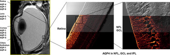
Müller cells have long been known to be involved in many retinal homeostatic functions including volume regulation (Newman & Reichenbach, Reference Newman and Reichenbach1996; Nagelhus et al., Reference Nagelhus, Veruki, Torp, Haug, Laake, Nielsen, Agre and Ottersen1998; Bringmann et al., Reference Bringmann, Reichenbach and Wiedemann2004, Reference Bringmann, Pannicke, Grosche, Francke, Wiedemann, Skatchkov, Osborne and Reichenbach2006). Müller cells span the width of the retina from the inner limiting membrane to the outer limiting membrane, with the cell soma lying within the inner nuclear layer (INL) (Dreher et al., Reference Dreher, Distler and Dreher1994; Newman & Reichenbach, Reference Newman and Reichenbach1996; Bringmann et al., Reference Bringmann, Pannicke, Grosche, Francke, Wiedemann, Skatchkov, Osborne and Reichenbach2006). AQP4 channels are found expressed on Müller cell membranes facing the retinal capillaries, abutting the vitreoretinal border and on membranes facing synapses in the plexiform layers in vascular mammalian retinae (Nagelhus et al., Reference Nagelhus, Veruki, Torp, Haug, Laake, Nielsen, Agre and Ottersen1998, Reference Nagelhus, Horio, Inanobe, Fujita, Haug, Nielsen, Kurachi and Ottersen1999) and also in similar areas in the avascular chick retina (Goodyear et al., Reference Goodyear, Junghans, Giummarra, Murphy, Crewther and Crewther2008) (Fig. 1).
(Color online) AQP4 in the retina. Image on the left is a magnetic resonance imaging photograph showing the known distribution of AQPs in the eye (photograph produced with the assistance of the Howard Florey Institute). The confocal micrographs (middle and right images) show localization of AQP4 in the avascularized chick retina during form-deprivation myopia. Expression of AQP4 has been localized to the endfeet of the Müller membrane at the vitreal interface, NFL, GCL, and the IPL, and shows similar localization to vascularized retinae, with the exception of endfeet abutting retinal capillaries. Upregulation of AQP4 during form deprivation occurs primarily in the NFL abutting the vitreal interface. This is thought to alter fluid flow into the vitreous cavity, leading to ocular enlargement. IPL, inner plexiform layer; NFL, nerve fiber layer; GCL, ganglion cell layer.

The expression of AQP4 on endfeet membranes of the Müller cell supports the notion that Müller cells contribute to transretinal fluid movements, particularly following the ion and osmotic changes induced by neuronal activity. Removal of excess water from the inner retina is achieved by facilitating water fluxes into the vitreous body and retinal capillaries (Nagelhus et al., Reference Nagelhus, Veruki, Torp, Haug, Laake, Nielsen, Agre and Ottersen1998). Expression of AQP4 channels may thus facilitate retinal signal transduction in both vascular (Nagelhus et al., Reference Nagelhus, Veruki, Torp, Haug, Laake, Nielsen, Agre and Ottersen1998) and avascular (Goodyear et al., Reference Goodyear, Junghans, Giummarra, Murphy, Crewther and Crewther2008) retinae (Fig. 1).
As noted earlier, AQP4 channels are not the only water transport mechanism implicated in fluid homeostasis of the retina. AQP1 has been found on photoreceptors and a subset of amacrine cells, predominantly in the outer retina. The authors consider this to reflect astrocytic expression rather than Muller cell expression (Iandiev et al., Reference Iandiev, Pannicke, Biedermann, Wiedemann, Reichenbach and Bringmann2006b). However, the altered expression of AQP1 was attributed to astrocytic expression rather than due to the expression of AQP1 on Müller cells.
Cotransport proteins known to facilitate transcellular ion movement elsewhere in the body also function as molecular water pumps in retina and in the absence of an osmotic gradient (Hamann & La Cour, Reference Hamann and la Cour2005). Such mechanisms would appear to predominate in the outer retina where the microvilli of the Müller cell facing the SRS are reported to be devoid of AQP4 channels (Nagelhus et al., Reference Nagelhus, Veruki, Torp, Haug, Laake, Nielsen, Agre and Ottersen1998). Furthermore, if AQP4 contributes to the development and efflux of edema from the brain and eye, surviving AQP4 knockout animals must rely on other mechanisms of fluid homeostasis such as cotransporter proteins. However, no changes in the expression of several other AQPs have been reported in AQP4 knockout animals (Ma et al., Reference Ma, Yang, Gillespie, Carlson, Epstein and Verkman1997). On the other hand, a downregulation of expression of the glutamate transporter 1 is seen in cultured astrocytes in AQP−/− mice. This could possibly lead to previously unforeseen alterations in ion and water homeostasis (Zeng et al., Reference Zeng, Sun, Gao, Fan, Ding and Hu2007).
AQP4 is yet to be localized to the RPE, and there is contradictory evidence whether other AQPs are present (Ruiz & Bok, Reference Ruiz and Bok1996; Hamann et al., Reference Hamann, Zeuthen, la Cour, Nagelhus, Ottersen, Agre and Nielsen1998; Stamer et al., Reference Stamer, Bok, Hu, Jaffe and McKay2003). It seems more likely that AQP4 in the inner retina will influence rapid fluid flow across the retina and in combination with the activity of ion cotransporters at the RPE will alter transport out of the retina and into the choroid (Hughes et al., Reference Hughes, Gallemore, Miller, Marmor and Wolfensberger1998).
A physiological role for AQP4 in retinal fluid homeostasis
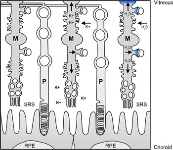
Given that neuronal activity is accompanied by the metabolic production of water and efflux of K+ out of cells, it is not surprising that Müller cells play a central role in the regulation of extracellular fluid homeostasis and tissue volume regulation (Newman et al., Reference Newman, Frambach and Odette1984; Newman, Reference Newman1985a, Reference Newman1987; Brew et al., Reference Brew, Gray, Mobbs and Attwell1986; Uckermann et al., Reference Uckermann, Vargova, Ulbricht, Klaus, Weick, Rillich, Wiedemann, Reichenbach, Sykova and Bringmann2004) (Fig. 2). Neuronal activity accompanying light onset results in a transient increase in potassium ([K+]) in the plexiform layers of the retina and a decrease in extracellular K+ ([K+]o) in the SRS, creating a need for the spatial buffering of the [K+]o (Karowski & Proenza, Reference Karowski and Proenza1977; Steinberg et al., Reference Steinberg, Oakley and Niemeyer1980). Such light activity–dependent increases in [K+] in inner retina require siphoning away from areas of high concentration by the Müller cells to help maintain the responsiveness of retinal cells to prolonged light stimulation (Newman, Reference Newman1984, Reference Newman1987; Newman et al., Reference Newman, Frambach and Odette1984; Newman & Reichenbach, Reference Newman and Reichenbach1996; Nagelhus et al., Reference Nagelhus, Veruki, Torp, Haug, Laake, Nielsen, Agre and Ottersen1998). The excess K+ is deposited into the vitreous (or where available into retinal blood vessels) or alternatively deposited back into the SRS to replenish the depletion of K+ there (Mori et al., Reference Mori, Miller and Tomita1976; Newman & Odette, Reference Newman and Odette1984; Newman et al., Reference Newman, Frambach and Odette1984; Newman, Reference Newman1985b, Reference Newman1987; Karwoski et al., Reference Karwoski, Lu and Newman1989; Frishman et al., Reference Frishman, Yamamoto, Bogucka and Steinberg1992). The predominant K+ channel in Müller cells primarily responsible for the spatial buffering of [K+]o, is the inward rectifying channel from the 4.1 subfamily (Kir4.1 channel) (Newman, Reference Newman1993; Ishii et al., Reference Ishii, Horio, Tada, Hibino, Inanobe, Ito, Yamada, Gotow, Uchiyama and Kurachi1997).
(Color online) Retinal K+ and fluid regulation by Müller cells. The high permeability of K+ into the Müller cell allows the light-induced excess K+ to be easily siphoned away from the extracellular space of the IPL and deposited in low-concentration areas such as the SRS and the vitreal interface and in vascular animals, into intraretinal blood vessels. Extracellular volume regulation is achieved in part by Müller cells using AQP4 channels located at the vitreoretinal border and on endfeet processes abutting retinal capillaries (blue shaded area on the Müller cell). This physical similarity has led to suggestions of a functional-dependent relationship between K+ siphoning and water transport by Müller cells. Figure adapted from Newman and Reichenbach (Reference Newman and Reichenbach1996). IPL, inner plexiform layer; M, Müller cell; P, photoreceptor cell.

Interestingly, Kir4.1 and AQP4 channels spatially colocalize on the endfeet processes of the Müller cell facing the vitreous and blood vessels of the mammalian retina. This has led to suggestions of a functional-dependent relationship (Nagelhus et al., Reference Nagelhus, Horio, Inanobe, Fujita, Haug, Nielsen, Kurachi and Ottersen1999). Typically, these retinal regions of colocalization of Kir4.1 and AQP4 channels play a role in creating K+ sinks for regulating high concentrations of [K+]o around active neuropil as described above (Newman, Reference Newman1993; Kofuji & Newman, Reference Kofuji and Newman2004).
A reduction in the electroretinogram (ERG) b-wave (the retinal response to light onset) in AQP null mice compared to controls suggests that the light onset–induced K+ changes are impaired in the absence of AQP4 (Li et al., Reference Li, Patil and Verkman2002). Li et al. (Reference Li, Patil and Verkman2002) argued that the reduced water permeability in AQP4 null mice could lead to changes in retinal hydration and consequently K+ levels, which in turn could influence K+ siphoning by Müller cells. However, it must be noted that the b-wave of the ERG has primarily a bipolar cell origin (Robson & Frishman, Reference Robson and Frishman1998; Sharma et al., Reference Sharma, Ball and Peachey2005), and hence, a reduction in the amplitude and latency of the b-wave in AQP4 null mice more than likely reflects a dampening of the depolarizing response of bipolar cell activation.
Investigations of ischemic insults targeting the retina of AQP4 knockout mice have also highlighted the involvement of AQP4 in regulating retinal neuronal activity. Da and Verkman (Reference Da and Verkman2004) found that elevated intraocular pressure induced significantly less impairment of retinal function as demonstrated by preservation of the b-wave of the ERG in ischemic retinae from AQP4−/− mice. Further, significantly less cell loss and better maintenance of the thicknesses of the INL and inner plexiform layers was seen in AQP4−/− mice compared to wild-type controls. However, no differences were seen in Müller cell swelling or extracellular space expansion. It should also be noted though as above that lower baseline b-wave amplitudes were found in one strain of AQP4−/− mice compared to control, thus confirming the earlier findings of Li et al. (Reference Li, Patil and Verkman2002) and raising the possibility that the presence of an already impaired b-wave amplitude may have masked any measurable impairment in AQP4−/− mice following the ischemic insult.
Pannicke et al. (Reference Pannicke, Iandiev, Uckermann, Biedermann, Kutzera, Wiedemann, Wolburg, Reichenbach and Bringmann2004) noted that ischemia induces a downregulation of Kir4.1 channels that is accompanied by decreased outward K+ currents from Müller cells. This downregulation led to glial cell swelling in the face of hypotonic stress (Pannicke et al., Reference Pannicke, Iandiev, Uckermann, Biedermann, Kutzera, Wiedemann, Wolburg, Reichenbach and Bringmann2004). Thus, given that AQP4 channel expression was preserved, the authors believe glial swelling results from the accumulation of intracellular K+ ions leading to a change in the osmotic gradient between the Müller cell and the extracellular space. Such glial cell swelling follows the reduced K+ efflux, generation of a reverse osmotic gradient, and drawing water into the glial cell through AQP4 channels.
The above-mentioned studies are suggestive of a functional association between the water transport and the spatial buffering of K+. The physical colocalization of AQP4 and Kir4.1 on endfeet of Müller cells in highly K+-permeable regions in the mammalian retina lends support for an association. However, an association does not necessarily imply a dependency in function, and indeed more recent evidence indicates that a functional relationship between Kir4.1 and AQP4 seems unlikely (Ruiz-Ederra et al., Reference Ruiz-Ederra, Zhang and Verkman2007). Ruiz-Ederra et al. (Reference Ruiz-Ederra, Zhang and Verkman2007) found no changes in Kir4.1 expression in AQP4−/− mice, nor did they observe any significant differences in K+ currents between wild-type and AQP4−/− mice in isolated Müller cells. Thus, further clarification of the relationship is needed both in the normal intact environment and after physiological stress is imposed. The functional coupling of Kir4.1 and AQP4 is further complicated as Na+ and bicarbonate transport have also been hypothesized to be implicated in the K+-mediated water influx through AQPs, presumably by creating an osmotic gradient for water to enter the Müller cell (Nagelhus et al., Reference Nagelhus, Mathiisen and Ottersen2004). Furthermore, the coenrichment of Kir4.1 and AQP4 is absent at the microvilli of the Müller cell where only Kir4.1 expression has been reported (Nagelhus et al., Reference Nagelhus, Horio, Inanobe, Fujita, Haug, Nielsen, Kurachi and Ottersen1999). As alluded to earlier, Müller cells are also known to clear excess K+ from the inner retina by deposition back into the SRS (Newman & Reichenbach, Reference Newman and Reichenbach1996; Kofuji & Newman, Reference Kofuji and Newman2004). Presumably, other mechanisms including other ion channels and cotransporters must work in concert with the apical membrane of the RPE for osmoregulation of the SRS.
AQP4, a potential target in myopia?
Myopic refractive errors are typically characterized by excessive elongation of the axial dimensions of the eye such that visual images cannot focus on the neural retina. Excessive accumulation of fluid in the vitreous is accompanied by reduced choroidal blood flow and decreased choroidal thickness in both the development of clinical myopia and the experimentally induced myopic refractive errors (Shih et al., Reference Shih, Fitzgerald, Norton, Gamlin, Hodos and Reiner1993; Crewther, Reference Crewther2000; Fitzgerald et al., Reference Fitzgerald, Wildsoet and Reiner2002). Yet, few studies have addressed the question of the origin and disposition of the fluid that is responsible for the abnormal axial elongation and ocular volume in myopia, despite the fact that at least 30% of the adult world population is affected by myopia (Seet et al., Reference Seet, Wong, Tan, Saw, Balakrishnan, Lee and Lim2001; Morgan & Rose, Reference Morgan and Rose2005). The public health and socioeconomic implications of such prevalence are enormous. One potential explanation for the disposition of fluid comes from evidence of changes to the normal pattern of ionic abundances from the retina to the choroid during recovery from experimentally induced myopia (Liang et al., Reference Liang, Crewther, Crewther and Junghans2004; Crewther et al., Reference Crewther, Liang, Junghans and Crewther2006). Liang et al. (Reference Liang, Crewther, Crewther and Junghans2004) and Crewther et al. (Reference Crewther, Liang, Junghans and Crewther2006) demonstrated that form-deprivation myopia in chicken is associated with a substantial increase in K+, Na+, and Cl− abundances in the outer retina and also prolonged increases in Na+ and Cl− in the inner retina. These changes are associated with axial elongation of the vitreous and thinning of the retina and choroid and hence volume changes in the eye.
AQP4 has been shown to play an important role in regulating fluid flow both in the brain and across the retina, with AQP4 expression up- or downregulating in certain pathological conditions as outlined above (Amiry-Moghaddam & Ottersen, Reference Amiry-Moghaddam and Ottersen2003; Bringmann et al., Reference Bringmann, Uckermann, Pannicke, Iandiev, Reichenbach and Wiedemann2005). Thus, we suggest that AQPs could offer an important mechanism for the transport of water across the retina during normal physiological conditions and hence would be expected to be involved in pathophysiological situations when fluid movement is affected, as in ischemia or volume changes during refractive error development. Given the reported changes to the osmotic environment during the development of experimentally induced myopia, it is likely that AQP4 serve in the movement of fluid across the retina. The involvement of AQP4 in myopia has recently been confirmed by our laboratory and demonstrates a temporal upregulation of AQP4 during the early increase in the rate of ocular growth that is concomitant with the induction of myopia in chickens (Goodyear et al., Reference Goodyear, Junghans, Giummarra, Murphy, Crewther and Crewther2008). AQP4 expression was found to significantly upregulate at the nerve fiber layer abutting the vitreoretinal border in chicks deprived of form vision (Fig. 1). The observation of a very early upregulation of AQP4 is not surprising, however, given the rapid elongation of the eye and enlargement of the vitreous chamber evident in form-deprived myopic eyes. Thus, the perturbation of AQP4 expression as a consequence of the changes to retinal signal transduction during reduced visual experience leads to excess fluid movement and deposition into the vitreous chamber (and possibly reduced fluid outflow into the choroid), resulting in ocular enlargement.
Conclusions
The eye is a unique encapsulated outgrowth of the brain where functioning requires exquisite fluid control. It is clear that AQP4 expression plays an important role in this maintenance of fluid homeostasis in both the eye and the brain. Experimentation that addresses the role of AQP4 in subtly altered physiological environments, such as during normal ocular growth or during the abnormal growth accompanying the development of refractive errors such as myopia and hyperopia, is likely to provide further understanding of ocular homeostasis and normal fluid movements. This will benefit not only our understanding of the role of AQP4 in central nervous system functioning and of the genesis and management of refractive error but also our understanding of the majority of acquired ocular pathologies (e.g., macular degeneration, diabetic retinopathy, and glaucoma) in which more permanently altered retinal fluid homeostasis leads to permanent retinal damage and ocular morbidity.