INTRODUCTION
Strongyloidiasis, caused by Strongyloides stercoralis, is a soil-transmitted helminthiasis distributed world-wide, affecting around 100 million people. However, this might be an underestimate due to the low sensitivity of the currently available diagnostic tools and a dearth of specialized surveys (Genta, Reference Genta1989; Olsen et al. Reference Olsen, van Lieshout, Marti, Polderman, Polman, Steinmann, Stothard, Thybo, Verweij and Magnussen2009). Endemic areas have been described in Southeast Asia, Latin America, sub-Saharan Africa, and parts of the southeastern United States (Siddiqui and Berk, Reference Siddiqui and Berk2001). Recent studies have identified endemic areas of chronic patients in the Valencian Mediterranean coastal region of Spain, resulting from environmental conditions (Oltra et al. Reference Oltra, Igual, Sanchez, Viñals, Andreu, Sarrion and Rodriguez-Calabuig2004). In this context, epidemiological studies in developed countries have identified endemic areas where a misdiagnosis, inadequate treatment and the facilitation of hyperinfection syndrome by immunosupression (i.e. by the administration of steroids) are too frequent and can cause a high mortality rate, ranging from 15 to 87% (Marcos et al. Reference Marcos, Terashima, Dupont and Gotuzzo2008).
Infection with S. stercoralis occurs transcutaneously and can be perpetuated by autoinfection, in which the filariform larva L3 is the infectious form (Keiser and Nutman, Reference Keiser and Nutman2004; Vadlamudi et al. Reference Vadlamudi, Chi and Krishnaswamy2006). The presentation of the infection is determined by the interaction between the host and the parasite. Such a long survival time of S. stercoralis in infected hosts could be due to diverse mechanisms of host immune response evasion, some of which should be dependent on the properties of the external surface of the parasite, as described in other parasitic nematodes (Page and Johnstone, Reference Page and Johnstone2007). Thus, the identification and characterization of S. stercoralis proteins present in the outermost surface is essential for a better understanding of the host–parasite relationship and for defining novel immunological, pharmacological and diagnostic targets (Page and Johnstone, Reference Page and Johnstone2007).
Advances in proteomics have now made possible the rapid and sensitive determination of the protein content of different parasitic helminth samples, including excretory-secretory products and tegumentary molecules (Bernal et al. Reference Bernal, Carpena, Espert, De la Rubia, Esteban, Toledo and Marcilla2006; Pérez-Sanchez et al. Reference Pérez-Sanchez, Valero, Ramajo-Hernandez, Siles-Lucas, Ramajo-Martin and Oleaga2008; Mulvenna et al. 2009; Bennuru et al. Reference Bennuru, Semnani, Meng, Ribeiro, Veenstra and Nutman2009; Robinson et al. Reference Robinson, Menon, Donnelly, Dalton and Ranganathan2009). Along with the traditional approach based on 1D or 2D gel electrophoresis in combination with in-gel digestion of bands or spots and analysis with mass spectrometry, technologies are now becoming available to analyse complex and low-abundance samples from helminths, like multi LC-MS and Offgel electrophoresis (OGE) (van Hellemond et al. Reference van Hellemond, van Balkom and Tielens2007; Audhya and Desai, Reference Audhya and Desai2008; Mulvenna et al. Reference Oltra, Igual, Sanchez, Viñals, Andreu, Sarrion and Rodriguez-Calabuig2009).
In the present work, we describe the first analysis of the proteome of S. stercoralis infective L3 larvae obtained from chronically infected, autochthonous patients from La Safor county in the Spanish province of Valencia. We have employed an experimental approach, based on controlled proteolytic cleavage and analysis by liquid chromatography tandem mass spectrometry, to attempt to identify potential surface-associated proteins. We performed 3 different treatments of the isolated larvae: a short digestion with trypsin, to enrich for surface-related proteins, the results of which we compared to an overnight digestion with trypsin of the partially digested larvae and finally samples of whole lysates obtained from detergent-treated larvae.
MATERIALS AND METHODS
Parasites
Fecal samples were obtained at the Francesc de Borja Hospital, Gandia, Valencia (Spain) from infected individuals from the endemic area of La Safor (Valencia, Spain) in compliance with Spanish ethical regulations (Oltra et al. Reference Oltra, Igual, Sanchez, Viñals, Andreu, Sarrion and Rodriguez-Calabuig2004). Only permanent inhabitants of the area in whom S. stercoralis or its larvae were identified and had never been treated previously for helminth infection were eligible for inclusion in the study.
Samples were processed on agar Petri dishes and L3 larvae were harvested and concentrated by centrifugation for 5 min at 1000 g, washed 3 times in 1 ml of phosphate buffered saline (PBS) pH 7·2 containing protease inhibitors (10 mm EDTA, and 1 mm PMSF) and samples were either processed immediately or stored in 70% EtOH at –20°C for further use.
Trypsin digestion of whole L3 larvae
Freshly obtained Strongyloides stercoralis L3 larvae were washed 3 times, 1 min per wash, with PBS and aliquots of 125 mg of larvae (w/w) were washed 3 times with 50 mm NH4HCO3 and incubated with 5 ng/μl (total volume 50 μl) of sequencing grade trypsin (Promega) in NH4HCO3 at 37°C for 30 min. The supernatant containing the released peptides was recovered by centrifugation, then reduced with DTT (10 mm) in 50 mm NH4HCO3 in a microwave oven for 3 min at 560 W, and alkylated with 55 mm iodoacetamide (IAM) in 50 mm NH4HCO3 for 30 min at room temperature in the dark. The reaction was stopped with 5 μl of 10% trifluoroacetic acid (TFA) and the peptides were preserved at −20°C. After the treatment, the integrity of larvae L3 worms was assessed microscopically.
Peptides were recovered by centrifugation and the partially digested L3 larvae were then reduced and alkylated with IAM as described above, and then digested with trypsin (10 ng/μl, vol. 50 μl) overnight at 37°C. The reaction was stopped with 5 μl of 10% TFA and the final solution was filtered using 22 μm filters.
Lysates of whole L3 larvae
Larval lysates were obtained by homogenizing the L3 larvae in 10 mm Tris-HCl pH 7·4, containing 1% NP40 detergent and a complete protease inhibitor cocktail (Roche). The amount of protein present in the supernatant (larval lysate) was estimated using the Bradford method (Bio-Rad). One mg of wet weight of larvae yielded approximately 7 μg of protein.
Liquid chromatography and tandem mass spectrometry (LC–MS/MS)
The resulting peptides from the above-mentioned assays were resuspended in 6 μl of 5% acetonitrile, 0·1% TFA and 5 μl of the sample were loaded onto a trap column (PepMap C18, 300 μm×5 mm, LC Packings) and desalted with 0·1% TFA at 40 μl/min during 5 min. The peptides were then loaded onto an analytical column (PepMap C18 3 μm 100Å, 75 μm×15 cm, LC Packings) equilibrated in 5% acetonitrile, 0·1% formic acid. Elution was carried out with a linear gradient 5–65% of solvent B (95% acetonitrile, 0·1% formic acid) during 120 min at a flow rate of 300 nl/min. The eluted peptides were analysed with a nanoESI-Q-TOF mass spectrometer (QSTAR-XL, Applied Biosystems) in an information dependent acquisition mode (IDA).
Identification of S. stercoralis proteins
MASCOT peak lists were generated by MASCOT DAEMON v 2.2.2 (Matrix Science) with Sciex Analyst import filter options. The peak list generation was done with default parameters for both engines. In both cases database searches were done with tryptic specificity without cys alkylation in the NCBInr 20081016 (7108540 sequences; 2453132438 residues) protein database using taxonomy restricted to Metazoa (Animals) for MASCOT. Additional searches of EST sequences available in the repositories (ftp://ftp.sanger.ac.uk/pub/pathogens/Strongyloides/ratti/ and http://www.nematode.net/FTP/cluster_ftp/index.php) did not provide additional information. Three missed cleavages and a tolerance in the mass measurement of 100 ppm in MS mode and 0·8 Da for MS/MS ions were allowed. Met oxidation and Asn/Gln deamination were set as variable modifications. Protein identification with confidence scores of >95% and mascot score >50 were considered significant. When the score was close to the limit, a manual inspection of the MS/MS spectra was done in order to assess the identification. A relative quantification of the proteins in the mixture was calculated using values obtained by MASCOT as raw emPAI (exponentially modified Protein Abundance Index). The PAI is defined as the number of observed peptides divided by the number of observable peptides per protein, which is proportional to individual protein content in a protein mixture (Ishihama et al. Reference Ishihama, Oda, Tabata, Sato, Nagasu, Rappsilber and Mann2005).
The molecular function and biological process were assigned to the identified proteins according to the gene ontology database (http://www.geneontology.org) and the Swiss-Prot/UniProt database (http://beta.uniprot.org).
RESULTS
Identification of proteins after short trypisinization of S. stercoralis L3 larvae
S. stercoralis L3 larvae were obtained from feces from chronic patients in the endemic area of La Safor (Valencia, Spain), which ensured that the identified proteins could correspond to actual proteins involved in the interaction with the human host.
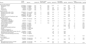
After checking distinct digestion protocols to study potential surface-associated proteins of S. stercoralis larvae, we used a short incubation period (30 min) with trypsin, which showed no morphological alteration of the larvae by light microscopy (data not shown). After that treatment, different peptides corresponding to 13 different proteins were identified in S. stercoralis L3 larvae (Table 1). Among these proteins, 5 were categorized as structural/cytoskeletal proteins and included different isoforms of myosin and actin, as well as paramyosin and tropomyosin. Actin exhibited a high raw emPAI value (0·41) indicating its relative high abundance in the extract, followed by paramyosin and tropomyosin with identical emPAI values of 0·11 (Table 1). Proteins corresponding to nuclear homologues were also identified after short trypsin treatment, including an elongation factor 1 alpha homologue (similar to the Onchocerca volvulus molecule) along with histone 2A and adenine nucleotide translocator (ant) protein homologues. Metabolic enzymes identified included phosphoenol pyruvate carboxykinase (PEPCK), succinate dehydrogenase, and glyceraldehyde 3-P dehydrogenase (GAPDH) homologues (Table 1). A stress-related 14-3-3 protein homologue was also identified after this short trypsin treatment (Table 1). Surprisingly, the only S. stercoralis protein previously identified was an IgG and IgE immunoreactive protein (Accession number 2290388), which exhibited the highest value of raw emPAI (0·52) (Table 1).
Proteins identified from Strongyloides stercoralis L3 larvae after short and long digestion, as well as from detergent extraction
(A protein was considered to be correctly identified if at least 2 different peptides were confidently matched with database sequences with an overall MASCOT score greater than 50. Protein identification with confidence scores of >95% were considered significant. When the score was close to the limit a manual inspection of the MS/MS spectra was done in order to accept the identification. The molecular function and biological process were assigned to the proteins identified according to the gene ontology database (http://www.geneontology.org) and the Swiss-Prot/UniProt database (http://beta.uniprot.org).

ND: not determined.
(a) Dirofilaria immitis.
(b) Trichinella spiralis.
(c) Onchocerca volvulus.
(d) Teladorsagia circumcinta.
In addition to these helminth proteins, some peptides corresponding to human proteins (including histones, ATP-synthase, sedlin, elastase and cytokeratins), as well as several peptides of bacterial origin, were also detected, suggesting their presence at the larvae surface (data not shown).
We next analysed the proteome of S. stercoralis L3 larvae after longer trypsin digestions (o/n) of the whole L3 larvae. Soluble proteins were subjected to LC-MS and the released peptides produced significant matches with a total of 20 different proteins (Table 1). After this treatment, we identified 9 additional proteins, probably due to harshness of the digestion protocol. Interestingly, 2 of the previously detected proteins after short digestion, myosin-N and the 14-3-3 homologues, were now absent after longer digestion (Table 1).
The protein homologues identified after prolonged treatments included galectin, Hsp70, arginine kinase, ATP-synthase, propionyl coenzyme A carboxylase alpha (pcca-1), vacuolar H ATPase family member (vha-13), a mitochondrial carrier protein, and a guanine nucleotide-binding protein G(0) (Table 1). Only 1 previously identified S. stercoralis protein, a metalloproteinase precursor (Accession number 17865392 [Gomez-Gallego et al. Reference Gomez-Gallego, Loukas, Slade, Neva, Varatharajalu, Nutman and Brindley2005]), was identified in this analysis (Table 1). The highest emPAI values corresponded to actin (0·67), the IgG and IgE immunoreactive protein (0·52), tropomyosin (0·38), ATP synthase (0·28) and galectin (0·25), suggesting their relatively high abundance in this fraction.
Identification of proteins in lysates of S. stercoralis L3 larvae
To compare the abundance of the proteins identified after short trypsin digestion with the proteins present in S. stercoralis larvae, we next performed a proteomic analysis of whole larval lysates obtained after detergent treatment. Most of the previously identified proteins in trypsin-treated larvae were again identified in lysates, with the exception of 5 proteins which were not present: pcca-1, vha-13, the mitochondrial carrier protein, the guanine nucleotide-binding protein G(0), and the S. stercoralis antigen recognized by IgG and IgE (Table 1). Four additional proteins were identified in lysates in comparison to trypsin digested samples; the homologues of the metabolic enzymes, citrate synthase and enolase, as well as the homologues of ATP: guanido phosphotransferase and histone 4 (Table 1). Again, actin showed the highest emPAI value (0·53) in lysates, followed by ATP synthase (0·36), histone 4 (0·35), galectin (0·25) and tropomyosin (0·24).
Although high-quality spectra were obtained, most of them were not assigned to actual proteins due to the unavailability of the S. stercoralis genome sequence and the low number of available ESTs.
DISCUSSION
Proteomic approaches are beginning to provide insights into helminth proteins involved in the interaction with the host (Bernal et al. Reference Bernal, Carpena, Espert, De la Rubia, Esteban, Toledo and Marcilla2006; van Hellemond et al. Reference van Hellemond, van Balkom and Tielens2007; Audhya and Desai, Reference Audhya and Desai2008; Pérez-Sanchez et al. Reference Pérez-Sanchez, Valero, Ramajo-Hernandez, Siles-Lucas, Ramajo-Martin and Oleaga2008; Mulvenna et al. 2009; Bennuru et al. Reference Bennuru, Semnani, Meng, Ribeiro, Veenstra and Nutman2009; Robinson et al. Reference Robinson, Menon, Donnelly, Dalton and Ranganathan2009). This work is the first study of the S. stercoralis proteome, where several isoforms corresponding to a total of 26 different proteins have been identified, similar to what has been reported for other related nematodes with uncharacterized genomes, such as Trichostrongylus colubriformis (Kiel et al. Reference Kiel, Josh, Jones, Windon and Hunt2007). In our study, we have used S. stercoralis larvae obtained from patients to be sure that the identifications could correspond to actual proteins involved in the interaction with the human host. Treatment with trypsin for a short period rendered peptides that could be assigned to 13 different proteins. We used this approach to identify proteins that are likely to be associated with the larval cell surface. However, more studies will be required to confirm this assumption and we cannot rule out the possibility that some secreted proteins could re-associate with the surface. Twenty different proteins were identified after re-digesting the remainder of the larvae, confirming an increase in the number of proteins due to the harshness of the digestion protocol, in agreement with results recently described for other helminths like Schistosoma bovis (Pérez-Sanchez et al. Reference Pérez-Sanchez, Valero, Ramajo-Hernandez, Siles-Lucas, Ramajo-Martin and Oleaga2008).
Proteins with the highest MASCOT scores corresponded to structural/cytoskeletal (or even motor) proteins. These proteins have also been identified in L3 materials from other nematodes like T. colubriformis (Kiel et al. Reference Kiel, Josh, Jones, Windon and Hunt2007), and very recently as components of the outermost layer of tegument of S. bovis (Pérez-Sanchez et al. Reference Pérez-Sanchez, Valero, Ramajo-Hernandez, Siles-Lucas, Ramajo-Martin and Oleaga2008).
Three typical nuclear protein homologues were also detected after short digestion of S. stercoralis larvae, including the elongation factor EF-1, an adenine nucleotide translocator and a histone 2A homologue. Histone-like molecules have been recently identified amongst the most abundant proteins in Brugia malayi and Ancylostoma caninum (Bennuru et al. Reference Bennuru, Semnani, Meng, Ribeiro, Veenstra and Nutman2009; Mulvenna et al. Reference Oltra, Igual, Sanchez, Viñals, Andreu, Sarrion and Rodriguez-Calabuig2009). The presence of elongation factors have been also detected recently in the tegument of S. bovis (Pérez-Sanchez et al. Reference Pérez-Sanchez, Valero, Ramajo-Hernandez, Siles-Lucas, Ramajo-Martin and Oleaga2008). We also identified 3 metabolic enzymes after short trypisinization of S. stercoralis L3 larvae: PEPCK, GAPDH and succinate dehydrogenase. These proteins were also identified after long trypsin digestion, as well as in detergent lysates. PEPCK was previously identified in the secretome of different nematodes like Brugia malayi (Bennuru et al. Reference Bennuru, Semnani, Meng, Ribeiro, Veenstra and Nutman2009; Hewitson et al. Reference Ishihama, Oda, Tabata, Sato, Nagasu, Rappsilber and Mann2008) and A. caninum (Mulvenna et al. Reference Oltra, Igual, Sanchez, Viñals, Andreu, Sarrion and Rodriguez-Calabuig2009), or even in the trematode Fasciola hepatica (Marcilla et al. Reference Marcilla, De la Rubia, Sotillo, Bernal, Carmona, Villavicencio, Acosta, Tort, Bornay, Esteban and Toledo2008). Future assays are needed to address its role in S. stercoralis larvae. Very recent studies in S. bovis have confirmed the localization of GAPDH in the tegument, which was the only glycolytic enzyme detected at the surface of female worms (Pérez-Sanchez et al. Reference Pérez-Sanchez, Valero, Ramajo-Hernandez, Siles-Lucas, Ramajo-Martin and Oleaga2008). Interestingly, GAPDH is a major protein present in total lysates, but absent in excretory-secretory products of the filarial nematode B. malayi (Bennuru et al. Reference Bennuru, Semnani, Meng, Ribeiro, Veenstra and Nutman2009; Hewitson et al. Reference Ishihama, Oda, Tabata, Sato, Nagasu, Rappsilber and Mann2008).
Enolase was identified only in lysates obtained after detergent treatment, suggesting that it is not one of the most abundant proteins at the surface of S. stercoralis. We identified this enzyme as a plasminogen-binding protein in the excretory/secretory products of trematodes (Bernal et al. Reference Bernal, De la Rubia, Carrasco-Abad, Toledo, Mas-Coma and Marcilla2004; Marcilla et al. Reference Marcilla, Perez-Garcia, Espert, Bernal, Muñoz-Antoli, Esteban and Toledo2007), confirming the results obtained in the nematode O. volvulus (Jolodar et al. Reference Jolodar, Fischer, Bergmann, Büttner, Hammerschmidt and Brattig2003). Enolase has been identified as being immunogenic in trematodes like S. bovis (Pérez-Sanchez et al. Reference Pérez-Sanchez, Valero, Ramajo-Hernandez, Siles-Lucas, Ramajo-Martin and Oleaga2008) and Echinostoma caproni (Sotillo et al. Reference Sotillo, Valero, Sanchez del Pino, Fried, Esteban, Marcilla and Toledo2008), and in nematodes like T. colubriformis (Kiel et al. Reference Kiel, Josh, Jones, Windon and Hunt2007), and very recently in Dirofilaria immitis (Oleaga et al. Reference Oleaga, Pérez-Sanchez, Pages, Marcos-Atxutegi and Simon2009). The enzyme arginine kinase has also been identified as being immunoreactive in T. colubriformis infection (Kiel et al. Reference Kiel, Josh, Jones, Windon and Hunt2007).
Two proteins, a lectin-binding protein, galectin, and a S. stercoralis metalloproteinase precursor (Gomez-Gallego et al. Reference Gomez-Gallego, Loukas, Slade, Neva, Varatharajalu, Nutman and Brindley2005), were identified only after long digestion and in detergent-extracted materials, but not after short trypsin digestion, suggesting their absence at the outermost surface of S. stercoralis. Galectins have been characterized as being secreted by other parasitic nematodes like Teladorsagia circumcincta (Craig et al. Reference Craig, Wastling and Knox2006), B. malayi (Hewitson et al. Reference Ishihama, Oda, Tabata, Sato, Nagasu, Rappsilber and Mann2008), or more recently in A. caninum (Mulvenna et al. Reference Oltra, Igual, Sanchez, Viñals, Andreu, Sarrion and Rodriguez-Calabuig2009) and D. immitis (Oleaga et al. Reference Oleaga, Pérez-Sanchez, Pages, Marcos-Atxutegi and Simon2009). This molecule was also identified as immunoreactive in T. colubriformis (Kiel et al. Reference Kiel, Josh, Jones, Windon and Hunt2007). The metalloproteinase precursor was cloned after its sequence identification in cDNA libraries from S. stercoralis L3 ESTs and its identification was accomplished by sequence homology to other related proteases (Gomez-Gallego et al. Reference Gomez-Gallego, Loukas, Slade, Neva, Varatharajalu, Nutman and Brindley2005).
Only 1 previously identified S. stercoralis protein, an IgG and IgE immunoreactive protein, was detected as highly abundant after short digestion. Our data suggest that this protein is likely to be associated with the larval surface and this could explain its reactivity with the host immune system.
The 14-3-3 and myosin-N homologues were identified after short digestion and also in detergent-extracted material, but not in long trypsin digestion. This could be due to their removal in short treatments. 14-3-3 molecules have been reported to be important for nematode larval development in plant-parasitic nematodes (Jaubert et al. Reference Jaubert, Laffaire, Ledger, Escoubas, Amri, Abad and Rosso2004), and very recently they have also been identified in the tegument of S. bovis (Pérez-Sanchez et al. Reference Pérez-Sanchez, Valero, Ramajo-Hernandez, Siles-Lucas, Ramajo-Martin and Oleaga2008). In addition, these molecules have been detected in the secretome of trematodes like F. hepatica (Robinson et al. Reference Robinson, Menon, Donnelly, Dalton and Ranganathan2009), and in nematodes like A. caninum (Mulvenna et al. Reference Oltra, Igual, Sanchez, Viñals, Andreu, Sarrion and Rodriguez-Calabuig2009).
Among the proteins identified only after long digestion, an HSP70 homologue was noteworthy. HSP70 proteins have been also widely detected as major parasite components in other helminth species (Bernal et al. Reference Bernal, Carpena, Espert, De la Rubia, Esteban, Toledo and Marcilla2006; Pérez-Sanchez et al. Reference Pérez-Sanchez, Valero, Ramajo-Hernandez, Siles-Lucas, Ramajo-Martin and Oleaga2008; Bennuru et al. Reference Bennuru, Semnani, Meng, Ribeiro, Veenstra and Nutman2009; Oleaga et al. Reference Oleaga, Pérez-Sanchez, Pages, Marcos-Atxutegi and Simon2009; Higon et al. Reference Higon, Monteagudo, Fried, Esteban, Toledo and Marcilla2008), being detected as a component of the tegument in trematodes like S. bovis (Pérez-Sanchez et al. Reference Pérez-Sanchez, Valero, Ramajo-Hernandez, Siles-Lucas, Ramajo-Martin and Oleaga2008) or E. caproni (Higon et al. Reference Higon, Monteagudo, Fried, Esteban, Toledo and Marcilla2008). Very recent studies have identified the Hsp70s, along with the myosin heavy chain, as possible drug targets in future treatments for S. mansoni (Caffrey et al. Reference Craig, Wastling and Knox2009).
In the present study, MS-BLAST results showed that a number of high-quality spectra could not be assigned to proteins identified using MS/MS searches, since they were either missed by MASCOT or were not represented in the database, due to incomplete nucleotide sequences. In fact, information regarding the annotated genome sequence and the number of EST data available for S. stercoralis are very limited (191 core nucleotide records, 122 protein sequences and 11392 ESTs at the time of the study, see also http://www.nematode.net/FTP/cluster_ftp/index.php). These unassigned spectra and the incomplete coverage of the S. stercoralis genome clearly suggests that novel proteins have yet to be identified, as has been proposed for A. caninum (Mulvenna et al. Reference Oltra, Igual, Sanchez, Viñals, Andreu, Sarrion and Rodriguez-Calabuig2009).
The kind of study described here is needed to expand the knowledge of the protein composition of parasites, like S. stercoralis. In this context, our results could help in the improvement and simplification of conventional diagnostic methods. Such advances constitute a major research priority for one of the most neglected tropical diseases (Olsen et al. Reference Olsen, van Lieshout, Marti, Polderman, Polman, Steinmann, Stothard, Thybo, Verweij and Magnussen2009), which is a public health problem not only in developing countries, but also in developed countries like Spain.
ACKNOWLEDGMENTS
Dr Lynne Yenush is acknowledged for critically reading the manuscript. The Servicio de Proteómica of the CIPF is a member of ProteoRed.
FINANCIAL SUPPORT
This work was supported by project PS09/02355 from the Fondo de Investigación Sanitaria (FIS) del Ministerio de Ciencia e Innovación (Madrid, Spain) and FEDER and project PROMETEO/2009/081 from Conselleria d'Educació, Generalitat Valenciana (Valencia, Spain). J.S. is a pre-doctoral fellow (FPI) from the Ministerio de Ciencia e Innovación (Madrid, Spain). A. P. was the recipient of fellowships BC06-284 and BC08-098 from the Universitat de València.