Pulmonary valve endocarditis is a rare entity accounting for 1.5 to 2% of the total cases of infective endocarditis. Reference Seraj, Gill and Sekhon1 It may occur as native pulmonary valve endocarditis or after prosthetic valve placement. Transcatheter pulmonary valve implantation is being used extensively as a less invasive alternative to surgical pulmonary valve replacement in residual right ventricular outflow tract disease either stenosis or regurgitation. Transcatheter pulmonary valve implantation has shown to provide satisfactory clinical outcomes with significant shorter length of hospital stay and decreased number of further surgical interventions. However, risk of prosthetic valve infective endocarditis remains a threat to valve function and patient outcome after transcatheter pulmonary valve implantation. The case being presented is a novel case of pulmonary valve endocarditis of Venus P valve with concomitant COVID-19 infection.
Case report
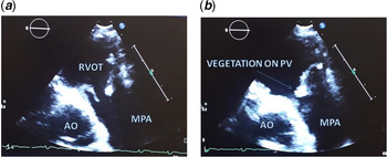
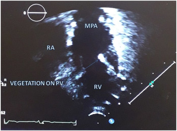
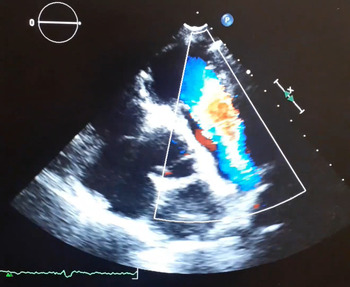
A 22-year-old male with past history of intracardiac repair with ventricular septal defect closure, infundibular resection with transannular patch for tetralogy of Fallot at 4 years of age and pulmonary valve replacement (Venus P valve 36 mm) at 18 years of age came with history of fever with chills, headache, myalgia, and vomiting. He also had difficulty in breathing with cough for 12 days before admission. Echocardiography on outpatient basis revealed moderate-sized vegetation on the pulmonary valve leaflet measuring 5.4 x 4.4 mm (Figures 1, 2) with mild pulmonary regurgitation (Figure 3). He was admitted with diagnosis of infective endocarditis and blood investigations including COVID-19 reverse transcription polymerase chain reaction and blood cultures were sent. On comprehensive medical examination, the patient was haemodynamically stable, febrile with temperature of 38.5 degree centrigrade, sinus rhythm with heart rate of 110 beats per minute, blood pressure of 115/75 mmHg, respiratory rate of 22 beats per minute, and SPO2 OF 97% on room air. On chest examination, bilateral vesicular breath sound was heard with no added sounds. On cardiovascular examination, first heart sound and second heart sound were normal, click was heard at left lower sternal border with grade 2 diastolic murmur at left upper sternal border. There were no petechiae, osler nodes, Janeway lesions, and splinter haemorrhages. Blood cultures were taken immediately and 12 hours apart. Along with blood cultures, COVID-19 reverse transcription polymerase chain reaction and inflammatory markers were sent. Reports revealed positive COVID-19 reverse transcription polymerase chain reaction, and blood reports showed leukocytosis with elevated C-reactive protein (7.18 mg/dL) and Erythrocyte sedimentation rate (ESR) (40 mm/hour), increased D-dimer (547 ng/ml), mild elevation of hepatic transminases, and normal renal parameters with sterile blood cultures. Chest X-ray showed prosthetic pulmonary valve in situ with no features suggestive of lung pathology. A 12-lead electrocardiography showed sinus rhythm with no features of right-sided volume and pressure overload. He was managed with intravenous antibiotics namely Ceftriaxone (100 mg/kg/day) and Vancomycin (40 mg/kg/day) and oral Rifampicin (15 mg/kg/day). He was shifted to COVID ward, and broad-spectrum oral antiviral Favipiravir (800 mg twice daily) and anticoagulation with low-molecular-weight heparin (10 IU/kg/hour) were also started. High-grade fever with chills remains persistent for 3 days. Repeat COVID-19 polymerase chain inflammatory markers namely reaction test after 5 days still was positive and C-reactive protein (1.52 mg/dL) and D-dimer (547 ng/ml) were still on the higher range. COVID-19 reverse transcription polymerase chain reaction test along with inflammatory markers were finally negative after 14 days of treatment with oral Favipiravir, and he was transferred back to non-COVID ward. Repeat transthoracic echocardiography was suggestive of decrease in size of vegetation. He was given total 6 weeks of intravenous antibiotics. Pre-discharge echocardiography after 6 weeks showed tiny echogenecity on the pulmonary valve. He was discharged after 6 weeks on dual antiplatelets (clopidogrel and aspirin) and oral antibiotics. However on follow-up, patient had spikes of fever for 1 day with echo suggestive of tiny echogenecity in the pulmonary valve and hence it is planned to keep the patient on oral antibiotics (Cefpodoxime and Rifampicin) for a total duration of 12 weeks.
Parasternal short-axis view showing vegetation on the prosthetic pulmonary valve during opening ( a ) and closing ( b ) of the valve leaflets. AO=aorta; MPA=main pulmonary artery; PV=pulmonary valve; RVOT=right ventricular outflow tract.

Apical four-chamber view in its most anterior tilt showing vegetation on pulmonary valve. MPA=main pulmonary artery; PV=pulmonary valve; RA=right atrium; RV=right ventricle.

Video of parasternal short-axis view showing prosthetic pulmonary valve with mild pulmonary regurgitation.

Discussion
Transcatheter pulmonary valve implantation is an appealing alternative to conventional surgical pulmonary valve replacement. Bonhoeffer et al. enlightened the feasibility of transcatheter pulmonary valve implantation for right ventricular outflow tract obstruction in 2000. Reference Bonhoeffer, Boudjemline and Saliba2 Since then in this modern era of transcatheter therapeutics, transcatheter pulmonary valve implantation has been used tremendously for patients with right ventricular outflow tract lesion after repair of CHD either stenotic or regurgitant. Pulmonary valve endocarditis is extremely rare in both normal and abnormal valves due to multiple factors like lower pressure within the right-sided chambers of the heart, lower incidence of congenital and acquired valvular abnormalities of right heart, lower venous oxygen content, and differences in endothelial lining and vascularisation of the pulmonary valve. Reference Seraj, Gill and Sekhon1 However, pulmonary valve endocarditis remains a dreadful complication of transcatheter pulmonary valve implantation and should not be overlooked. The first reported case of pulmonary valve endocarditis was 5 years after the first transcatheter pulmonary valve implantation in a patient who underwent dental procedure without antibiotic prophylaxis. Reference Khambadkone, Coats and Taylor3 The risk factors for infective endocarditis in pulmonary valve are predominantly male patient with nonnative right ventricular outflow tract anatomy, prior history of infective endocarditis, discontinuation of antiplatelets, longer procedure, higher number of stents, and higher residual right ventricular outflow tract gradient. There has been hypothesis of transcatheter implantation technique involving crimping and balloon expansion that cause alternation of anatomy of the valve causing favourable site for bacterial colonisation. Reference Sadeghi, Wadia and Lluri4 MelodyTM valve (Medtronic, Inc., Minneapolis, MN), Edwards Sapien valve (Edwards life sciences, Irvine, CA), and Venus P valve (Venus Medtech, Hangzhou, China) are commonly available valves being used for transcatheter pulmonary valve implantation. The incidence of infective endocarditis was reported higher in MelodyTM valve due to increased propensity of valves with bovine jugular tissue for bacterial adherence. Reference Abdelghani, Nassif and Blom5,Reference Hascoet, Mauri and Claude6 The lesser incidence of infective endocarditis in Sapien valve consisting of bovine pericardial tissue leaflets may be due to limited number of implantation and duration of follow-up as compared to Melody valve. Furthermore, Venus P valve is a new generation device made of porcine pericardium leaflets mounted in self-expanding nitinol stent with an additional advantage of implantation in a dilated and distensible right ventricular outflow tract. More than 100 Venus P valves have been implanted worldwide. Reference Mounir, Hak Lee and Matthew7 The initial experience with this valve is promising with excellent patient outcomes. Reference Garay, Pan, Zhang, Wang and Springmuller8 The reported complications after Venus P valve placement were mainly stent migration and stent fractures. There are minimal literatures that report the occurrence of infective endocarditis after Venus P valve implantation. One of the reported literature mentioned about infective endocarditis in a 49-year-old female patient 2 months after transcatheter Venus P valve. Reference Wang, Li, Ma and Pan9 Our case was much different from the previous reported case as infective endocarditis was reported late after 2 years of transcatheter pulmonary valve implantation and there was concomitant COVID-19 infection. The hypothesis of thrombus formation in the prosthetic valve that may form a nidus for bacterial colonisation and causing delayed presentation of infective endocarditis is well correlated with the fact that the patient was kept on single antiplatelet regime after transcatheter pulmonary valve implantation. This highlights the importance of role of dual antiplatelet regimen in such patients to prevent delayed infective endocarditis. The management of culture negative infective endocarditis in right-sided endocarditis further poses challenges for the clinician regarding the choice of antibiotics and total duration of antibiotics. The role of long-term oral antibiotics in culture-negative infective endocarditis is of doubtful benefit due to limited data to support or refute this regimen. Our case had spikes of fever for a day at follow-up hence we planned to continue antibiotics for a total of 12 weeks duration. Nonetheless, the clinical manifestations of COVID-19 and infective endocarditis are overlapping with features like high-grade fever with chills and rigour, dyspnoea, fatigue, cough, and myalgia. Muzakkie et al. reported a 61-year-old male with the diagnosis of infective endocarditis of the mitral valve concomitant with COVID-19 infection. Reference Amir, Djaharuddin, Sudharsono and Ramadany10 Though there are very few literatures on infective endocarditis with concomitant COVID-19 infection, the probability of both infection occurring simultaneously should be kept in mind which will aid in timely diagnosis and management of patient. To the best of our knowledge, this case report happens to be the first case of COVID-19 infection and infective endocarditis of the pulmonary valve and also the first case of delayed presentation of infective endocarditis in Venus P valve.
Conclusion
This case emphasises that there can be delayed presentation of infective endocarditis after transcatheter pulmonary valve implantation highlighting the importance of dual antiplatelet regimen after transcatheter pulmonary valve implantation. Furthermore, due to limited literatures on management of concomitant COVID-19 infection and infective endocarditis, this case report may give an insight regarding management of such patients.
Supplementary material
To view supplementary material for this article, please visit https://doi.org/10.1017/S1047951121002080
Acknowledgements
Covid team and the technical staff of Max hospital saket.
Financial support
This research received no specific grant from any funding agency, commercial, or not-for-profit sectors.
Conflict of interest
None.
Ethical standards
The authors assert that all procedures contributing to this work comply with the ethical standards of the relevant national guidelines on human experimentation (please name) and with the Helsinki Declaration of 1975, as revised in 2008, and has been approved by the institutional committees.