Background
Congenital biventricular cardiac diverticula are extremely rare and usually associated with other complex cardiac and thoracoabdominal anomalies. This report describes biventricular diverticula as a part of incomplete Cantrell’s syndrome in a 1-day-old newborn.
Case presentation
A 1-day-old, term newborn baby was referred from the delivery room with upper abdominal pulsatile mass. The prenatal course, delivery, and family history were unremarkable. The 1st and 5th minute Apgar scores 7 and 9, respectively. On physical examination, the baby appeared well with oxygen saturation 95% on room air, blood pressure 67/38 mmHg, heart rate 135 bites per minute, and respiratory rate 40 per minute. On inspection, a pulsatile mass originating from the subxyphoid region and extending to the slightly enlarged umbilicus was seen [Supplementary Video 1]. The abdominal wall appeared intact. The otherwise systemic examination was normal.
Sonographic evaluation of the abdomen revealed thick-walled tubular channels arising from apical parts of both ventricles and pulsating synchronously with hearth compatible with biventricular diverticula. While the right ventricular diverticulum was smaller, the left ventricular diverticulum was extending up to the umbilicus, and an anatomical continuity was present between the left ventricular diverticulum and obliterated umbilical vein. A small abdominal wall defect containing the left ventricular diverticulum and obliterated umbilical vein was seen at the umbilical region. The relationships of the other abdominal vascular structures were normal [Supplementary Video 2].
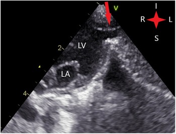
Echocardiography depicted a left ventricular apical wall defect continuing with a diverticulum (Fig 1), a patent ductus arteriosus and a secundum type atrial septal defect.
Modified apical two chamber echocardiography view of the patient showing the left ventricular apical wall defect and the diverticulum (arrow). I: Inferior, L: Left, R: Right, S: Superior. LV: Left ventricle, LA: Left atrium.

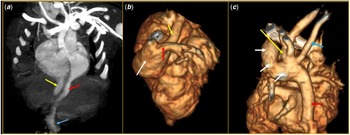
For further evaluation of associated anomalies, a nongated CT angiography was performed using a 16-channel CT scanner under mild sedation. Besides biventricular diverticula, the CT angiography showed crossing pulmonary arteries (the left pulmonary artery ostium originates from the main pulmonary artery above and to the right of the right pulmonary artery), right-sided descendent aorta, the aberrant course of the right subclavian artery, large patent ductus arteriosus, and slightly enlarged and horizontally orientated right ventricle. In addition to that, deficient sternal ossification (only three sternal ossification centres existed) was seen (Fig 2). The patient was referred to a tertiary heart centre and successfully operated for the biventricular diverticula. One month after surgical repair, the patient was admitted to our outpatient clinic with normal echocardiography scan despite a small-to-moderate secundum type atrial septal defect.
Oblique Maximum Intensity Projection (a) and 3D Volume Rendered CT angiography images. a Thick-walled left (red arrow) and right ventricular (yellow arrow) diverticula arising from apical parts of both ventricles. While right ventricular diverticulum is smaller, left ventricular diverticulum extends up to the umbilicus (blue arrow). b Crossing pulmonary arteries- the left pulmonary (red arrow) artery ostium originates from the main pulmonary artery (white arrow) above and to the right of the right pulmonary artery (yellow arrow). c Large patent ductus arteriosus (white arrows), right-sided descendent aorta (red arrow), the aberrant course of the right subclavian artery (blue arrow) and left sided arcus aorta (yellow arrow) are shown.

Discussion
Congenital ventricular diverticulum usually occurs (70%) as a part of complex cardiac syndromes, most commonly with the Pentalogy of Cantrell, and frequently associated with sternal, pericardial, diaphragmatic, and thoracoabdominal wall defects. Reference Edgett, Nelson, Hall, Fishback and Jahnke1 It can originate from both ventricles while the left ventricular diverticulum is more frequently reported. Reference Di Sessa, Howard and Salim2–Reference Ashraf, Gingell, Pieroni, Dhar and Subramanian4 Ventricular diverticula could be classified as fibrous or muscular. A muscular diverticulum is an outpouching originating usually from the apical part of the ventricle, contracting synchronously with it and containing all layers of heart tissue. Reference Hoffman, Menon, Everitt and Burch3,Reference Ashraf, Gingell, Pieroni, Dhar and Subramanian4
While the exact aetiology of this complex condition is not very clear, different theories have been proposed. A developmental failure in the differentiation of somatic and splanchnic mesoderm, the failure of ventral folding of the transverse septum, partial fusion of the ventral part of the septum transversum which is forming pericardium, and the primitive cardiac loop giving rise to the left ventricle are some of the theories explaining the possible causes of these complex defects. Reference Ashraf, Gingell, Pieroni, Dhar and Subramanian4–Reference Pirasteh, Carcano, Kirsch and Mohammed6
Syndromic association of ventricular diverticulum was described by Cantrell et al. Reference Cantrell, Haller and Ravitch5 in 1958, and the syndrome was named as Pentalogy of Cantrell or Cantrell syndrome. The estimated incidence of this syndrome is approximately 1 per 65,000–200,000 live births Reference Carmi and Boughman8 and classically consists of five congenital anomalies: intracardiac defect, lower sternal anomalies, pericardial defect, diaphragmatic defect, and epigastric omphalocele. Reference Cantrell, Haller and Ravitch5 Intracardiac defects may include ventricular septal defect (most common), atrial septal defect, pulmonary atresia, ectopia cordis, patent ductus arteriosus, tetralogy of Fallot, and diverticulum. Reference Mallula, Sosnowski and Awad9 The defects of the abdominal wall also vary from milder anomalies as umbilical hernia to the separation of recti, and even omphalocele. Therefore, the prognosis of the patient depends on the complexity of abnormalities.
As the complexity and expression of symptoms vary widely between patients, Toyama classified Cantrell’s syndrome into three different groups: class I presents with all five defects and is a definite diagnosis; class II is a probable diagnosis and presents with four defects, and class III is considered as an incomplete expression and presents with varying combinations of defects. Reference Toyama10
Differential diagnosis of ventricular diverticula may include ventricular aneurysms, ventricular clefts, and pseudoaneurysms. Congenital ventricular aneurysms are akinetic or dyskinetic ventricular out pouches, with no organised -or thin myocardium, connecting with a wide neck to the ventricular chamber. The main diagnostic features differentiating diverticulum from aneurysm are its synchronous contraction with heart, narrow connection to the ventricle, and composement of all myocardial layers. A myocardial cleft is an asymptomatic contractile protrusion within the myocardium that does not reach the epicardium and usually is accompanied with hypertrophic cardiomyopathy. Pseudoaneurysms are akinetic protrusions composed from pericardium and are caused by acute rupture of the ventricle wall due to infections or trauma. Therefore, pseudoaneurysms – being the potentially fatal conditions – must be differentiated from other pathologies of the ventricular wall.
In the present case, presence of both right and left ventricular diverticulum, large Patent ductus arteriosus (PDA), crossing pulmonary arteries, aberrant course of right subclavian artery, right-sided descending aorta, alterations in the ossification of the distal sternum; the presence of umbilical hernia leads to the diagnosis of a congenital biventricular diverticula as a part of the class III Cantrell’s syndrome. The patient was successfully operated at another heart centre for the surgical repair of both ventricular diverticula.
Supplementary material
For supplementary material accompanying this paper visit https://doi.org/10.1017/S1047951122001238
Acknowledgements
None.
Financial support
This research received no specific grant from any funding agency, commercial, or not-for-profit sectors.
Conflict of interest
None.
Ethical standards
All procedures performed were in accordance with the ethical standards of the institutional research committee and with the 1964 Helsinki Declaration and its later amendments or comparable ethical standards.
Informed consent
Informed written consent from parents was obtained.