The relationship between hypercholesterolemia and cardiovascular disease is firmly established. Familial hypercholesterolemia has been reported to cause early and progressive cardiovascular disease, which is clinically evident even in childhood and adolescence. Reference Maliachova and Stabouli1,Reference Gautschi, Pavlovic and Nuoffer2 Low-density lipoprotein-cholesterol is among the best-known atherogenic substances and is a major cause of mortality and morbidity with the development of atherosclerotic plaque and myocardial infarction in the coronary arteries. Reference Goonasekara, Balse, Hatem, Steele and Fedida3 Ischemic heart is prone to arrhythmia and atherogenic agents can worsen cardiac ischemia by promoting atherosclerotic plaque formation in the coronary arteries.
There are few studies in the literature about the development of other cardiac conditions such as atrial fibrillation due to hypercholesterolemia, and different results have been reported in these studies. Reference Li, Du and Guo4,Reference Roh, Chung and Lee5 However, the relationship between hypercholesterolemia and cardiac atrial and/or ventricular arrhythmia has not been clearly elucidated.
P wave and P wave dispersion are the most important noninvasive electrocardiography markers used to evaluate the risk of atrial arrhythmia. Reference Dilaveris, Batchvarov, Gialafos and Malik6 Previously, P wave dispersion has been studied in different disease groups in childhood, Reference Pérez-Riera, de Abreu, Barbosa-Barros, Grindler, Fernandes-Cardoso and Baranchuk7 while there are no studies in familial hypercholesterolemia children in the literature.
Long QT interval is a risk factor for life-threatening arrhythmias and sudden death with its interaction in genetic and environmental factors. Reference Van der Bijl, Heradien, Doubell and Brink8 QT dispersion measurement is an assessment of ventricular myocardial repolarisation in electrocardiography and indicates the risk of ventricular arrhythmia and coronary artery disease. In different diseases (cardiomyopathy, mitral valve prolapse, ischemic heart disease), QT dispersion may increase the risk of arrhythmia and sudden death. Reference Elming, Holm and Jun9,Reference Akpinar, Ocal and Irdem10
In addition to QT dispersion, the Tpeak-end interval is a marker of transmural dispersion of polarisation in electrocardiography. Reference Kors, Ritsema van Eck and van Herpen11 Prolongation of Tpeak-end interval is associated with ventricular arrhythmia in different situations (such as long QT syndrome, hypertrophic cardiomyopathy). Reference Yan and Antzelevitch12
In electrocardiography, JT and corrected JT parameters are more specific measurements of ventricular repolarisation than QT and corrected QT, eliminating QRS time variability. It is a more useful marker of repolarisation abnormalities. It is especially important in the diagnosis of patients with long QT syndrome who have borderline or normal corrected QT measurements on electrocardiography. Reference Berul, Sweeten, Dubin, Shah and Vetter13
In this study, we aimed to compare the risk of atrial and/or ventricular arrhythmia with the control group in children with familial hypercholesterolemia by examining specific parameters on electrocardiography.
Material and methods
In total, 85 cases between 3 and 17 years old who were diagnosed with familial hypercholesterolemia in the Pediatric Metabolism and Pediatric Cardiology outpatient clinic between January 2020 and March 2020 were included in our study. Total cholesterol was >200 mg/dl and low-density lipoprotein-cholesterol was >130 mg/dl in all of these patients.
As a control group, 83 children aged 3–17 years without any hypercholesterolemia (total cholesterol < 200 mg/dl, low-density lipoprotein-cholesterol < 100 mg/dl) who were directed to the Child Metabolism and Pediatric Cardiology outpatient clinic were included.
Age, gender, weight, body mass index information of case, and control groups were recorded. Total cholesterol, low-density lipoprotein-cholesterol, high-density lipoprotein-cholesterol, total cholesterol/high-density lipoprotein-cholesterol ratio values of the case group were recorded. Systolic blood pressure, electrolyte, and hormonal tests were normal in the case and control groups. Echocardiographies were also normal, and they were not using drugs.
Electrocardiography was taken from all children. Electrocardiographies were performed at 12 lead, 25 mm/second speed, and 10 mm/mV calibration.
In electrocardiographies, heart rate, P, QRS, T wave, PR interval (from the beginning of the P wave to the beginning of the QRS complex), QT interval (from the beginning of the Q wave to the end of the T wave), P wave dispersion (difference between the minimal and maximal P intervals measured in all leads in each patient’s electrocardiography), corrected QT interval (calculated with Bazett formula), QT dispersion (difference between the minimal and maximal QT intervals measured in all leads in each patient’s electrocardiography), corrected QT dispersion (measured in all leads of electrocardiography and calculated with Bazett formula), T peak-end (the interval between the peak and end of the T wave), JT interval (between the end of the S wave and end of the T wave), corrected JT (calculated with Bazett formula) were measured and compared to the control group. Reference Roguin14 All measurements were taken by two blind observers using a magnifying glass and ruler. In measurements, intraobserver and interobserver variability was determined as <5%.
Average, standard deviation, median lowest, median highest, frequency, and ratio values were used in the descriptive statistics of the data. The distribution of variables was measured by Kolmogorov–Simirnov test. In the analysis of quantitative independent data, Mann–Whitney U-test was used. Chi-square test was used in the analysis of qualitative independent data. Spearman correlation analysis was used in the correlation analysis. SPSS 26.0 program was used in the analysis.
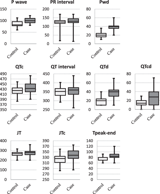
Comparison of P wave, PR interval, P wave dispersion, QT interval, corrected QT, QT dispersion, corrected QT dispersion, JT, corrected JT, Tpeak-end interval in the case and control groups.

Results
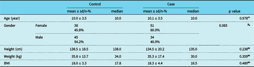
The demographic characteristics of the case and control group (age, gender, height, weight) and p values are given in Table 1.
Case and control group demographic characteristics (age, gender, height, weight) and p values

mMann–whitney U-test/X²Chi-square test, sd:standard deviation, BMI:body mass index
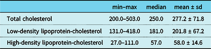
Total cholesterol, low-density lipoprotein-cholesterol, high-density lipoprotein-cholesterol minimum–maximum values, median, mean, and standard deviations in the case group are given in Table 2. In the control group, total cholesterol is <200 mg/dl, low-density lipoprotein-cholesterol is <100 mg/dl.
Case group total cholesterol, low-density lipoprotein-cholesterol, high-density lipoprotein-cholesterol values

sd:standard deviation
In the electrocardiography rhythm evaluations of all children, there was sinus rhythm in 68.5% (n = 115) and left inferior axis in 93.5% (n = 157).
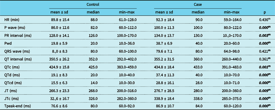
In the case group, P wave, PR interval, and P wave dispersion values were significantly higher than the control group (p ˂ 0.05). Heart rate, QRS wave, QT interval did not differ significantly (p ˃ 0.05) in the case and control groups. Corrected QT, QT dispersion, corrected QT dispersion, JT interval, corrected JT, Tpeak-end interval values were significantly higher (p ˂ 0.05) in the case group (Table 3 and Fig 1). T wave negativity rate did not differ significantly (p ˃ 0.05) in the case and control groups (Table 3).
Comparison of electrocardiography parameters and p values of the case and control group

HR:heart rate, Pwd:P wave dispersion, QTc:corrected QT, QTd:QT dispersion, QTcd:corrected QT dispersion, JTc:corrected JT. mMann–Whitney U-test, sd:standard deviation, ms:milliseconds, min:minutes
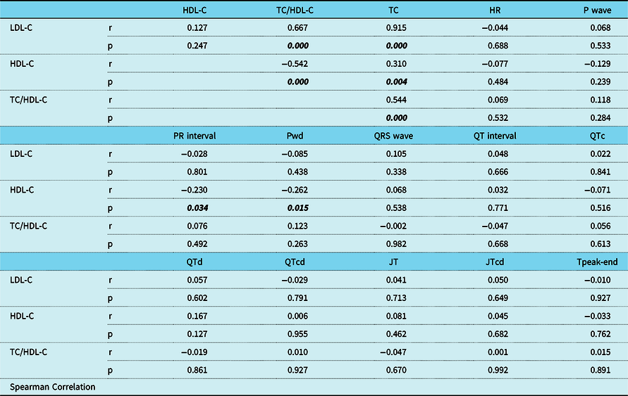
There was a significant (p ˂ 0.05) positive correlation between low-density lipoprotein-cholesterol value and total cholesterol, total cholesterol and high-density lipoprotein-cholesterol ratio. There was a significant (p ˂ 0.05) negative correlation between high-density lipoprotein-cholesterol value and total cholesterol/high-density lipoprotein-cholesterol, PR interval, P wave dispersion value. A significant (p ˂ 0.05) positive correlation was observed between high-density lipoprotein-cholesterol value and total cholesterol value (Table 4). There was no correlation between other parameters (Table 4). There was no significant correlation (p> 0.05) between low-density lipoprotein-cholesterol, high-density lipoprotein-cholesterol values and age, height, weight, BMI, P wave dispersion, corrected QT, QT dispersion, corrected QT dispersion, JT interval, Tpeak-end.
Sperman Correlation analysis between cholesterol and electrocardiography parameters

TC:total cholesterol, LDL-C:low-density lipoprotein-cholesterol, HDL-C:high-density lipoprotein-cholesterol, HR:heart rate, Pwd:P wave dispersion, QTc:corrected QT, QTd:QT dispersion, QTcd:corrected QT dispersion, JTc:corrected JT.
Discussion
Familial hypercholesterolemia is an autosomal dominant genetic disorder known by high plasma levels of low-density lipoprotein-cholesterol. There are “heterozygous” and “homozygous” forms and they are formed as a result of mutation in the low-density lipoprotein receptor gene. Homozygous familial hypercholesterolemia is a rare disease affecting one in a million people worldwide. Heterozygous familial hypercholesterolemia is common and occurs in every 200–250 people, and it is estimated that 6.8–8.5 million children and adolescents may be affected worldwide. Reference Maliachova and Stabouli1
In this study, we compared the parameters of children with familial hypercholesterolemia and children without hypercholesterolemia on electrocardiography. For the first time, we found statistically significant changes in electrocardiography parameters in children with familial hypercholesterolemia compared to the control group.
P wave duration, PR interval, P wave dispersion value were significantly higher (p < 0.05) in the electrocardiography in children with familial hypercholesterolemia.
P wave dispersion is a noninvasive electrocardiographic marker for atrial remodeling and is a predictor for atrial fibrillation. P wave dispersion is defined as the difference between the widest and narrowest P wave duration recorded from 12 electrocardiography leads. Increased P wave duration and P wave dispersion are indicative for atrial arrhythmias and especially paroxysmal atrial fibrillation. Reference Pérez-Riera, de Abreu, Barbosa-Barros, Grindler, Fernandes-Cardoso and Baranchuk7
P wave dispersion has been investigated in many diseases such as coronary artery disease, heart failure, hypertrophic cardiomyopathy, diabetes mellitus, hypothyroidism, and renal disease. Reference Pérez-Riera, de Abreu, Barbosa-Barros, Grindler, Fernandes-Cardoso and Baranchuk7 There is no study in the literature in children with familial hypercholesterolemia. P wave duration and P wave dispersion were found to be significantly different in children with familial hypercholesterolemia compared to the control group, and this supports that atrial arrhythmias can be seen in children with familial hypercholesterolemia.
As the heart rate increases (the RR interval becomes shorter), the QT interval becomes shorter. Therefore, the corrected QT interval accurately determines the QT interval for the heart rate using the previous RR interval. Reference Kors, Ritsema van Eck and van Herpen11,Reference Malik15 The QT dispersion, corrected QT dispersion, and Tpeak-end interval, used to show ventricular conduction and disruption of myocardial oxygenation as a result of autonomic dysfunction, are considered as markers that reflect abnormal ventricular repolarisation associated with arrhythmogenesis. In many diseases, QT dispersion, corrected QT dispersion, and Tpeak-end intervals have been investigated, Reference Yan and Antzelevitch12,Reference Castro Hevia, Antzelevitch and Tornés Bárzaga16 but there is no study in children with familial hypercholesterolemia. In addition, the corrected QT, QT dispersion, corrected QT dispersion, Tpeak-end intervals in the electrocardiography were significantly higher (p < 0.05) in the electrocardiography than the control group in children with familial hypercholesterolemia. This situation reveals the risk of ventricular arrhythmia in children with familial hypercholesterolemia.
In conclusion, we showed that the elevation of total cholesterol and low-density lipoprotein-cholesterol changed some parameters in electrocardiography and that these changes were related to the risk of atrial and ventricular arrhythmia.
There are many studies in the literature on hypercholesterolemia and atherosclerotic cardiovascular disease, heart failure. In recent years, the European Atherosclerosis Society Consensus report made it clear that low-density lipoprotein-cholesterol caused atherosclerotic cardiovascular disease, with evidence from numerous and many different clinical and genetic studies. In this report, it is said that low-density lipoprotein-cholesterol is not only an increased risk biomarker but also a causal factor in the pathophysiology of atherosclerotic cardiovascular disease and is a central determinant for the onset and progression of the disease. In addition, it is recommended to lower low-density lipoprotein-cholesterol earlier in individuals at high cardiovascular risk, especially in familial hypercholesterolemia patients. Reference Ference, Ginsberg and Graham17
Some studies have shown that high plasma cholesterol is associated with cardiac arrhythmias. In animal models, it has been found that there is a direct relationship between post-infarction plasma high-density lipoprotein-cholesterol and low-density lipoprotein-cholesterol levels and ischemia-reperfusion-related ventricular arrhythmias in rat heart preparations, and high plasma high-density lipoprotein-cholesterol has a strong protective effect. Reference Mochizuki, Okumura and Tanaka18 Similarly, another study showed that high-density lipoprotein-cholesterol reduced the incidence rates of ventricular arrhythmia after ischemia-reperfusion in rats. Reference Imaizumi, Miura and Nakamura19 Conversely, rabbits fed with a high cholesterol diet showed a higher rate of abnormal heart rhythm and greater sensitivity to ventricular fibrillation than those fed with a low cholesterol diet. Reference Liu, Wu and Lu20 These experimental data are consistent with clinical findings.
Circulating high-density lipoprotein-cholesterol levels have been found to decrease in many patients with atrial fibrillation, Reference Annoura, Ogawa, Kumagai, Zhang, Saku and Arakawa21 and high triglyceride and low-density lipoprotein-cholesterol levels are associated with a risk of ventricular tachyarrhythmia. Reference Liu, Wu and Lee22
In obese patients with high cholesterol levels, the prolongation of the QT interval has been observed. Reference El-Gamal, Gallagher and Nawras23 It is not known whether the prolonged action potential is associated with cholesterol level or other diseases accompanying obesity (such as hypertension). It has been observed that the QT interval is shortened in the treatment with atorvastatin, and it has been suggested that there is a causal relationship between serum cholesterol levels and action potential prolongation. Reference Vrtovec, Okrajsek, Golicnik, Ferjan, Starc and Radovancevic24
Roh et al revealed that high total cholesterol variability is associated with increased risk of atrial fibrillation by multivariate analysis using a nationwide cohort database. He stated that total cholesterol variability can be used as a new marker to classify the risk of atrial fibrillation and lowering total cholesterol variability below certain thresholds may reduce atrial fibrillation risk. Reference Roh, Chung and Lee5
In addition to the well-known risk of coronary heart disease in patients with heterozygous familial hypercholesterolemia, Hovland et al have shown that the risk of heart failure and atrial fibrillation/flutter doubles. He emphasised that this may have an important prognostic effect on patients and an economic effect on the society. Reference Hovland, Mundal and Igland25
In the light of these findings, children of parents with high total cholesterol and low-density lipoprotein-cholesterol should be screened for hypercholesterolemia. Children with hypercholesterolemia should be thoroughly evaluated by the paediatric cardiologist. Parameters in the electrocardiography should be evaluated more carefully in terms of atrial and ventricular arrhythmia.
The main limitation of our study was the lack of long-term follow-up of the patients. Electrocardiography pathologies may be observed in the follow-up.
Conclusion
As a result, we found that high total cholesterol and low-density lipoprotein-cholesterol variables are associated with an increased risk of cardiac atrial and/or ventricular arrhythmia. These findings suggest that total cholesterol and low-density lipoprotein-cholesterol variability can be used as a new marker for the risk of cardiac arrhythmia. In this case, decreasing total cholesterol and low-density lipoprotein-cholesterol variability below certain thresholds may decrease the risk of cardiac arrhythmia.
Acknowledgements
Thanks to the patients for cooperation and thanks to Murat Çakar for his contribution to translating the article.
Financial support
This research received no specific grant from any funding agency, commercial or not-for-profit sectors.
Conflict of interest
All authors declare that they have no conflict of interest.
Ethical standards
This study was approved by the hospital Ethics Committee of Okmeydanı Training and Research Hospital and carried out according to the Declaration of Helsinki protocol. All participants provided written informed consent forms for participation in the study (Approval number 48670771-514.10).