Congenital pulmonary vein stenosis is a rare disease that is mostly seen in young patients. Patients with the congenital form of pulmonary vein stenosis have a very guarded prognosis. Those with the involvement of most or all pulmonary veins nearly always have relentless progression and long-term survival is rare.Reference Latson and Prieto1 Breinholt et alReference Breinholt, Hawkins and Minich2 found a mortality rate of 83% in patients with three or four stenosed pulmonary veins. Balloon dilation, stent implantation, or surgical treatment have unfortunately had limited success, mainly owing to a high rate and severity of restenosis.Reference Driscoll, Hesslein and Mullins3–Reference Tomita, Watanabe and Yazaki5
Paclitaxel (Taxol) is a microtubule-stabilising compound with potent anti-tumour activity. It induces cellular modifications that result in reduced proliferation, migration, and signal transduction.Reference Herdeg, Oberhoff and Baumbach6 Paclitaxel-eluting balloons deliver a homogenous drug concentration to the vascular wall.Reference Scheller, Speck and Bohm7 Pre-clinical studies have shown the efficiency of paclitaxel-eluting balloons in inhibiting neo-intimal proliferation.Reference Scheller, Speck and Romeike8 Paclitaxel-eluting balloons are a new and innovative method in the treatment of in-stent stenosis and small vessel disease in adult cardiac pathology.
To our knowledge, we report the first treatment of severe neonatal pulmonary vein stenosis with a paclitaxel-eluting balloon.
Case report
We report the case of a newborn with a gestational age of 38 plus 5 weeks, presenting with respiratory distress to a local paediatric hospital. On the second day, pulmonary bleeding occurred, requiring NO- ventilation, antibiotic therapy, and catecholamine support.
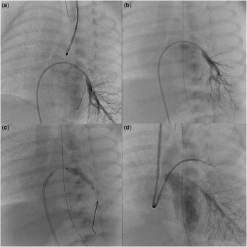
At the age of 3 weeks, the child was transferred to our tertiary referral cardiac centre. Echocardiography was performed, revealing severe stenoses of all pulmonary veins entering the left atrium. Further cardiac anatomy was normal. Angiography with selective injections in the pulmonary veins showed severe stenoses of all the four pulmonary veins at their entrance into the left atrium. The left inferior pulmonary vein showed a stenosis, with a narrowing from 3.2 millimetres to 1.1 millimetres distally (Fig 1) and a pressure gradient of 37 millimetres of mercury. Initial conventional balloon dilation of the left inferior pulmonary vein stenosis with a Savvy balloon of 4 × 20 millimetres resulted in immediate relief of the stenosis – vascular calibre 2.6 millimetres – and a significant reduction of the pressure gradient to 15 millimetres of mercury. On account of haemodynamic instability, further treatment of other pulmonary veins was not possible. After only 1 week, the clinical symptoms deteriorated again.
(a) Stenosis of left inferior pulmonary vein. (b) Pulmonary vein restenosis 1 week after conventional balloon dilation. (c) Balloon dilation of left inferior pulmonary vein restenosis by paclitaxel-eluting balloon. (d) Results 2 weeks after redilation with a paclitaxel-eluting balloon.

The second angiography revealed severe restenosis of the pre-dilated left inferior pulmonary vein, from 2.6 millimetres to 1.5 millimetres (Fig 1). During the second catheter intervention the left inferior and the right superior pulmonary vein stenoses were dilated with a 4-millimetre conventional balloon. Thereafter, the left inferior pulmonary vein restenosis was dilated with a 4 × 20-millimetre paclitaxel-eluting balloon (SeQuent Please, B. Braun Melsungen AG, Vascular Systems, Berlin, Germany). The balloon was placed through a long sheath to avoid stripping of paclitaxel from the balloon. The inflation time was 60 seconds with a pressure of 12 bar. Directly after redilation the vascular calibre increased to 2.8 millimetres.
A third angiography after 2 weeks showed an effective redilation of the left inferior pulmonary vein with the paclitaxel-eluting balloon (Fig 1). No signs of restenosis occurred; vascular calibre was 2.7 millimetres. The right-sided pulmonary vein, which was treated without paclitaxel, showed severe restenosis. Redilation was performed with a 3 × 20-millimetre paclitaxel-eluting balloon. The immediate angiography and the short-time echocardiographic follow-up presented good results.
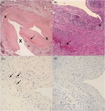
The plasma levels of paclitaxel 30 minutes, 1 hour, and 4 hours after intervention (Table 1) were below the systemic effective level of 85 nanograms per millilitre.Reference Huizing, Keung and Rosing9 Adverse systemic effects of paclitaxel did not occur. Blood count, liver enzymes, and retention parameters remained normal. Unfortunately, the child died 16 days after the third intervention due to respiratory failure in case of pulmonary infarction. The autopsy analysis of the pre-dilated left inferior pulmonary vein did show a focal rupture and a few fibrin deposits within the intima. Proliferation or immunohistochemical reaction due to balloon dilation with a paclitaxel-eluting balloon did not occur (Fig 2).
Paclitaxel plasma levels 30 min, 1 h and 4 h after balloon dilation by paclitaxel-eluting balloon.

Vein of lower lobe of left lung with congenital stenosis 4 weeks after re-dilatation with a drug-eluting balloon a (H&E) and (b) Elastica van Gieson – focal rupture (R) and few fibrin deposits (arrow) within the intima. Only modest mainly pre-existing intimal fibrosis. Artificial lesion due to autopsy (X). (c) Slight resorptive cellular reaction with brown staining of only few macrophages (arrows) in the immunohistochemical reaction with CD68. (d) In immunohistochemical reaction with the proliferation antibody MIB1 absence of positive brown staining of proliferating cells.

Discussion
We report a neonatal patient with severe stenoses of all pulmonary veins. The prognosis of this disease is universally dismal.Reference Breinholt, Hawkins and Minich2 Previous studies have shown limited success of balloon and cutting balloon dilation of pulmonary vein stenoses.Reference Driscoll, Hesslein and Mullins3, Reference Seale, Daubeney, Magee and Rigby4 Stent implantation due to restenosis is required in nearly all cases, but also leads to unsatisfactory results.Reference Tomita, Watanabe and Yazaki5 Many surgical therapies have been attempted to treat pulmonary vein stenosis, but mortality due to restenosis remains high.
Latson and PrietoReference Latson and Prieto1 prefer the term “primary” pulmonary vein stenosis in contrast to congenital pulmonary vein stenosis because this disease is progressive and may not even be evident at birth. In most patients there is no evidence of inflammation. This suggests a neo-proliferative progress. Sadr et alReference Sadr, Tan, Kieran and Jenkins10 found proliferative myofibroblastic cells in a small number of autopsy specimens. The effect of anti-proliferative therapy on these cells in patients with pulmonary vein stenoses is unknown. Provided that the neo-proliferative myofibroblastic cells are responsible for the large amount of restenoses, the local application of paclitaxel by a balloon could be an alternative strategy in the therapy of this frustrating disease. Paclitaxel is reported to reduce proliferation, migration, and signal transduction.Reference Scheller, Speck and Bohm7
After conventional balloon dilations without paclitaxel, restenoses occurred early in our patient. In contrast, the dilation of restenosis by the paclitaxel-eluting balloon showed good results in the short-time follow-up and the histological examination of the pulmonary veins after paclitaxel treatment showed only slight resorptive cellular reaction and absence of proliferating cells (Fig 2). This indicates that dilation of pulmonary vein restenosis by drug-eluting balloon results in an improved increase of the diameter as shown in our patient in whom conventional balloon dilation and drug-eluting balloon dilation were compared in two pulmonary veins (Fig 1).
Although the patient died at the age of 2 months due to pulmonary infarction, the results of dilation by a drug-eluting balloon showed good anatomic results in the short-time follow-up.
Paclitaxel is administered only during the short inflation time of the balloon, and is subject to rapid dilution and elimination. Adverse effects of paclitaxel were not observed in our patient. The plasma levels of paclitaxel remained below the systemic effective level known for adult patients.Reference Huizing, Keung and Rosing9 Levels in neonatal patients have not yet been reported.
This first experience of dilation of severe pulmonary vein restenosis with a paclitaxel-eluting balloon seems to be an innovative procedure for this frustrating disease. The short-time follow-up showed good initial results. Further studies are required to establish or refute the benefit of this attempt in treating pulmonary vein stenosis or restenosis.