Introduction
Reactive oxygen species (ROS) cause oxidative stress in a variety of cell lines, including reproductive cells. ROS are believed to influence physiological changes, male infertility and viability in mammalian spermatozoa (de Lamirande et al., Reference de Lamirande, Jiang, Zini, Kodama and Gagnon1997; Baker & Aitken, Reference Baker and Aitken2005; Koppers et al., Reference Koppers, De Iuliis, Finnie, McLaughlin and Aitken2008). Sea urchin spermatozoa also generate at least two types of ROS (Kazama & Hino, Reference Kazama and Hino2012). The first of these, hydrogen peroxide (H2O2), is believed to be generated spontaneously at the initiation of motility, while the second, superoxide (O2−), is detected by egg jelly treatment. Nevertheless, the effects and functions of H2O2 and O2− remain to be elucidated.
In general, mitochondrion respiration is responsible for the generation of ROS (de Lamirande et al., Reference de Lamirande, Jiang, Zini, Kodama and Gagnon1997; Liu et al., Reference Liu, Fiskum and Schubert2002; Andreyev et al., Reference Andreyev, Kushnareva and Starkov2005). Sea urchin spermatozoa represent an excellent model for reproductive biology (Vacquier & Hirohashi, Reference Vacquier and N. Hirohashi2004; Vacquier, Reference Vacquier2011) and details of their respiration and physiology are well known. The initiation of motility is known to require extracellular Na+ (Nishioka & Cross, Reference Nishioka, Cross, Dirksen, Prescott and Fox1978). Intracellular pH is elevated by Na+/H+ exchange, which in turn activates motility and respiration (Christen et al., Reference Christen, Schackmann and Shapiro1982). The energy for flagellar bending is derived primarily from mitochondrion respiration, for which the major substrates are intracellular phospholipids (Mita & Nakamura, Reference Mita and Nakamura1998). Phosphocreatine kinase (Tombes et al., Reference Tombes, Brokaw and Shapiro1987; Tombes & Shapiro, Reference Tombes and Shapiro1989; Dorsten et al., Reference Dorsten, Wyss, Wallimann and Nicolay1997) and adenylate kinase (Kinukawa et al., Reference Kinukawa, Nomura and Vacquier2007) contribute to the ATP supply. Flagellar bending is tightly coupled to respiration (Shapiro et al., Reference Shapiro, Cook, Quest, Oberdorf and Wothe1990).
In the present study, we focused on the generation of H2O2 by two species of sea urchin, Anthocidaris crassispina and Hemicentrotus pulcherrimus. To investigate the relationship between ROS generation and respiration, a fluorescent indicator CM-H2DCFDA, 5- and 6-chloromethyl-2′,7′-dichlorodihydrofluorescein diacetate was used. CM-H2DCFDA was previously used to detect ROS generation in living cells (Liu et al., Reference Liu, Athar, Lippai, Waldren and Hei2001; Duan et al., Reference Duan, Gross and Sheu2007; Kazama & Hino, Reference Kazama and Hino2012). This membrane-permeable indicator accumulates in intracellular compartments, such as mitochondria, where it is cleaved by esterases and converted into its fluorescent form through oxidation (Kirkland & Franklin, Reference Kirkland and Franklin2001; Myhre et al., Reference Myhre, Andersen, Aarnes and Fonnum2003).
In the ROS-detected condition and by the treatment with H2O2, we estimated sperm fertilizability using the ratio of formation of the fertilization envelope (FE) after insemination. In this paper, we discuss ROS generation, the implications of oxidative stress and also the validity of sperm preservation methods for sea urchin spermatozoa.
Materials and methods
Artificial seawaters and chemicals
We used two types of artificial seawater: normal artificial seawater (NSW) and sodium-free artificial seawater (NaFSW). NSW consisted of 420 mM NaCl, 9.0 mM KCl, 10 mM CaCl2, 24.5 mM MgCl2, 25.5 mM MgSO4 and 2.15 mM NaHCO3. In NaFSW, choline chloride and KHCO3 were substituted for NaCl and NaHCO3, respectively. All chemicals were purchased from Wako Pure Chemical Industries Ltd (Osaka, Japan). The artificial seawaters were buffered with 10 mM HEPES (Dojindo Laboratory, Kumamoto, Japan) and adjusted to pH 8.0 with NaOH (NSW) or KOH (NaFSW).
The respiratory inhibitor antimycin A (AMA) was purchased from Sigma Chemical Co. (St. Louis, MO, USA). Stock solutions of AMA (20 mg/ml) dissolved in dimethyl sulfoxide (DMSO; Wako Pure Chemical Industries Ltd, Osaka, Japan) were stored at –20°C. The concentration of hydrogen peroxide (H2O2; Wako Pure Chemical Industries Ltd, Osaka, Japan) was adjusted with NSW immediately prior to use.
Sea urchin gametes
Two species of sea urchins, Anthocidaris crassispina and Hemicentrotus pulcherrimus were collected from Sagami Bay, Kanagawa, Japan and used during each breeding season. Spermatozoa and unfertilized eggs were obtained by injecting 0.5 M KCl into the coelom to induce spawning. Condensed sea urchin spermatozoa, termed ‘dry sperm’, were stored on ice until required. Motility ratios were estimated prior to use in experiments.
Unfertilized eggs were washed twice with fresh NSW and the number was adjusted to 5000 ± 500 eggs/ml. The egg suspension was stored at room temperature (22–26°C) until required. The eggs were used for insemination within 6 h of spawning. When the duration of an experiment exceeded 6 h, fresh eggs were collected from new female batches.
Sperm preparation
The ‘dry sperm’ were diluted to various concentrations and the diluents were stored at several different temperatures. The spermatozoa were used for motility and fertilizability assays, or for measurements of oxygen consumption and ROS generation. For treatment with H2O2, spermatozoa were diluted 100-fold into NSW that contained H2O2.
Spermatozoa motility assay
Spermatozoa motility was estimated by two methods. Firstly, the motility rate was scored and classified into five groups as follows: 0–20, 20–40, 40–60, 60–80 or 80–100%, denoting spermatozoa motility under a microscope as ‘±’, ‘+’, ‘++’, ‘+++’ or ‘++++’ respectively. This method was applied to the selection of male batches prior to use in experiments and also during some experiments.
Secondly, the travelling distance assay (Turner & Giles, Reference Turner and Giles1982; Mita et al., Reference Mita, Hino and Yasumasu1984) was used. Briefly, the distance in which spermatozoa travelled through a glass capillary (diameter = 1 mm, length = 90 mm, Drummond Scientific Co., PA, USA) was assessed. The sperm suspension was transferred into a Petri-dish and the glass capillary (pre-filled with NSW) was inserted and rested at a 20° angle in this Petri-dish. After 30 min, the distance travelled by spermatozoa in the glass capillary was observed and measured by light microscopy.
Spermatozoa fertilizability assay
An FE is formed on a sea urchin egg within a few minutes after insemination. Hence, in the present study, we defined a fertilized egg by the formation of an FE. The treated spermatozoa were added and mixed into the unfertilized egg suspension at room temperature. At insemination, the respective concentrations were set at 30 × 105 spermatozoa/ml and 5000 ± 500 eggs/ml in Anthocidaris crassispina and 25 × 105 spermatozoa/ml and 5000 ± 500 eggs/ml in Hemicentrotus pulcherrimus. After 10 min, eggs were fixed with 2% glutaraldehyde. Fertilized and unfertilized eggs were then scored under an invert microscope (Eclipse TE300, Nicon Co., Tokyo, Japan). After scoring more than 100 eggs, the ratio of the formation of an FE was determined as the number of eggs forming an FE per total scored eggs.
Measurement of respiratory rate
The oxygen consumption of spermatozoa was estimated using a Clark-type oxygen electrode, as described previously (Hino et al., Reference Hino, Fujiwara and Yasumasu1980a; Kazama et al., Reference Kazama, Asami and Hino2006). Briefly, the oxygen electrode was connected to a closed vessel containing 2 ml of artificial seawater and maintained at 20°C by means of a covered jacket connected to a water bath. Dry sperm were added to the vessel. The dissolved oxygen concentration of the artificial seawater was measured over 5 min. Where necessary, we added 4 μl of AMA stock solution or DMSO, or 20 μl of 4 M NaCl to the sperm suspensions. Respiratory rates were determined by oxygen consumption and expressed as μM O2/min. Respiratory rates per unit number of spermatozoa were expressed as nmol O2/min/106 spermatozoa, after determination of sperm concentrations with a hemocytometer.
Measurement of ROS levels
A previous study showed that the CM-H2DCFDA-detectable ROS from sea urchin spermatozoa was H2O2 (Kazama & Hino, Reference Kazama and Hino2012). The fluorescent ROS indicator CM-H2DCFDA (C6827, Molecular Probes Inc., Eugene, OR, USA) was used to determine the ROS level in spermatozoa. CM-H2DCFDA is cell permeable and reacts with H2O2, ONOO− and hydroxyl radicals (Myhre et al., Reference Myhre, Andersen, Aarnes and Fonnum2003; Kazama & Hino, Reference Kazama and Hino2012) and is therefore commonly used to detect ROS. The stock solution of CM-H2DCFDA, at a concentration of 0.5 mM, was prepared in DMSO immediately prior to use. To detect ROS, dry sperm were suspended in a cuvette containing 2 ml of artificial seawater, with adjustment to 1 μM CM-H2DCFDA. The relative fluorescence unit (RFU) of each sperm suspension was measured at 5-s intervals for 10 min, using a VersaFluor Fluorometer System (Bio-Rad Laboratories Inc., Hercules, CA, USA), equipped with a 485–495 nm excitation filter and a 515–525 nm emission filter, as described previously (Kazama & Hino, Reference Kazama and Hino2012). The ROS generation rates were determined as the increasing rate of RFU for CM-H2DCFDA and were expressed as RFU/min and RFU/min/106 spermatozoa. After measurement, the sperm suspensions were diluted 100-fold with NSW and observed using a fluorescence microscope system (BX 51 equipped with a 100-W Hg lamp and mirror units of U-MWIB3; Olympus Co., Tokyo, Japan).
Measurement of adenine nucleotide contents
Content of three adenine nucleotides (ANPs) – ATP, ADP and AMP – was measured in A. crassispina sperm by reverse phase high-performance liquid chromatography (HPLC), as described previously (Kazama et al., Reference Kazama, Asami and Hino2006). The ANP standards were purchased from Sigma Chemical Co. (St. Louis, MO, USA). The sperm ANPs were extracted as described by Hino et al. (Reference Hino, Hiruma, Fujiwara and Yasumasu1980b). Sperm suspensions were centrifuged (3000 g for 10 min at 4°C) to obtain a pellet. The pellet and dry sperm were homogenized with a hand-driven glass homogenizer, after addition of the same volume of ice-cold 7% perchloric acid. The samples were deproteinized by centrifugation (3000 g for 10 min at 4°C). The supernatants were subjected to HPLC analysis within 1 h of starting the extraction.
Data analysis
All data are represented as mean ± standard error of mean (SE). Statistical analyses were performed using Student's t-test. Formal statistical significance was defined as P < 0.05.
Results
Respiratory activity and ROS levels in sea urchin spermatozoa
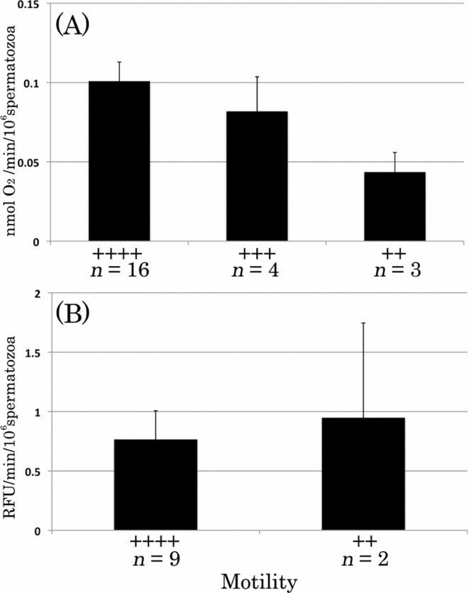
We measured oxygen consumption and CM-H2DCFDA fluorescence in A. crassispina spermatozoa after determining the motility ratio of each batch. The respiratory rate per unit number of spermatozoa increased in line with the motility ratio (Fig. 1A). The respiratory rate of batches with a motility ratio of ‘++’ was significantly lower than that of batches with a motility ratio of ‘++++’ (P = 0.031). In accordance with several previous studies (Christen et al., Reference Christen, Schackmann and Shapiro1982; Tombes & Shapiro, Reference Tombes and Shapiro1989; Shapiro et al., Reference Shapiro, Cook, Quest, Oberdorf and Wothe1990), our data indicated that the respiratory activity of sea urchin spermatozoa is strongly linked to motility. However, the difference in ROS level between ‘++’ and ‘++++’ batches was unclear (Fig. 1B) and two batches that were classified as ‘++’ showed a marked variation (0.69 RFU/min/106 spermatozoa and 7.94 RFU/min/106 spermatozoa). Motility rate was lower for a few cases of sperm just after they were obtained. Hence, all subsequent data were collected using the ‘++++’ spermatozoa just after they were obtained.
Rates of oxygen consumption and CM-H2DCFDA fluorescence in sea urchin spermatozoa. The rates of oxygen consumption (A) and CM-H2DCFDA fluorescence (B) were measured over 200 s, per unit number of Anthocidaris crassispina spermatozoa, after determining the motility of dry sperm. Spermatozoa were suspended at a concentration of 5 × 10⁶ spermatozoa/ml.

Oxygen consumption and ROS levels of sea urchin spermatozoa in NSW and NaFSW
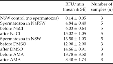
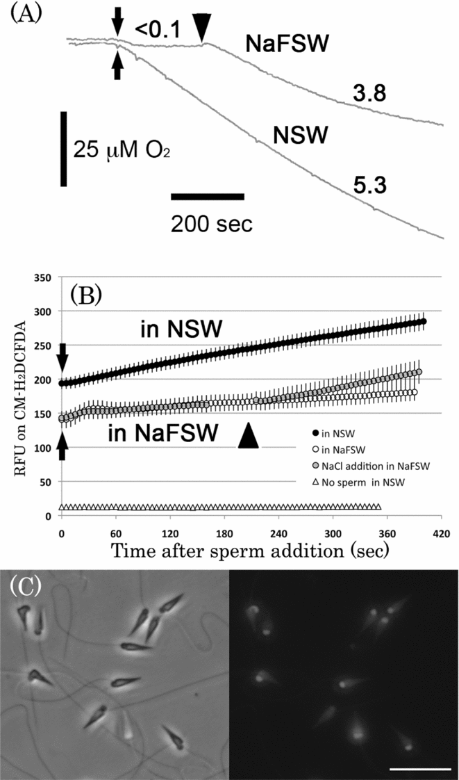
The oxygen consumption and ROS level of spermatozoa were greater in NSW than in NaFSW (Fig. 2A). Following the addition of NaCl solution, both the rates of oxygen consumption and ROS generation recovered to the same level as those suspended in NSW (Table 1). NaCl addition significantly affected oxygen consumption for spermatozoa suspended in NaFSW. Under the same conditions, the sperm motility ratio in NaFSW increased from less than 10% to 80% or more.
Reactive oxygen species (ROS) generation rates for A. crassispina spermatozoa in NSW and NaFSW
ROS generation rates were measured as relative fluorescence units (RFU)/min in sperm suspensions (15×107 spermatozoa/ml) with 1 μM CM-H2DCFDA.
Rates of oxygen consumption and RFU on CM-H2DCFDA in sea urchin spermatozoa. Typical rates of oxygen consumption (A) and relative CM-H2DCFDA fluorescence intensities (B) of Anthocidaris crassispina spermatozoa suspended in normal artificial seawater (NSW) and sodium-free artificial seawater (NaFSW) are shown. The arrows in (A) indicate the addition of spermatozoa. Arrowheads in both panels show the point of addition of 20 μl of 4 M NaCl solution. The rate of oxygen consumption in (A) is represented as μM O2/min. Closed and open plots in (B) represent the means of five relative fluorescence unit (RFU) measurements in NSW and NaFSW, respectively. The half-tone plots show the RFU in NaFSW following NaCl addition. The open triangles indicate no spermatozoa. Following RFU measurements, spermatozoa in NSW were observed by fluorescence microscopy, at a 100-fold dilution. The left (bright-field) and right (fluorescence) images in (C) are shown from the same view. Scale bar: 10 μm.
ROS generation (as assessed using CM-H2DCFDA) increased in NSW after the addition of sperm (to a final concentration of 15 × 107 sperm/ml). The ROS level of sperm suspensions in NSW (13.58 ± 1.03 RFU/min; Table 1) was significantly higher than that of sperm suspensions in NaFSW (4.84 ± 0.40 RFU/min), however the rate recovered to approximately the same level (15.0 ± 1.0 RFU/min) as that of sperm suspensions in NSW after NaCl addition (Fig. 2B and Table 1). Moreover, a stronger fluorescence signal was detected at the mid-piece of the spermatozoa, after measurements were performed in NSW (Fig. 2C).
In the absence of dry sperm, we observed no difference in ROS generation rates between NaFSW and NSW (0.14 ± 0.05 RFU/min). Suspensions that contained no CM-H2DCFDA showed no change in RFU (<0.1 RFU/min; data not shown). Thus, the RFU cannot be attributed to auto-oxidation of CM-H2DCFDA or to self-fluorescence of spermatozoa.
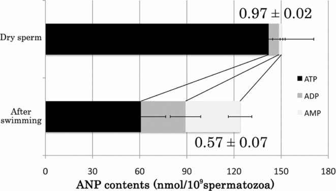
Mitochondrion respiration was previously reported to change from state 4 (absence of ADP) to state 3 (presence of ADP) via supplementation with ADP through flagellar motion (Christen et al., Reference Christen, Schackmann and Shapiro1982, Reference Christen, Schackmann and Shapiro1983). In the present study, the ANP content and Atkinson's energy charge (Atkinson, Reference Atkinson1968) for dry and motile sperm suggested a change of respiratory state (Fig. 3). Thus, ROS generation by sea urchin spermatozoa is activated at the initiation of motility (ADP production), with a rise in respiratory activity (ATP production). Our data indicate that ROS generation is derived from mitochondrial respiratory activity and that this mitochondrion respiratory activity is triggered by motility.
Adenine nucleotides in sea urchin spermatozoa. The contents of three adenine nucleotides (ANPs) – ATP, ADP and AMP – were measured in Anthocidaris crassispina spermatozoa by reverse phase high-performance liquid chromatography (HPLC). Spermatozoa were used before and after swimming, as dry sperm and spermatozoa suspended in normal artificial seawater (NSW) for 10 min, respectively. The values for each column showed Atkinson's energy charge, a parameter of available ANPs. All measurements were replicated five times independently.
Effect of antimycin A on ROS levels
Sea urchin sperm possess cytochromes b, c 1, c, a and a 3, at the same levels as those found in rat liver (Wilson & Epel, Reference Wilson and Epel1968). Treatment of sea urchin spermatozoa with the respiratory inhibitor AMA was shown previously to result in decreased oxygen consumption (Hiruma et al., Reference Hiruma, Hino, Fujiwara and Yasumasu1982; Kazama et al., Reference Kazama, Asami and Hino2006; Kazama & Hino, Reference Kazama and Hino2012). Spectral analyses revealed accompanying alterations in the redox state of cytochromes, suggesting that AMA treatment inhibits complex III of the respiratory chain (Hiruma et al., Reference Hiruma, Hino, Fujiwara and Yasumasu1982; Kazama et al., Reference Kazama, Asami and Hino2006).
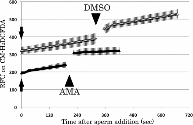
We measured RFU on CM-H2DCFDA in sperm suspensions that contained DMSO (final concentration of 0.2%) or AMA (final concentration of 4 μg/ml). After AMA addition the ROS generation rates of spermatozoa suspended in NSW were decreased (Fig. 4). By contrast, the addition of DMSO (as a co-solvent) did not cause a change in the ROS level.
Effect of antimycin A (AMA) on reactive oxygen species (ROS) generation in sea urchin spermatozoa. The relative fluorescence unit (RFU) on CM-H2DCFDA was measured in Anthocidaris crassispina spermatozoa before and after the addition of AMA or dimethyl sulfoxide (DMSO). The arrowheads correspond to AMA or DMSO addition. The RFU/min data are shown in Table 1.
The ROS generation rates for all treatments are shown in Table 1. The ROS generation rates before and after AMA addition (13.78 ± 3.50 RFU/min and 3.40 ± 1.74 RFU/min, respectively) differed significantly. Significant differences in the measurements were observed before and after NaCl addition (6.03 ± 0.64 RFU/min and 15.02 ± 1.05 RFU/min, respectively); however, no differences were observed after the addition and in NSW without any addition.
Dependency of respiratory and ROS generation rates on sperm concentration
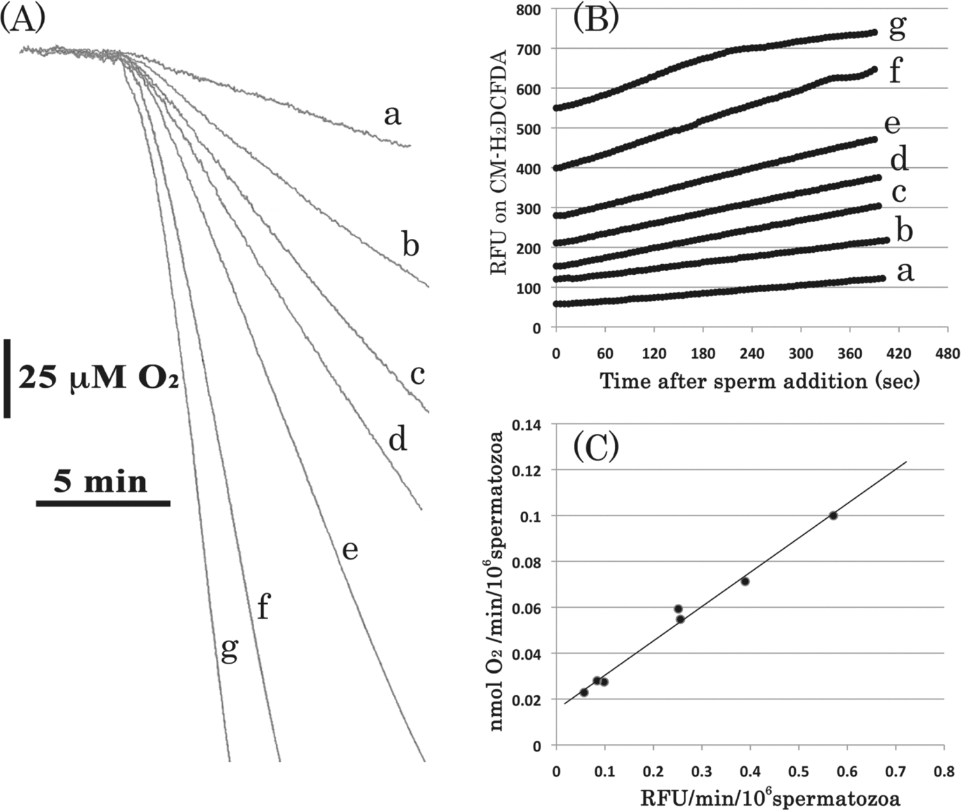
Figure 5 shows the typical respiratory and ROS generation rates for various concentrations of a single batch of A. crassispina with ‘++++’ motility in NSW. At higher sperm concentrations, the steepness of the oxygen consumption (Fig. 5A) and ROS generation slopes (Fig. 5B) increased. The ROS generation rate showed the similar increasing to the respiratory rate dependently on sperm concentration. Our data suggest that, for A. crassispina suspended in NSW, the respiratory rate (μmol O2/min) and ROS generation rate (RF/min) increase in a sperm concentration-dependent manner. Figure 5C shows the respiratory rate per unit number of spermatozoa (nmol O2/min/106 spermatozoa) versus the ROS generation rate per unit number of spermatozoa (RFU/min/106 spermatozoa). We observed a strong relationship between the both rates.
Rates of oxygen consumption and CM-H2DCFDA fluorescence at various concentrations of spermatozoa. Typical measurements of oxygen consumption (A) and relative fluorescence unit (RFU) on CM-H2DCFDA (B), using various concentrations of the same male batch of Anthocidaris crassispina, are shown. The letters (a–g) correspond to the following sperm concentrations (×106 spermatozoa/ml): (a), 18; (b), 39; (c), 91; (d), 101; (e), 309; (f), 482; (g), 744. The respiration rate in (A) and reactive oxygen species (ROS) generation rate in (B) were calculated as nmol O2/min/106 spermatozoa and RFU/min/106 spermatozoa, respectively, as shown in (C).
Effect of H2O2 on spermatozoa fertilizability
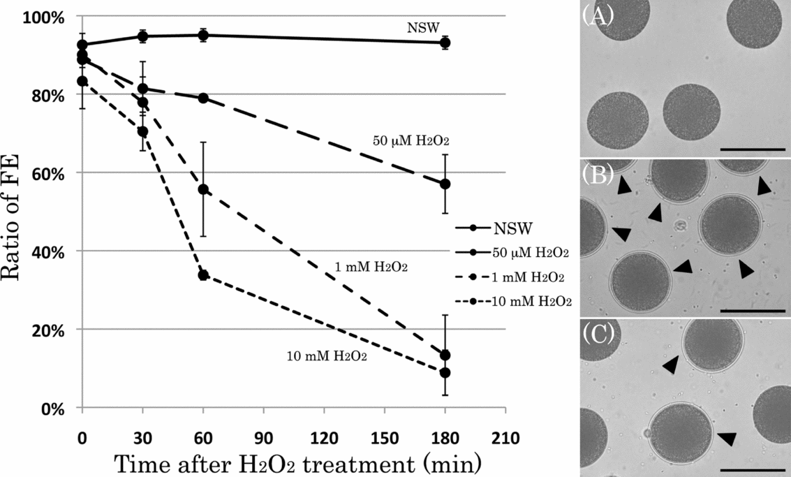
The CM-H2DCFDA-detectable ROS from spermatozoa of the sea urchin Pseudocentrotus depressus was H2O2 (Kazama & Hino, Reference Kazama and Hino2012), therefore H2O2 should be detectable in other species of sea urchin. In Figs. 6 and 7A, we determined the fertilizability of A. crassispina spermatozoa following H2O2 treatment on ice, by observing the formation of egg FEs. Unfertilized eggs were inseminated with H2O2-treated spermatozoa for various time periods (Fig. 6). After 10 min, we measured the ratio of FEs. In the absence of insemination, eggs showed no FE (Fig. 6A). For spermatozoa suspended in NSW alone, almost all inseminated eggs formed an FE (Fig. 6B). By contrast, for spermatozoa treated with 10 mM H2O2, only some eggs formed an FE (Fig. 6C). The ratio of FE (%) decreased in a manner that was dependent on the concentration of H2O2 and also on the treatment time.
Changes in the ratio of formation of a fertilization envelope (FE) following H2O2 treatment of spermatozoa. The ratio of formation of an FE was assessed in Anthocidaris crassispina spermatozoa treated with H2O2 at 50 μM, 1 mM and 10 mM on ice (n = 4). The H2O2-treated spermatozoa were added into the egg suspension (30 × 105 spermatozoa/ml and 5000 ± 500 eggs/ml). After 10 min for insemination, the suspension was fixed with 2% glutaraldehyde and FE-forming eggs were scored (left). The photographs (right) show eggs without insemination (A) and following insemination with spermatozoa suspended in normal artificial seawater (NSW) (B) and treated with 10 mM H2O2 (C) for 30 min. Arrowheads indicate the FE-forming eggs. Scale bar: 10 μm. FE (%) was scored after fixation, as described in the Materials and methods.
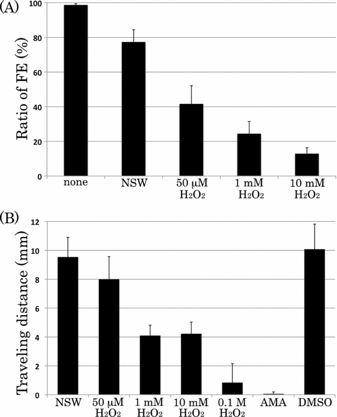
Effect of H2O2 on sperm fertilizability and motility in Anthocidaris crassispina. The sperm fertilizability was estimated fertilization envelope (FE) (%) in (A). The suspension of gametes was fixed with 2% glutaraldehyde, after 10 min for insemination with H2O2-treated spermatozoa. The motility of H2O2-treated A. crassispina spermatozoa was estimated by the travelling distance assay (B). The assays in (A) and (B) were each replicated five times independently.
Spermatozoa were subsequently treated with H2O2 for 30 min at room temperature. The treatment was terminated by centrifugation (3000 g for 10 min at 4°C) and eggs were washed twice with fresh NSW to remove the H2O2. The ratio of FE (%) for eggs inseminated with spermatozoa suspended in NSW alone was significantly higher than that for eggs inseminated with spermatozoa treated with 1 mM H2O2 or 10 mM H2O2 (P = 0.0035 and P = 0.0009, respectively; Fig. 7A). The ratio of FE (%) for eggs inseminated with spermatozoa treated with 50 μM H2O2 did not differ significantly (P = 0.0329) from that of eggs inseminated with spermatozoa suspended in NSW alone. Thus, the decrease in the ratio of FE (%) was caused by an H2O2 effect on spermatozoa, but not on the eggs.
Effect of H2O2 on spermatozoa motility
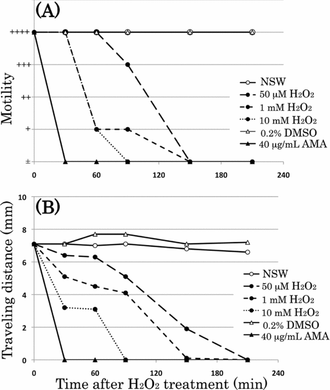
Next, we used the travelling distance assay to determine the motility of spermatozoa treated with H2O2 (Figs. 7B and 8). We observed that spermatozoa not treated with H2O2 travelled significantly further in 30 min than did spermatozoa treated with 1 mM H2O2 or 10 mM H2O2 (P < 0.001). AMA-treated spermatozoa were immotile and constituted a negative control. The travelling distance of DMSO-treated spermatozoa differed significantly from that of AMA-treated spermatozoa, but not from that of the control (no H2O2).
Effects of H2O2 on sperm motility. Spermatozoa were assessed using the motility rate (A) and traveling distance assay (B). (A) and (B) show typical data sets from a single batch of Anthocidaris crassispina.
The typical travelling distance for an individual batch of male spermatozoa is shown in Fig. 8B. During the 210 min of treatment, NSW- and DMSO-treated spermatozoa travelled 6–8 mm through the glass capillary. AMA-treated spermatozoa were immotile and failed to travel through the capillary. For H2O2-treated spermatozoa, the travelling distance decreased in a manner that was dependent on the H2O2 concentration and also on the treatment time. Measurement of the motility ratio yielded similar results (Fig. 8A). AMA-treated spermatozoa showed ‘±’ motility after 30 min. The motility ratios of spermatozoa treated with DMSO and NSW were ‘++++’ after 180 min. Spermatozoa treated with high concentrations of H2O2 showed lower motility ratios than did those treated with low concentrations of H2O2. As with the travelling distance, the sperm motility decreased in a manner that was dependent on the H2O2 concentration and also on the treatment time.
Effect of temperature on ROS generation and spermatozoa fertilizability
Given the negative effect of H2O2 on spermatozoa fertilizability, we investigated whether the conditions that induced a higher ROS generation rate would lead to a reduction in spermatozoa fertilizability.
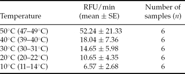
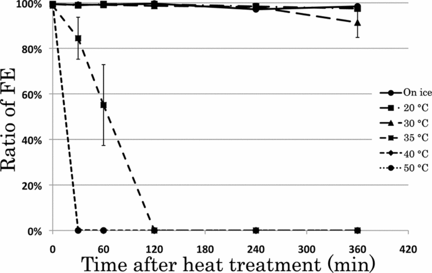
First, we investigated the effect of a higher seawater temperature on ROS generation and spermatozoa fertilizability (Table 2). Between 10°C and 40°C, the ROS generation rate increased linearly, in a manner that was dependent on the temperature. At 50°C, the rate showed the most rapid increase, with substantial differences among individual batches. Preservation at higher temperatures led to a reduction in spermatozoa fertilizability, in a manner that was dependent on the temperature and also on the time of preservation (Fig. 9). At a preservation temperature of <30°C, the ratio of FE for eggs inseminated with spermatozoa was close to 100%. Storage at 35°C led to a decrease in the ratio of FE (%). Eggs inseminated with spermatozoa stored for 30 min at 40°C or 50°C failed to form an FE.
Reactive oxygen species (ROS) generation rates for H. pulcherrimus spermatozoa at various temperatures
ROS generation rates were measured as relative fluorescence units (RFU)/min. Temperatures were confirmed in sperm suspensions (13 × 107 spermatozoa/ml) with 1 μM CM-H2DCFDA after RFU measurement.
Ratio of formation of a fertilization envelope (FE) after insemination with spermatozoa stored at various temperatures. Hemicentrotus pulcherrimus spermatozoa were diluted 500-fold (50 × 106 spermatozoa/ml) and stored at various temperatures. They were then added into the egg suspension (25 × 105 spermatozoa/ml and 5000 ± 500 eggs/ml). The suspension was fixed with 2% glutaraldehyde, after 10 min for insemination and the FE (%) was scored. In the case of treatment at 40°C and 50°C, plots show experiment time. Each plot shows the mean and SE of five independent experiments.
Our data indicate that higher temperatures induce a higher rate of ROS generation in sea urchin spermatozoa and that preservation at such temperatures leads to a decrease in spermatozoa fertilizability.
Effect of sperm dilution on ROS generation and fertilizability
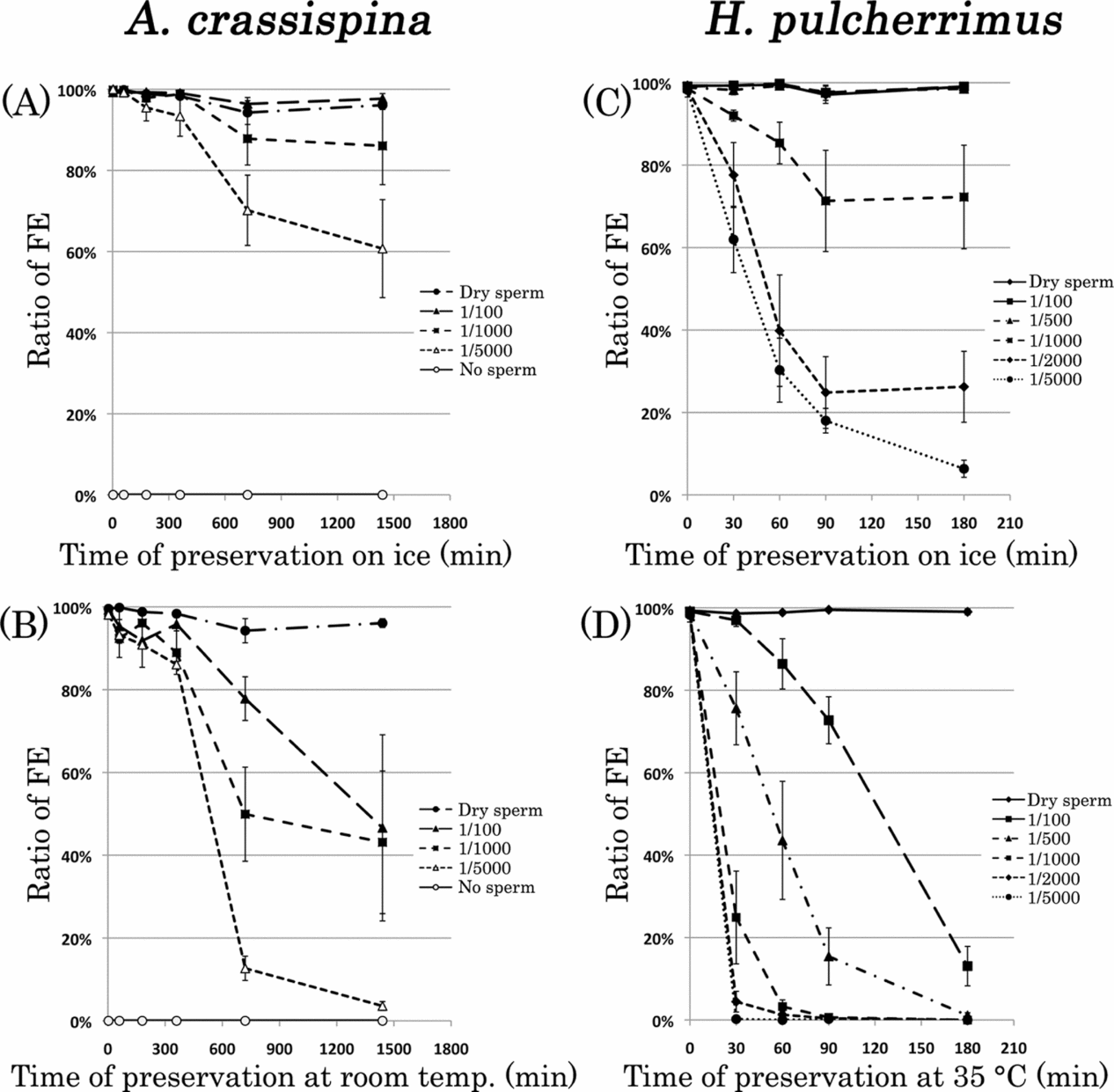
Next, we investigated the effect of sperm concentration on ROS generation and spermatozoa fertilizability. As shown in Fig. 5B, the ROS generation rate increased in a manner that was dependent on sperm concentration. Figure 10 shows the ratio of FE (%) derived using spermatozoa stored at various concentrations in two sea urchins, A. crassispina and H. pulcherrimus.
Ratio of formation of a fertilization envelope (FE) after insemination with stored Anthocidaris crassispina spermatozoa at various concentrations. The FE (%) was used to access the fertilizability of spermatozoa stored on ice (A, C), at room temperature (B) and at 35°C (D) at various dilution rates. (A, B) A. crassispina; (C, D) Hemicentrotus pulcherrimus. Unfertilized eggs for the FE (%) assay were stored at room temperature after adjustment to 5000 ± 500 eggs/ml and used until 6 h after spawning. The preservation times of the spermatozoa diluents are shown on the x-axes. Each plot shows the mean and standard error (SE) of four independent experiments.
For both species of sea urchin, eggs inseminated with spermatozoa stored at higher concentrations showed a higher ratio of FE (%) than did eggs inseminated with spermatozoa stored at lower concentrations. Following insemination with stored spermatozoa, the ratio of FE decreased in a manner that was dependent on the preservation time. This decrease was more marked in eggs inseminated with spermatozoa stored at a higher dilution. In the case of insemination with ‘dry sperm’ (stored on ice without dilution), the ratio of FE showed only a small decrease.
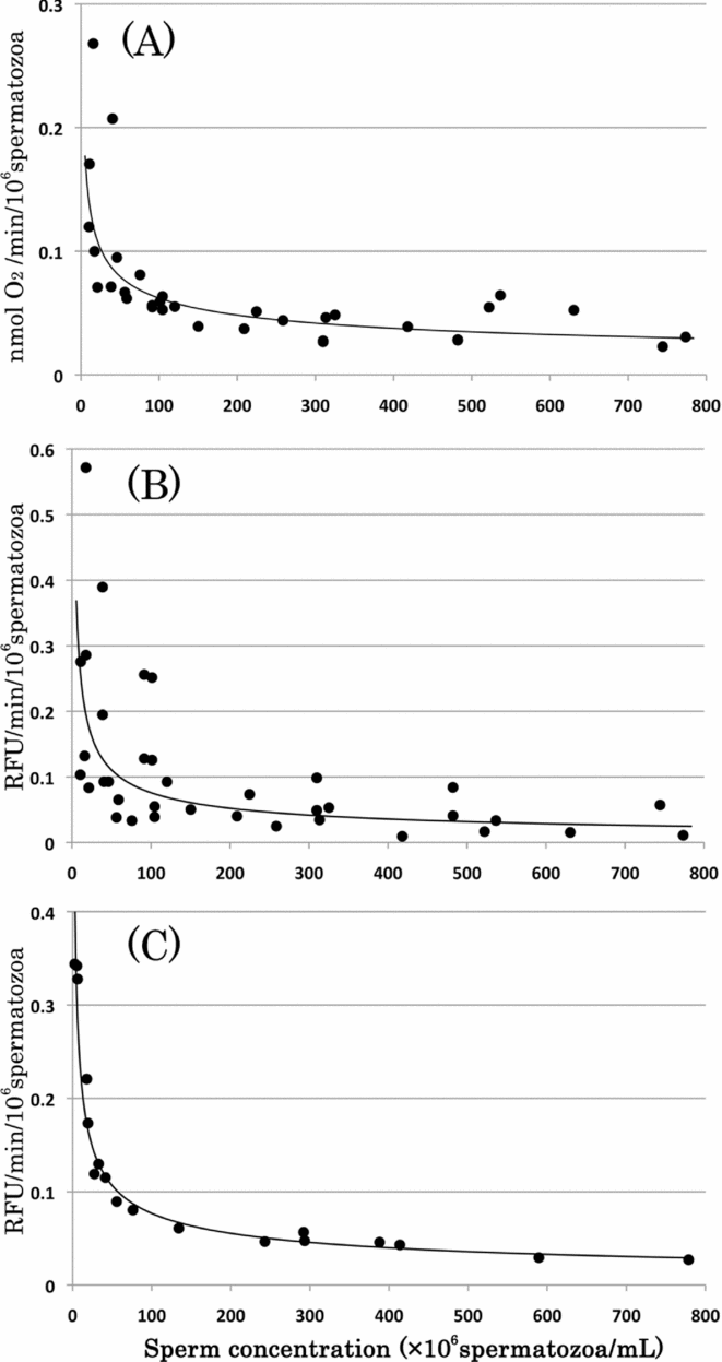
Next, we investigated the ‘sperm dilution effect’ (Gray, Reference Gray1928; Rothschild, Reference Rothschild1948; Mohri & Yasumasu, Reference Mohri and Yasumasu1963) by plotting the ROS generation rate per unit number of spermatozoa (RFU/min/106 spermatozoa) and the rate of respiration per unit number of spermatozoa (nmol O2/min/106 spermatozoa) against the sperm concentration (Fig. 11). We observed that each rate decreased in a manner that was dependent on the sperm concentration, although there were differences among the four different batches of A. crassispina.
Rates of oxygen consumption and CM-H2DCFDA fluorescence at various concentrations of sea urchin spermatozoa. The rates of oxygen consumption (A) and reactive oxygen species (ROS) generation (B) per unit number of spermatozoa were derived from 36 measurements, using four different batches of Anthocidaris crassispina. In the case of Hemicentrotus pulcherrimus, the ROS generation rate per unit number of spermatozoa (C) was obtained from a single male batch.
We further observed that the decrease in the ratio of FE (%) was accelerated at higher preservation temperatures. This decrease was most marked in the case of A. crassispina spermatozoa stored at room temperature (Fig. 10A, B). For H. pulcherrimus, the ratio of FE (%) derived using spermatozoa stored on ice was higher than that obtained using spermatozoa stored at 35°C, for all dilution concentrations (Fig. 10C, D). However the decrease in the rates for the two sea urchins were different at lower (Fig. 10A, C) and higher (Fig. 10B, D) temperature, and spermatozoa in both sea urchins had higher FE (%) after preservation at a lower temperature. Our data indicate that optimum sea urchin fertilizability is maintained by storage of undiluted spermatozoa on ice, in order to minimize ROS generation and maximize longevity.
Discussion
In the present study, sea urchin spermatozoa generated CM-H2DCFDA-detectable ROS, immediately following suspension in aerobic NSW and concomitantly with spontaneous initiation of motility and increased respiration. Although, in a few cases, the ROS levels in spermatozoa with lower motility was unclear, this phenomenon was demonstrated markedly in high motility batches. When sea urchin spermatozoa are motile and dispersed in seawater, CO2 decreases and O2 increases in the surrounding medium (Mohri & Yasumasu, Reference Mohri and Yasumasu1963). Thus, ROS generation is stimulated by the change from an anaerobic to an aerobic environment.
Sea urchin spermatozoa suspended in aerobic NaFSW exhibited no respiration and no ROS generation. Such spermatozoa were immotile and showed higher values of Atkinson's energy charge. Our present findings suggest that ROS generation is dependent on sperm motility via mitochondrion respiration in the male selected for high motile ability. Following the addition of Na+, or an increase in pH, the spermatozoa became motile, with increasing ADP and activation of respiration (Nishioka & Cross, Reference Nishioka, Cross, Dirksen, Prescott and Fox1978; Christen et al., Reference Christen, Schackmann and Shapiro1982). ADP supply is dependent on motility. Thus, ROS generation should initiate a change from an idle to an activated state of mitochondrion respiration in sea urchin spermatozoa.
The ROS generation rate per unit number of spermatozoa increased in a manner that was dependent on sperm concentration. Higher dilutions of dry sperm allow the activation of motility and respiration – a phenomenon known as the ‘sperm dilution effect’ (Gray, Reference Gray1928; Rothschild, Reference Rothschild1948). Our present findings are in accordance with those of Byrne et al. (Reference Byrne, Soars, Selvakumaraswamy, Dworjanyn and Davis2010), who reported that sperm densities are effective for successful fertilization. The increase in ROS generation at lower sperm concentrations can also be attributed to the ‘sperm dilution effect’, with ROS causing a decrease in fertilizability. Furthermore, given that ROS generation is dependent on respiratory activity, the ‘sperm dilution effect’ may also reflect a loss of sperm fertilizability caused by excess ROS generation per unit number of spermatozoa. Hence, the ‘sperm dilution effect’ acts not only as a trigger for sperm activation factors such as motility and respiration, but also as a constraint factor for viability, with ROS generation causing a decrease in the motility and fertilizability of sea urchin spermatozoa.
The influence of ROS generation on the ‘sperm dilution effect’ may not be present during natural oceanic fertilization. H2O2 can diffuse freely among cell membrane systems (Mathai & Sitaramam, Reference Mathai and Sitaramam1994) and must also be able to diffuse freely within the ocean simultaneously with sperm dispersion. Thus, sea urchin spermatozoa in natural oceans may not be exposed to high H2O2 concentrations.
In the case of reproductive and developmental biology investigations, sea urchin spermatozoa must be protected from ROS during preservation in the laboratory. The results of our present study suggest that storage of high sperm concentrations at low temperatures is an effective method to maintain low respiratory activities and low ROS levels and thereby prevent oxidative damages. However, effects of temperature differed between the two sea urchins. This difference may be caused by a different temperature sensitivity, because the reproductive seasons of A. crassispina and H. pulcherrimus are summer and winter, respectively. In fact, a lower temperature also effectively lowers ROS levels. Hence, the traditional storage method for sea urchin spermatozoa, such as ‘dry sperm’ on ice, is reasonable for sperm viability and longevity.
In natural fertilization, the negative effects of ROS on sperm fertilizability may only be observed at the point of contact with the egg. Sea urchin eggs release H2O2 during cortical exocytosis to ‘hard’ FEs after contact with the spermatozoon (Foerder et al., Reference Foerder, Klebanoff and Shapiro1978; Boldt et al., Reference Boldt, Schuel, Schuel, Dandekar and Troll1981; Coburn et al., Reference Coburn, Schuel and Troll1981). Thus, H2O2 release from the egg should be a mechanism for blocking polyspermy, such that later spermatozoa are affected by H2O2 release and loose viability (Boldt et al., Reference Boldt, Schuel, Schuel, Dandekar and Troll1981; Coburn et al., Reference Coburn, Schuel and Troll1981). Our present findings support the hypothesis that H2O2 causes a loss of sperm fertilizability. Sea urchin spermatozoa could be exposed to H2O2 when close to the egg and this event would be positive way to prevent polyspermy and enable successful fertilization of the egg.
In conclusion, sea urchin spermatozoa generate CM-H2DCFDA-detectable ROS immediately upon initiation of motility. The ROS generation rate was dependent on the mitochondrion respiratory activity, temperature and sperm dilution ratio. Hence, the RFU on CM-H2DCFDA can be considered to represent the activation of mitochondrion respiration, derived via ADP through flagellar motility. ROS generation reduces the motility and fertilizability of spermatozoa. Thus, storage of high concentrations of spermatozoa at low temperatures is required to minimize oxidative damage and to maximize longevity.
Acknowledgements
We thank the Manazuru Marine Laboratory, Faculty of Education and Human Sciences, Yokohama National University for assistance with material collection. Part of this work was supported by a Grant-in-Aid for Young Scientists (B) (No. 17770198) from the Ministry of Education, Culture, Sports, Science and Technology, Japan and also by a grant from the Research Institute for Integral Science, Kanagawa University.