1. Introduction
The animal fossil record is heavily biased towards the preservation of biomineralized tissues such as bone and shell. Non-biomineralized tissues exhibit considerable variation in their susceptibility to decay (Briggs, Reference Briggs2003; Butterfield, Reference Butterfield2003). The most labile organs, such as muscle and skin, are readily metabolized by bacteria, and require exceptional sedimentological and diagenetic conditions for their preservation (Seilacher Reference Seilacher1970; Briggs, Reference Briggs2003; Butterfield, Reference Butterfield2003). This normally involves their rapid burial and early replication by authigenic minerals (e.g. Briggs et al. Reference Briggs, Kear, Martill and Wilby1993; Wilby, Reference Wilby1993; Briggs, Reference Briggs2003). In contrast, more refractory (structural) tissues, such as cuticle, are significantly more decay-resistant and may be preserved organically (e.g. Butterfield, Reference Butterfield1990; Baas et al. Reference Baas, Briggs, van Heemst, Kear and de Leeuw1995).
Under favourable depositional conditions, arthropods and graptolites may be organically preserved in mudrocks (e.g. Underwood, Reference Underwood1992; Orr, Briggs & Kearns, Reference Orr, Briggs and Kearns2008). The organic exo- and endocuticle of arthropods consist of tanned or sclerotized complexes of protein and chitin (Stevenson, Reference Stevenson, Bliss and Mantel1985; Maddocks, Reference Maddocks, Harrison and Humes1992). In the fossil record, these complexes may, at least in part, be found both structurally (Briggs, Evershed & Stankiewicz, Reference Briggs, Evershed and Stankiewicz1998; Briggs et al. Reference Briggs, Stankiewicz, Meischner, Bierstedt and Evershed1998; Lingham-Soliar, Reference Lingham-Soliar1999) and chemically intact (e.g. Stankiewicz et al. Reference Stankiewicz, Briggs, Evershed, Flannery and Wuttke1997; Briggs, Evershed & Stankiewicz, Reference Briggs, Evershed and Stankiewicz1998; Briggs et al. Reference Briggs, Stankiewicz, Meischner, Bierstedt and Evershed1998), and such refractory organic complexes are capable of resisting decomposition long enough to be diagenetically transformed into more stable biomacromolecules (Briggs, Reference Briggs1999; Gupta et al. Reference Gupta, Tetlie, Briggs and Pancost2007). Likewise, the organic preservation of graptolite periderm may reflect the fact that it comprised a form of collagen that was tightly held together by interconnecting ‘rods’ (Crowther, Reference Crowther1981). In addition, the tanning of collagen may significantly increase its decay resistance (Briggs & Kear, Reference Briggs and Kear1993b; Aufderheide, Reference Aufderheide2003; Lastowka, Brown & Maffia, Reference Lastowka, Brown and Maffia2005). The notable recalcitrance of graptolite periderm is reflected in the abundance of these fossils in laminated Early Palaeozoic hemipelagites deposited on anoxic seafloors (Underwood, Reference Underwood1992; Briggs et al. Reference Briggs, Kear, Bass, de Leeuw and Rigby1995; Page at al. Reference Page, Gabbott, Wilby and Zalasiewicz2008); their soft-parts, however, have seldom been preserved (Loydell, Orr & Kearns, Reference Loydell, Orr and Kearns2004). The preservation of other soft-bodied organisms in this facies is also rare, although worms (e.g. Ruedemann, Reference Ruedemann1934), arthropods (see Vannier, Reference Vannier, Williams, Haywood, Gregory and Schmidt2007) and problematica (e.g. Page et al. Reference Page, Wilby, Mellish, Williams and Zalasiewicz2009) have been reported. These provide evidence of the existence of a more diverse biota in addition to the graptolites and rare shelly fossils that are typically preserved.
In this paper we report carbonaceous soft-tissues in a non-biomineralized arthropod from a Katian (Late Ordovician, mid-Caradocian equivalent), graptolite-bearing, hemipelagic mudstone in Wales, UK. Although the organophosphatic carapaces of Caryocaris (Churkin, Reference Churkin1966) may occur in large numbers alongside graptolites (Vannier et al. Reference Vannier, Racheboeuf, Brussa, Williams, Rushton, Servais and Siveter2003), non-biomineralized arthropods have not to our knowledge been reported from graptolitic mudrocks. The occurrence reported here augments the known diversity of Early Palaeozoic zooplankton and highlights the potential of anoxic hemipelagic facies for capturing soft-tissues other than graptolite periderm.
2. Material and stratigraphical setting
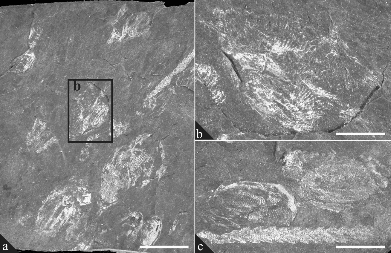
The arthropods are clustered on a single lamina in a block of hemipelagic mudstone (counterpart rock slabs: British Geological Survey MWL5229 and MWL5230) that was collected loose from the cliffs at Catch-y-Mitsiwr [National Grid Reference SN 0024 4025] on the western side of Dinas Island, southwest Wales (see Davies et al. Reference Davies, Waters, Wilby, Williams and Wilson2003). At least 23 arthropod ‘valves’ are visible, along with a few diplograptid graptolites. The arthropods form a moderately dense (about 1/cm2 on average), monospecific accumulate (Fig. 1), the full lateral extent of which is unknown. The collection locality exposes the Cwm-yr-Eglwys Mudstone Formation, part of the Late Ordovician succession of north Pembrokeshire and south Cardiganshire (Davies et al. Reference Davies, Waters, Wilby, Williams and Wilson2003), and yields the biozonal index graptolite species Dicranograptus clingani (Williams et al. Reference Williams, Davies, Waters, Rushton and Wilby2003), which has an exclusively Katian age (equivalent to a mid-Caradocian age in British Ordovician terminology).
Arthropod accumulate. All figured specimens are from BGS slab MWL 5229 and its counterpart slab MWL 5230. (a, c) Carapaces and accompanying graptolites apparently flow-aligned although the hemipelagic mudstone matrix indicates sea-bottom conditions were generally quiescent; (b) detail of carapaces indicating the thinness of the structure and its distortion. The spaces between the cuticle are filled by the clay mineral illite (white areas), which displays a tectonic fabric. Scale bar: (a) 1.2 cm; (b) 3.5 mm; (c) 5 mm.

3. Environmental setting
The Cwm-yr-Eglwys Mudstone Formation forms part of the thick sedimentary fill of the ensialic Welsh Basin, an area of enhanced Early Palaeozoic subsidence on the Eastern Avalonia palaeocontinent. It consists of dark grey turbiditic mudstone with abundant thin beds and laminae of siltstone, fine-grained sandstone and black, pyritic, organic-rich, hemipelagic mudstone (Davies et al. Reference Davies, Waters, Wilby, Williams and Wilson2003). The thickness of the formation varies across a number of major NE–SW faults, which define a series of deep syndepositional graben with steep, canyon-like walls (Davies et al. Reference Davies, Waters, Wilby, Williams and Wilson2003). To the north of the Newport Sands Fault, the Cwm-yr-Eglwys Mudstone Formation passes laterally into the sandstone turbidite-dominated Dinas Island Formation (Davies et al. Reference Davies, Waters, Wilby, Williams and Wilson2003). Graptolites are common throughout the Cwm-yr-Eglwys Mudstone Formation and include Dicellograptus clingani, Orthograptus ex gr. calcaratus, Lasiograptus cf. harknessi, Climacograptus antiquus s.l., Ensigraptus cf. caudatus and Corynoides sp. These indicate an open marine biotope for the overlying water column (Williams et al. Reference Williams, Davies, Waters, Rushton and Wilby2003). The local presence of Amorphognathus conodonts suggests oceanic influences and the upwelling of cool, nutrient-rich water along the adjacent slope (see Armstrong & Owen, Reference Armstrong, Owen, Crame and Owen2002). The only other fauna preserved in the unit are rare phosphatic linguliformean brachiopods, which are thought to have settled from the plankton (see Botting & Thomas, Reference Botting and Thomas1999).
There is no indication of any benthonic faunal activity in the hemipelagic mudstones, and sea-bottom conditions are believed to have been anoxic (Davies et al. Reference Davies, Waters, Wilby, Williams and Wilson2003). Weak, likely intermittent current activity on (or near) the sea bottom is indicated by flow-aligned graptolites throughout much of the formation, as well as by the alignment of the arthropods themselves (Fig. 1a, c).
4. Restoration of the arthropod specimens
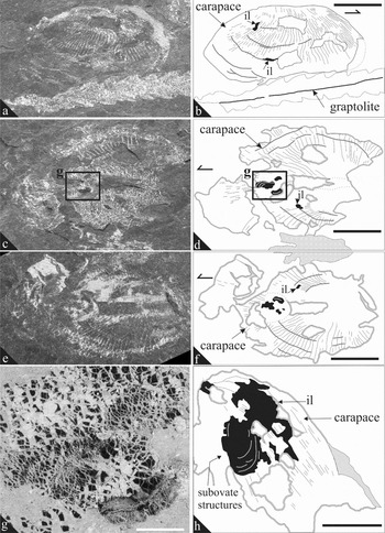
The arthropod specimens have undergone extensive bedding-parallel strain, and their carapaces are comprehensively fractured into a distinctive ‘checkerboard’ microfabric (Fig. 2g). Fibrous synkinematic intergrowths of kaolin, chlorite, illite and monazite fill the interstices between fragments (Wilby et al. Reference Wilby, Page, Zalasiewicz, Milodowski, Williams and Evans2007; Page et al. Reference Page, Gabbott, Wilby and Zalasiewicz2008) and occur in strain shadows behind ribs on the external surfaces of the valves (see Section 8, Fig. 2g, h). Additionally, they occur on the internal surfaces of the valves, behind the anterior and dorsal free margins.
Non-biomineralized arthropod tissues. All figured specimens are from BGS slab MWL 5229 and its counterpart slab MWL 5230. (a–f) Photographs with accompanying camera-lucida interpretations. (g, h) Backscattered scanning electron micrographs. (a, b) Right lateral view of carapace (arrow points to anterior), with straight dorsal hinge and small patches of carbonaceous tissue, interpreted to be remnants of the inner lamella, visible where the carapace has peeled away (arrowed ‘il’). The ribs on the lateral surface are more closely spaced towards anterior, where the carapace is also more convex. (c–f) Carapace preserved in ‘butterfly’ aspect, anterior to the left; (e) and (f) are the counterpart of (c) and (d). There is no distinct hinge. Articulation may be by means of a thin band of weakly mineralized cuticle, though there is no preserved evidence for this. The carapace shows a concentration of carbonaceous material towards the anterior dorsal region, which might represent remnants of the eyes or other indeterminate labile tissues, and which preserve a concentric structure. (g) Close-up of sub-ovate carbonaceous structures near the anterior dorsal margin and clearly ‘pressed’ against the inner surface of the carapace cuticle. At least 2, incomplete structures with a concentric pattern are preserved. (h) Inner surface of anterior dorsal region of right valve, with sub-ovate carbonaceous tissue possibly representing the remnants of the eye or other labile organic tissues, and a thinner film of carbonaceous tissue probably representing the inner lamella (il). Scale bar: (a–f) 4 mm; (g) 500 μm; (h) 2.5 mm.

The original outlines of the arthropods have been restored using a modified version of the retrodeformation technique described by Rushton & Smith (Reference Rushton and Smith1993). This technique applies a two-dimensional vector transform on the major and minor axes of the strain ellipse; implicit is the assumption that deformation is homogeneous and was orthogonal to bedding.
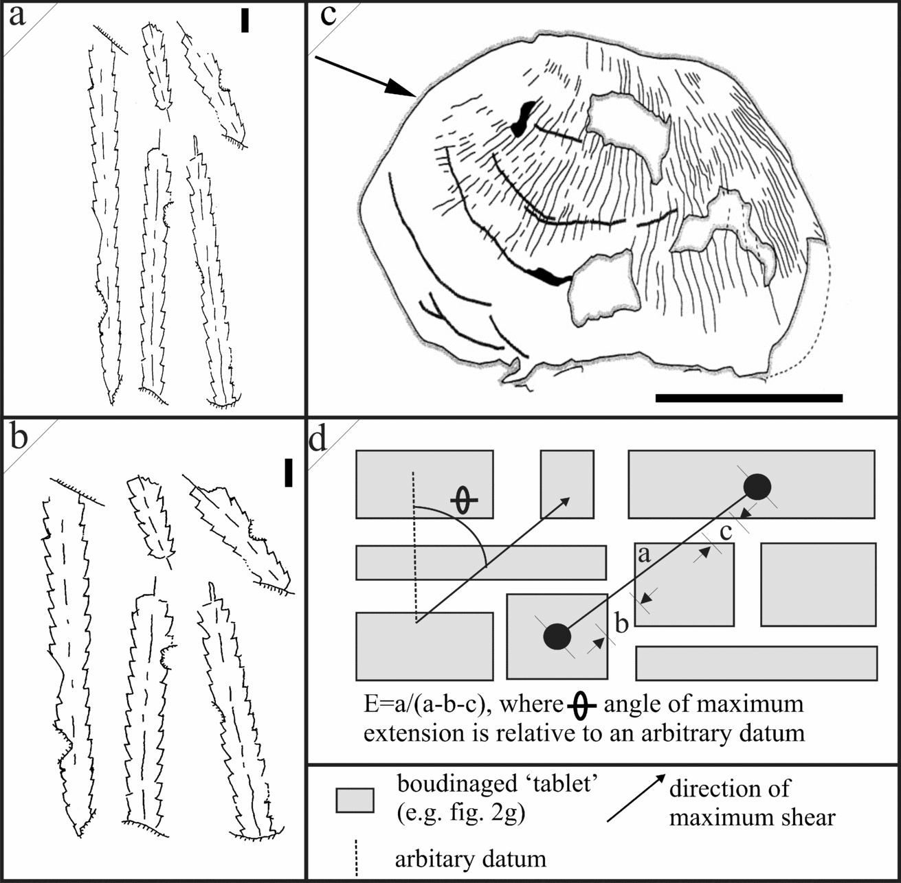
We determined a strain ellipse for the bivalved arthropods by estimating the direction (θ) and magnitude (E) of maximum extension (Fig. 3d) from which the major and minor axes can be interpolated. The direction of maximum extension was measured relative to an arbitrary datum; this lies parallel to the long axes of the synkinematic phyllosilicates as they formed parallel to shear (Underwood, Reference Underwood1992; Wilby et al. Reference Wilby, Page, Zalasiewicz, Milodowski, Williams and Evans2007). The magnitude of maximum extension was determined by calculating the extension revealed by the checkerboard boudinage. Over 200 measurements were taken on SEM images of two arthropod carapaces and three associated graptolites. These all yielded similar values for the magnitude and direction of strain, with respective values of 1.67 ± 0.08 and 79 ± 6° (expressed as means +/− standard deviations). Restoration was performed on digitized camera lucida interpretations of the fossils with Adobe Photoshop® image manipulation software. The images were rotated such that their maximum extension direction lay parallel to the y axis. As the arthropods are preserved in checkerboard boudinage with authigenic phyllosilicates filling the spaces in between the carapace boundins (Fig. 2g), there is no need to conserve volume in retrodeformation. As such they were transformed by a factor of 0.6 in the y axis alone. When applied to graptolites, this technique produces notably more symmetrical outlines (cf. Fig. 3a, b), confirming its veracity for producing retrodeformed images of the bivalved arthropod (Fig. 3c).
Reconstructed (retrodeformed) outline for the bivalved arthropod. (a, b) Camera lucida drawing of graptolites before (a) and after (b) retrodeformation. (c) Restored outline of bivalved arthropod, arrow points to anterior (see Fig. 2b for outline before retrodeformation). (d) Mechanism for retrodeformation (see text for details). Scale bar: (a, b) 5 mm; (c) 4 mm.

5. Arthropod morphology
5.a. Carapace
The carapace is up to 1.0 cm long and 0.8 cm high (restored dimensions, Fig. 3c) and is preserved as a thin (a few tens of microns thick), black, structureless, organic film; it shows no evidence of having been originally biomineralized. In lateral view it is sub-ovate (Fig. 3c). The anterior margin is more inflated and more robust than the posterior margin, which tapers slightly. The dorsal margin is straight and is about half the full length of the carapace. A lack of evidence for displacement of the two halves of the carapace suggests that it was not hinged; instead, it appears to have been folded along the dorsal mid-line to form two identical, nominal ‘valves’. Narrow ribs extend between the dorsal and free margins of the valves, and probably acted to strengthen them. Similar ribbing is known from Cambrian bivalved arthropods (Melnikova, Siveter & Williams, Reference Melnikova, Siveter and Williams1997, pl. 1, fig. 5). Over the mid- and posterior regions the ribs are spaced at 0.2–0.3 mm (measurements from deformed specimens), but towards the anterior margin they are much more closely spaced. Crudely concentric narrow folds (e.g. Fig. 2a, b) are interpreted as post-mortem creases, rather than genuine zones of relief. There are no lobes, sulci or spines.
Both open (‘butterfly’ or dorso-ventral aspect) and closed (lateral aspect) carapaces are present (Fig. 2a–f), indicating that articulation along the dorsal margin was possible. This is inferred to have been by means of a narrow band of flexible cuticle in the same manner as other bivalved arthropods such as Isoxys (see Williams, Siveter & Peel, Reference Williams, Siveter and Peel1996), although direct evidence of this structure in the Cardigan arthropod specimens is invariably concealed by sediment.
5.b. Soft integument: putative inner lamella
A thin (a few microns thick), black, amorphous layer of carbonaceous material (confirmed by EDX analysis of uncoated specimens) is preserved on the inner surface of several carapaces and adheres to internal moulds (e.g. Fig. 2a, b, h). It is less lustrous than the carapace and the rock matrix, but otherwise shows little contrast with either. The material appears to form a continuous, even film over the carapace, extending to the posterior free margin of the dorsum. In no specimen, however, is it possible to see whether it extends fully to the ventral margin or to the dorsal mid-line. Conceivably, this layer may represent a refractory organic residue left by the decay of the animal. However, there are no apparent variations in its thickness or distribution that might correspond to the positions of specific appendages or body divisions. Further, there is no evidence of it extending beyond the margins of the carapace, where remnants of appendages might reasonably be expected. Instead, the layer is interpreted as the inner lamella.
Inner lamellae are widespread in crustaceans and crustacean-like arthropods bearing a carapace, including a range of fossil taxa (e.g. Hinz, Reference Hinz1992; Williams, Siveter & Peel, Reference Williams, Siveter and Peel1996; Siveter, Waloszek & Williams, Reference Siveter, Waloszek and Williams2003). They consist of weakly or non-sclerotized chitin and remain uncalcified except along the carapace margins in some ostracods (Maddocks, Reference Maddocks, Harrison and Humes1992). Rather than being a simple marginal infold of the carapace, the inner lamella continues into the soft-bodied integument (Okada, Reference Okada1982; Maddocks, Reference Maddocks, Harrison and Humes1992). Experimental data for the preservation potential of inner lamellae are lacking, but the occurrence of other carbonized examples in the fossil record (e.g. Braun, Reference Braun1997) implies a degree of resilience. Indeed, they are among the most frequently preserved tissues in phosphatocopids from the Cambrian Orsten of Sweden, although they are often torn, creased or wrinkled (e.g. see Maas, Waloszek & Müller, Reference Maas, Waloszek and Müller2003, pls 27A, 30A, 33C, 40C), presumably as a result of decay prior to their replacement in phosphate. Rarely, inner lamellae are preserved in the Orsten fauna even when other non- or weakly sclerotized soft integument, such as the appendages and the trunk, are absent (e.g. see Maas, Waloszek & Müller, Reference Maas, Waloszek and Müller2003, pl. 4E).
5.c. Antero-dorsal soft anatomy
Discrete, substantial masses of carbonaceous material, several tens of microns thick, are present in the antero-dorsal region of two of the arthropod carapaces from Cardigan (Fig. 2g, h). In both cases, these structures are sub-ovate, about 2.5 mm long, and consist of a series of closely spaced, concentrically arranged, narrow bands (each about 30 μm wide). They are noticeably duller than the carapaces themselves (e.g. Fig. 2c, e) and have undergone a different pattern of fracture in strain (Fig. 2g). In one of the specimens, the sub-ovate structure can be demonstrated to lie internal to the putative inner lamella (Fig. 2h). In the other, there is the suggestion that the preserved sub-ovate structure consists of two slightly offset or superimposed masses, one sub-oval and the other reniform (Fig. 2g). The nature of these structures is uncertain, but their similar positioning in two specimens, one preserved in open attitude, and the other in closed, suggests that they are in situ and have not been displaced from elsewhere within the carapace (cf. Schmidt & Sellmann, Reference Schmidt and Sellmann1966). We postulate that they may represent one of three alternative tissues, each potentially located in an antero-dorsal position. These are discussed in turn in Sections 5.c.1 to 5.c.3.
5.c.1. Remnants of musculature
Many bivalved arthropods (e.g. ostracods, conchostracans) possess a set of bilaterally symmetrical, transverse adductor muscles that connect the body to the exoskeleton (Vannier, Wang & Coen, Reference Vannier, Wang and Coen2001) and act in opposition to the dorsal articulating structure (ligament, hinge, etc). The muscles are generally round or oval in cross-section and are often marked externally by a tubercle or sulcus, and internally by a group of taxonomically diagnostic scars (e.g. see Wilkinson et al. Reference Wilkinson, Williams, Siveter and Wilby2004, figs 1.5, 3).
Adductors are not, however, present in all bivalved arthropods (e.g. phyllocarids, phosphatocopids, bradoriids), and the Cardigan specimens lack any expression of them on the carapace. Further, the preserved structures appear to be positioned too far anterodorsally to have been able to function effectively in closing the carapace, although similarly positioned nodes on Perspicaris have been inferred as muscle attachment points (Briggs, Reference Briggs1977).
Muscles are considered to be a labile tissue (Briggs & Kear, Reference Briggs and Kear1993b, Reference Briggs and Kear1994; Briggs, Reference Briggs2003) with low potential for organic preservation (Butterfield, Reference Butterfield1990). As such, it seems unlikely that muscles would be preserved in these specimens.
5.c.2. Carapace attachment
All bivalved arthropods are attached to their carapace by a modified tergite situated on the dorsal surface of their body, presumably derived from a head segment (Waloszek et al. Reference Waloszek, Chen, Maas and Wang2005). For example, in recent ostracods a sheet of chitinous fibres connects the body and both valves across the uncalcified dorsal isthmus where the inner lamella connects to the body cuticle (e.g. see Kornicker, Reference Kornicker and Neale1969; Maddocks, Reference Maddocks, Harrison and Humes1992; Vannier, Wang & Coen, Reference Vannier, Wang and Coen2001). This attachment is contiguous with the outer lamella of both valves and does not play an active role in opening or closing the carapace. The extent of the dorsal attachment to the carapace is often ill-defined in fossil material, even when soft-parts are preserved, as for example in the Cambrian Isoxys (see Vannier & Chen, Reference Vannier and Chen2000, fig. 10C). In Cambrian phosphatocopids (see Müller, Reference Müller, Bate, Robinson and Sheppard1982), a long and thin attachment ligament extends along the entire dorsal mid-line, anchoring the head and the majority of the trunk to the carapace (see Maas, Waloszek & Müller, Reference Maas, Waloszek and Müller2003, fig. 23). In conchostracans, ostracods and phyllocarids, the attachment ligament is cephalic (e.g. McLaughlin, Reference McLaughlin1980, fig. 7B). In the latter groups, it bears a superficial resemblance to the structure present in the Cardigan fossils, particularly in the specimen preserved in open attitude. This is largely a taphonomic artefact; the closed specimen indicates the structure to be positioned too ventrally to be the site of attachment.
The preservation of such anatomy, but the absence of evidence of more decay-resistant structures, such as appendages, is at odds with the findings of actualistic taphonomic experiments (e.g. Briggs & Kear, Reference Briggs and Kear1993a,Reference Briggs and Kearb, Reference Briggs and Kear1994; Hof & Briggs, Reference Hof and Briggs1997; Orr, Briggs & Kearns, Reference Orr, Briggs and Kearns2008). Furthermore, the preservation of these anterodorsal structures differs notably from that of the chitinous carapace (Fig. 2c–h), suggesting they were not composed of chitin in vivo (Page et al. Reference Page, Gabbott, Wilby and Zalasiewicz2008), so are unlikely to represent an attachment tergite.
5.c.3. Eyes
Many extant and extinct bivalved arthropods possess sub-oval or reniform compound eyes. In recent phyllocarids and Isoxys they are stalked and protrude beyond the anterior end of the carapace (e.g. Briggs et al. Reference Briggs, Sutton, Siveter and Siveter2003; Vannier & Chen, Reference Vannier and Chen2000; Garcia-Bellido, Vannier & Collins, in press), but in other groups, such as myodocopid ostracods (see Vannier, Abe & Ikuta, Reference Vannier, Abe and Ikuta1998; Oakley & Cunningham, Reference Oakley and Cunningham2002), they lie within the carapace. Numerous bivalved arthropods have spherical eyes (e.g. Hou et al. Reference Hou, Aldridge, Bergstrom, Siveter, Siveter and Feng2004) and these are generally of comparable size, shape and position to the paired structures in the Cardigan arthropods. Commonly, such eyes are associated with structures in the overlying carapace. In some ostracods the carapace is locally thinned and more translucent (visual windows) (Vannier & Abe, Reference Vannier and Abe1992; Perrier, Vannier & Siveter, Reference Perrier, Vannier and Siveter2007, and in press), in certain podocopid ostracods ‘lens-like’ structures are present (see Kontrowitz & Myers, Reference Kontrowitz and Myers1984), and in certain ostracods such as Silurian mydocopes (V. Perrier, unpub. Ph.D. thesis, Univ. Lyon, 2007) their position is defined by ‘eye lobes’. However, such differentiation of the carapace is not universally present and it has not been observed in the Cardigan arthropods.
Recent bivalved arthropods such as myodocopid ostracods have compound eyes that bear more or less densely packed ommatidia. In some cylindroleberidids (Vannier, Abe & Ikuta, Reference Vannier, Abe and Ikuta1996) the ommatidia form well-ordered rows of lenses. Evidence for the arrangement of such structures in fossil examples is rare, but pits formerly accommodating lens-like ommatidia have been reported in Triassic myodocopids (Weitschat, Reference Weitschat1983). Although ommatidia are usually arranged in a polygonal manner, it is conceivable that the concentric patterns preserved in the oval structures within the Cardigan arthropods could represent the boundaries between adjacent rows of lenses in a paired compound eye, or perhaps post-mortem products of tissue shrinkage and/or creasing (cf. Duncan, Titchener & Briggs, Reference Duncan, Titchener and Briggs2003, p. 261; Maas, Waloszek & Müller, Reference Maas, Waloszek and Müller2003).
In the Burgess Shale, the eyes of Isoxys are frequently the best preserved of all of its soft anatomy, occurring in 90% of specimens (Garcia-Bellido, Vannier & Collins, in press), indicating a relatively high preservation potential for the eyes of certain bivalved arthropods. This, along with the morphological and preservational arguments against these structures representing muscles or carapace attachment tergites detailed (see Sections 5.c.1 and 5.c.2, above), suggests the antero-dorsal structures most likely represent eyes.
6. Affinities
Disparate arthropods may possess a superficially similar bivalved carapace; taxonomic assignment therefore relies heavily on soft-part morphology and, in particular, on cephalic appendages (Briggs, Reference Briggs and Schram1983; Siveter, Williams & Waloszek, Reference Siveter, Williams and Waloszek2001; Siveter, Waloszek & Williams, Reference Siveter, Waloszek and Williams2003). With the possible exception of the eyes (see Section 5.c.3), no head, trunk or appendage anatomy is preserved in the Cardigan specimens. As a result, their affinities remain uncertain.
Extremely thin, weakly or non-biomineralized, bivalved carapaces were adopted by several Early Palaeozoic arthropods such as, for example, Isoxys (Williams, Siveter & Peel, Reference Williams, Siveter and Peel1996; Vannier & Chen, Reference Vannier and Chen2000; Garcia-Bellido, Vannier & Collins, in press). The absence of a discrete dorsal hinge in the Cardigan material recalls the situation in some phyllocarids (Rolfe, Reference Rolfe and Moore1969), although the specimens lack evidence of the articulating rostral plate that is present in some, but not all, species (Rolfe, Reference Rolfe and Moore1969, p. R299; Vannier et al. Reference Vannier, Racheboeuf, Brussa, Williams, Rushton, Servais and Siveter2003). An inner lamella is possessed by several crustacean and crustacean-like arthropods and is of little taxonomic significance (cf. Briggs, Reference Briggs and Schram1983).
In terms of overall morphology, the Cardigan specimens do not resemble the carapace of any known Early Palaeozoic bivalved arthropod of comparable size. Marine pelagic myodocopid ostracods have a fossil record extending back to the latest Ordovician (Gabbott et al. Reference Gabbott, Siveter, Aldridge and Theron2003) and they sometimes attain centimetre scale, but their carapaces have well-defined rostral notches and adductor muscle scars, neither of which are seen in the Cardigan arthropods. Coeval marine benthonic ostracods are typically much smaller, have a calcified carapace with strong ventral overlap, and an articulating hinge (e.g. Williams & Vannier, Reference Williams and Vannier1995). The carapaces of some leperditicopid arthropods are of a similar size (typically 5 to 50 mm) to the Cardigan specimens, but they are heavily calcified, asymmetric, have pronounced ventral overlap and a complex pattern of muscle scars on the internal surface (Vannier, Wang & Coen, Reference Vannier, Wang and Coen2001). Additionally, they are entirely benthonic and typically occupy shallow marine settings (e.g. Berdan, Reference Berdan1984; Vannier, Wang & Coen, Reference Vannier, Wang and Coen2001), contrary to that inferred for the Cardigan arthropods (see Section 7). Similarly, the Cardigan specimens show no resemblance to the elongate and spinose Ordovician bivalved arthropod Caryocaris (see Vannier et al. Reference Vannier, Racheboeuf, Brussa, Williams, Rushton, Servais and Siveter2003), which though common in graptolitic mudrocks, is of an organophosphatic composition (Churkin, Reference Churkin1966; Page et al. Reference Page, Wilby, Mellish, Williams and Zalasiewicz2009).
Some Cambrian svealutid bradoriids, such as Anabarochilina, Liangshanella and Tsunyiella, may have had a pelagic mode of life and possessed only weakly mineralized carapaces devoid of an articulating hinge structure (see Siveter & Williams, Reference Siveter and Williams1997; Hou et al. Reference Hou, Siveter, Williams and Feng2002). One cambriid bradoriid from the Lower Cambrian of Kazakhstan figured by Melnikova, Siveter & Williams (Reference Melnikova, Siveter and Williams1997, pl. 1, fig. 5) also has dorsal to ventrally orientated ribbing on the valve surface in a similar manner to the Cardigan specimens, but its overall morphology is different. As such, these bradoriids superficially resemble the Cardigan arthropod specimens, although no specific bradoriid form shows a strong morphological similarity. Equally, the Cardigan arthropods superficially resemble (and are within the size range of) some smaller thylacocephalans, an arthropod group with a fossil record that extends back to the Silurian and possibly earlier (Lange et al. Reference Lange, Hof, Schram and Steeman2001; Vannier et al. Reference Vannier, Chen, Huang, Charbonnier and Wang2006). They possess non-biomineralized carapaces with a surface sculpture of alternating ribs and furrows, the ribs running ventrally from the dorsal margin (see Lange et al. Reference Lange, Hof, Schram and Steeman2001). However, thylacocephalan carapaces differ by having a well-developed rostrum and an arched dorsal margin.
More comparable with the Cardigan specimens are the many ‘featureless’ pelagic bivalved arthropods that are frequent in Early Cambrian black shales of the Yangtze platform (Steiner et al. Reference Steiner, Mehl, Reitner and Erdtmann1993, Reference Steiner, Wallis, Erdtmann, Zhao and Yang2001; Zhu et al. Reference Zhu, Zhang, Steiner, Yang, Li, Erdtmann, Zhu and Steiner2004; Vannier et al. Reference Vannier, Caron, Yuan, Briggs, Collins, Zhao and Zhu2007; Vannier, Reference Vannier, Williams, Haywood, Gregory and Schmidt2007), and these show a similar style of preservation to our specimens. Some resemble Isoxys volucris (see Williams, Siveter & Peel, Reference Williams, Siveter and Peel1996), while others have a more circular carapace outline, but all possess a weak carapace that is prone to wrinkling and tearing.
7. Palaeoecology
The bivalved arthropods most likely had a pelagic ecology, as indicated by the depositional setting, their ‘light-weight’ carapaces, and the associated planktonic fauna of graptoloids, conodonts and brachiopods (Williams et al. Reference Williams, Davies, Waters, Rushton and Wilby2003). Indeed the lack of bioturbation in the laminated host mudrock matrix, as well as the absence of a benthic or nektobenthic fauna, indicate inhospitable, anoxic bottom waters (Wignall & Hallam, Reference Wignall, Hallam, Tyson and Pearson1991), consistent with the presence of relatively abundant organic carbon and disseminated pyrite in the mudrock matrix.
Similar ‘light-weight’ carapaces were adopted by other Early Palaeozoic pelagic arthropods such as Isoxys and Caryocaris, which dominated midwater niches prior to their colonization by ostracods in mid-Silurian times (Siveter, Vannier & Palmer, Reference Siveter, Vannier and Palmer1991; Vannier & Chen, Reference Vannier and Chen2000; Vannier et al. Reference Vannier, Racheboeuf, Brussa, Williams, Rushton, Servais and Siveter2003). This guild likely fulfilled an important ecological role in distal shelf to proximal continental slope settings, forming a link in the food web between low trophic level microphytoplankton (e.g. acritarchs) and secondary consumers (e.g. trilobites, orthoconic nautiloids, conodonts) (Vannier et al. Reference Vannier, Racheboeuf, Brussa, Williams, Rushton, Servais and Siveter2003). Like many Early Palaeozoic pelagic arthropods (e.g. Vannier et al. Reference Vannier, Racheboeuf, Brussa, Williams, Rushton, Servais and Siveter2003), the Cardigan arthropods occupied an area of oceanic upwelling and high productivity (Williams et al. Reference Williams, Davies, Waters, Rushton and Wilby2003), in this case situated above a complex subaqueous topography (Davies et al. Reference Davies, Waters, Wilby, Williams and Wilson2003). Their preserved density suggests that they may have locally formed dense schools, perhaps analogous to those of certain phyllocarids and euphausiids (krill) which congregate diurnally in modern submarine canyons (e.g. Greene et al. Reference Greene, Wiebe, Burczynski and Youngbluth1988), although alternatively, they could have been more widely dispersed and have been channelled post-mortem into detritus traps formed by steep-sided depressions (cf. Vetter, Reference Vetter1995).
8. Taphonomy
The taphonomy of the arthropods is complex and has a strong tectonic overprint. They are preserved en masse on a single lamina (Fig. 1a) and are exposed at its intersection with a gently inclined cleavage plane, along which the rock split. Most of the fossils have been transgressed by the cleavage plane and are only partially revealed, the remainder of their carapaces lying concealed in the sediment either above or below it with their valves either open in dorso-ventral (‘butterfly’) aspect or closed in lateral aspect. Approximately a third of the specimens are preserved in lateral aspect; in such specimens, the plane of splitting tends to pass either through the carapace, revealing either internal details or the external surface of one of the valves. A single specimen is preserved in ‘butterfly’ aspect. In this specimen (Fig. 2c–f) the plane of splitting passes from the external surface of the left valve to the internal surface of the opposing valve. The remaining specimens are preserved in oblique aspect and have varying relationships to the cleavage.
The carapaces show substantial, largely concentric, creasing (Fig. 2a–f). This is generally considered indicative of an originally flimsy carapace (e.g. see Siveter, Rushton & Siveter, Reference Siveter, Rushton and Siveter1995; Williams, Siveter & Peel, Reference Williams, Siveter and Peel1996; Hou et al. Reference Hou, Siveter, Williams and Feng2002), homologous to that of the modern phyllocarid Nebaliopsis typica (see Vannier, Boissy & Racheboeuf, Reference Vannier, Boissy and Racheboeuf1997). Incipient decay of the Cardigan arthropods, involving the loss of the most labile constituents from their carapaces, may have further enhanced their flexibility (cf. Briggs & Kear, Reference Briggs and Kear1993b), although no direct evidence of microbial activity (cf. Hof & Briggs, Reference Hof and Briggs1997, fig. 2) is observed. Most of the specimens are compressed. Some, however, especially those preserved in lateral aspect, retain a degree of three-dimensionality, particularly in their anterior and antero-central regions. Sediment infill of such carapaces has acted to reduce the effects of compaction, although in several cases their external ribs have become superimposed on the infill (that is, they are compound fossils).
Actualistic taphonomic data are lacking for bivalved arthropods. However, data are available for decapod, branchiopod and stomatopod crustaceans (Briggs & Kear, Reference Briggs and Kear1993a,Reference Briggs and Kearb, Reference Briggs and Kear1994; Hof & Briggs, Reference Hof and Briggs1997; Orr, Briggs & Kearns, Reference Orr, Briggs and Kearns2008), and these can be used to interpret the preservation of the Cardigan arthropods.
Only the arthropods’ most integral tissues (those most strongly connected to the carapace) are preserved. As such, their preservation differs notably from arthropods thought to have been buried alive or shortly after death (cf. Orr, Briggs & Kearns, Reference Orr, Briggs and Kearns2008). Thin-valved pelagic arthropods, like those described here from Cardigan, are likely to endure protracted periods of post-mortem drift (Racheboeuf, Vannier & Ortega, Reference Racheboeuf, Vannier and Ortega2000) and might be expected to arrive at the sediment surface in an advanced state of disarray. Even if the part-decayed bodies of these arthropods were present at the sediment–water interface, they would probably have been disarticulated and washed away by the currents that caused the graptolites to align (cf. Orr, Briggs & Kearns, Reference Orr, Briggs and Kearns2008). This seems consistent with the preservation of the putative inner lamella (firmly attached), which is only weakly sclerotized (Okada, Reference Okada1982; Maddocks, Reference Maddocks, Harrison and Humes1992), but the absence of more heavily sclerotized anatomy such as limbs (relatively weakly attached). Their eyes, in contrast, are likely to have a relatively high preservation potential because of the head's fusion to the carapace. Cephalic structures may have been among the last to separate from the carcass, reflected in their apparent preponderance in other fossil bivalved arthropods (e.g. see Dechaseaux, Reference Dechaseaux1951; Gramann, Reference Gramann1962; Siveter, Rushton & Siveter, Reference Siveter, Rushton and Siveter1995; Braun, Reference Braun1997).
While some histological details have been reported in other examples of carbonaceous soft-part preservation (e.g. Lingham-Soliar, Reference Lingham-Soliar1999), suggesting a lack of microbial intervention and the retention of direct molecular sister products (Briggs, Evershed & Stankiewicz, Reference Briggs, Evershed and Stankiewicz1998; Briggs et al. Reference Briggs, Stankiewicz, Meischner, Bierstedt and Evershed1998), the organic preservation described here is amorphous.
The preservation of anatomy such as eyes and inner lamellae may have been afforded by: (1) persistence of a more recalcitrant residue of the originally more labile tissues (e.g. Briggs, Reference Briggs1999; Vinther et al. Reference Vinther, Briggs, Prum and Saranathan2008); (2) the tissues being lightly sclerotized and therefore having an enhanced preservation potential (e.g. Butterfield, Reference Butterfield1990; Briggs, Reference Briggs2003); (3) pseudomorphing by microbes, their more resistant cell walls being what is preserved (e.g. fig. 2a of McNamara et al. Reference McNamara, Orr, Kearns, Alcalá, Anadón and Peñalver-Mollá2006); (4) decay having been short-circuited by the high organic carbon content of the surrounding hemipelagic sediment which, through microbial action, would have released high concentrations of self-poisoning microbial waste products; or (5) contact with the sediment; Martill (Reference Martill1987a,Reference Martillb) noted that organic preservation only occurred on the underside of ichthyosaurs. There is no evidence for bacteria-inhibiting clays having been adsorbed to the surfaces of the organics (cf. Butterfield, Reference Butterfield1990).
Subsequent to the initial, organic preservation of these fossils, they have undergone a complex metamorphic history, which saw them experience considerable strain in tectonism (see Section 4) and become the locus of authigenic mineralization. As is common in such mudrocks, these fossils served as a site for phyllosilicate formation in very low-grade metamorphism (Underwood, Reference Underwood1992; Page et al. Reference Page, Gabbott, Wilby and Zalasiewicz2008), during which authigenesis of kaolin, illite and chlorite occurred (Page et al. Reference Page, Gabbott, Wilby and Zalasiewicz2008). These phyllosilicates display an anatomy-specific distribution, with kaolin only occurring in association with the putative eyes, and chlorite only occurring in association with the carapaces (see Page et al. Reference Page, Gabbott, Wilby and Zalasiewicz2008). Phyllosilicate authigenesis likely began during burial and continued during cleavage formation with the phases growing in spaces created during their deformation, at which time synkinematic monazite also developed (Wilby et al. Reference Wilby, Page, Zalasiewicz, Milodowski, Williams and Evans2007).
9. Conclusions
The record of Ordovician planktonic communities is overwhelmingly dominated by graptolites. Thin-valved arthropods from the Cwm-yr-Eglwys Mudstone Formation represent an important addition to this community and highlight the potential of anoxic hemipelagic mudstones for capturing soft-bodied organisms. The depositional setting, along with the preserved density of these fossils, suggests shoaling of these arthropods in an area of high productivity.
Acknowledgements
We thank Nick Butterfield (Cambridge) and an anonymous referee for their constructive reviews. We are grateful to Paul Shepherd and Antoni Milodowski (BGS) for assistance with photography and electron microscopy, respectively. PRW and JRD publish with the permission of the Executive Director, BGS. Cambridge Earth Sciences contribution number ES.9420.