Introduction
Since the early 1900s, during middle cranial fossa surgery, the superior aspect of the petrous pyramid has been approached extradurally in order to correct petrositis.Reference House1 In 1904, Parry described a middle fossa approach for sectioning the VIIIth nerve, to treat Ménière's disease.Reference Parry2
In 1954, Clerc and Batisse revived this approach to the petrous pyramid in order to perform destructive labyrinthectomy through the superior canal. In 1958, William House began searching for a method to remove osteosclerotic foci from the region of the internal auditory canal. In 1959, he performed the first operation using diamond drill and operating microscope.Reference House3, Reference Lampert4 Today, the middle cranial fossa approach is used to access many different types of pathology. Many variations of the original exposure have been described: an enlarged middle cranial fossa approach; an extended approach through the middle cranial fossa; a translabyrinthine-transtentorial approach; and a translabyrinthine-transtentorial approach via the middle cranial fossa.Reference Wigand, Haid, Berg and Rettinger5–Reference Morrison9
The middle cranial fossa approach is a technically demanding procedure, with little margin for error. This is mainly due to limitations in exposure and difficulty in identifying landmarks.Reference House and Shelton10, Reference Shelton, Brackmann and House11 This study describes a different landmark, the processus cochleariformis, which the author has used in middle cranial fossa surgery for 10 years.Reference Ulug and Ulubil12–Reference Ulug and Ulubil14 Using 10 cadaver specimens, 20 measurements were made to relate the processus cochleariformis to other critical structures such as the vertical crest, and to give an impression of the inner-ear structures in the intact bone.
Materials and methods
This anatomical study was performed on 10 cadaveric specimens fixed in formaldehyde solution. All of the skulls were from male adult Caucasians. There were no variations or pathological findings on the skulls. The right side of the skulls was dissected.
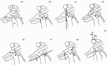
Dissection was performed with an operating microscope, utilising cutting and diamond burs with continuous irrigation. In contrast to the classical middle cranial fossa approach, the tegmen tympani was opened first (Figure 1a). After the malleolar head, incus body and processus cochleariformis had been exposed, bone was drilled in a lateromedial direction, with the centre of drilling located directly medial to the processus cochleariformis. This was the anticipated position of the vertical crest. During drilling, continuous irrigation was used to help identify the blue lines of the ampulla of the superior semicircular canal (SSC), the basal turn of the cochlea and the labyrinthine segment of the facial nerve, and therefore to prevent damage to these structures. The dissection was continued freely beyond 7–8 mm, as neither the cochlea nor the vestibulum extends medially at this point, but the anticipated position of the critical structures was always borne in mind.
Temporal bone measurements. See text for explanation of figure parts. M = malleolar head; I = incus body; PC = processus cochleariformis; GSPN = greater superifical petrosal nerve; TFN = tympanic facial nerve; BTC = basal turn of cochlea; LFN = labyrinthine facial nerve; VC = vertical crest; V = vestibulum; SVN = superior vestibular nerve; IAC = internal auditory canal; SCC = superior semicircular canal

After the delicate anatomical dissection had been completed, all the relevant underlying anatomical structures were unroofed and measurements were made using small size callipers accurate to 0.1 mm and a goniometer accurate to 1°. Twenty reproducible measurements were defined on the 10 cadaver specimens.
The first six of these measurements defined the relation of the processus cochleariformis to the inner ear and related structures. These included measurements of the distances from the processus cochleariformis to the vertical crest, to the cochlea and to the vestibulum, and measurement of the angles between the same structures and the coronal plane (Table I, Figure 1b and 1c).
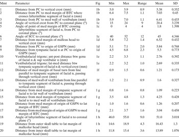
Temporal bone measurements

Mmt = measurement; Min = minimum; Max = maximum; SD = standard deviation; PC = processus cochleariformis; med = medial; BTC = basal turn of cochlea; n = nerve; SCC = superior semicircular canal; GSPN = greater superficial petrosal n; ant = anterior; post = posterior; btw = between; sup = superior; lat = lateral
The other 14 measurements defined the exact location of the inner ear and adjacent structures within the bone (Table I, Figure 1d to 1h). These included measurements of the distances from the malleus to the vertical crest, from the processus cochleariformis to the greater petrosal nerve, and from the facial nerve at the processus cochleariformis to the greater petrosal nerve. Measurements were also made of the diameters of the vestibulofacial trigone, of the distances of the cochlea and the vestibulum to an imaginary line passing through the vertical crest, of the distances of the vestibulum and the cochlea to the tympanic facial nerve and the superficial petrosal nerve, of the angle of the labyrinthine segment of the facial nerve to the coronal plane, and of the distances from the outer and inner skull tables to the malleus.
After all these measurements had been noted, the minimum, maximum, range, mean and standard deviation of each were defined.
Results
The results of the measurements are summarised in Table I. The following text gives mean data values.
The measurements relating the processus cochleariformis to the inner ear and related structures revealed consistent results. The distance from the processus cochleariformis to the vertical crest was found to be 5.38 mm. The distance from the processus cochleariformis to the medial margin of the basal turn of the cochlea where it crosses the labyrinthine segment of the facial nerve was found to be 7.03 mm. The distance from the processus cochleariformis to the medial wall of the vestibulum was found to be 6.41 mm (Figure 1b).
The angle of the vertical crest from the processus cochleariformis to the coronal plane was measured as 20.4º. The angle of the point at which the medial margin of the basal turn of the cochlea crosses the labyrinthine segment of the facial nerve, from processus cochleariformis to coronal plane, was measured as 1.8°. The angle of the superior semicircular canal to the coronal plane was found to be 45° (Figure 1c).
The measurements relating inner ear related structures to various other structures also revealed consistent results. The distance from the medial margin of the malleus head to the vertical crest was 6.52 mm. The distance from the processus cochleariformis to the origin of the greater superficial petrosal nerve was 5.84 mm. The distance from the tympanic facial nerve at the processus cochleariformis to the origin of the greater superficial petrosal nerve was 5.3 mm (Figure 1d). The anterior–posterior dimension of the vestibulofacial trigone was found to be 2.76 mm. The lateral–medial dimension of the vestibulofacial trigone was found to be 2.69 mm (Figure 1e).
The distance from the medial margin of the basal turn of the cochlea to an imaginary line running parallel to the tympanic segment of the facial nerve and passing through the vertical crest was measured as 1.21 mm. The distance of the medial wall of the vestibulum from an imaginary line running parallel to the tympanic segment of the facial nerve and passing through the vertical crest was measured as 1.6 mm (Figure 1f).
The distance from the medial margin of the tympanic segment of the facial nerve to the lateral wall of the vestibulum was found to be 1.09 mm. The distance from the medial margin of the tympanic segment of the facial nerve to the medial wall of the vestibulum was found to be 4.25 mm. The distance from the medial margin of the origin of the greater superficial petrosal nerve to the lateral margin of the basal turn of the cochlea was found to be 1.26 mm. The distance from the medial margin of the origin of the greater superficial petrosal nerve to the medial margin of the basal turn of the cochlea was found to be 3.04 mm (Figure 1g).
The angle of the labyrinthine segment of the facial nerve to the coronal plane was found to be 51°. The distance from the outer skull table to the lateral margin of the malleolar head was measured as 16.43 mm. The distance from the inner skull table to the lateral margin of the malleolar head was measured as 13.89 mm (Figure 1h).
Discussion
A review of the literature concerning non-classical middle cranial fossa landmarks revealed only a few studies. Most of these attempted to find a correlation between distant structures and the inner-ear area; however, the distant structures chosen cannot be used as exact surgical guides in this complex area. Chopra et al. assessed the distance from the inner table of the craniotomy to the superior semicircular canal for use as a landmark, and found 22 mm on average.Reference Chopra, Fergie, Mehta and Liew15 Miller and Pensak proposed use of the root of the zygoma and the foramen spinosum as landmarks, and demonstrated that the distance from the root of the zygoma to the head of the malleus was 18.7 mm and the distance from the foramen spinosum to the head of the malleus was 19.2 mm.Reference Miller and Pensak16 In the present author and colleagues' previous study, Henle's spine was proposed for use as the main landmark to determine other structures of the skull base, including the inner-ear area.Reference Ulug, Ozturk and Sahinoglu17 Similarly, in other studies the zygomatic root and foramen spinosum were used as landmarks for more medially located structures.Reference Lee, Kim and Lee18, Reference Todd19
The first six measurements assessed by the present study indicate that the distances and angles of the major inner-ear structures to the processus cochleariformis are constant and reliable. This means that the processus cochleariformis can be used as a landmark in middle cranial fossa surgery. The main structure, the vertical crest, is at a distance of 5–6 mm and an angle of 20° from the processus cochleariformis to the coronal plane. The important point at which the medial margin of the basal turn of the cochlea crosses the labyrinthine segment of the facial nerve is 6.5 to 7.5 mm from the processus cochleariformis and lies at an angle of approximately 0° from the processus cochleariformis to the coronal plane. The superior semicircular canal lies at a 45° angle from the processus cochleariformis to the coronal plane, and the medial wall of the vestibulum is 6–7 mm from the processus cochleariformis in this direction.
House's original description of the middle cranial fossa approach emphasised the initial identification of the greater superficial petrosal nerve (facial hiatus) as the only landmark to follow to the geniculate ganglion, labyrinthine segment and internal auditory canal.Reference House1–Reference Lampert4 Thus, the overall dissection proceeds in a lateral to medial direction. In this technique, damage to the cochlea, vestibule and the superior semicircular canal is mainly avoided by staying close to the facial nerve. Although the eminentia arcuate was mentioned, neither this structure nor the middle meningeal artery were used as landmarks in House's original middle cranial fossa approach. However, the use of these points as landmarks does appear in House's later publications.Reference House and Shelton10, Reference Graham20, Reference Meyerhoff21 The most important modification of this technique was introduced by Ugo Fisch. In his transtemporal supralabyrinthine approach, he describes use of the blue line of the superior semicircular canal as a landmark and notes that the internal auditory canal lies within a 60° angle from this line.Reference Lampert4, Reference Fisch and Mattox22, Reference Fisch23 Another technique, described by Garcia Ibanez and Garcia Ibanez, uses the arcuate eminence as a landmark; however, the superior semicircular canal is not described as blue-lined. In this technique, bone removal begins medially near the porus acousticus.Reference Lampert4, Reference Garcia Ibanez and Garcia Ibanez24
These are the three most frequently used methods for exposing the internal auditory canal, and can be used separately or in combination.Reference Lampert4, Reference Meyerhoff21 However, a search for additional landmarks continues because of the surgical difficulty in this area. Recently, Kobayashi and Nakao proposed using illumination through the meatus acusticus externus as an aid during middle cranial fossa surgery.Reference Kobayashi and Nakao25 The author's proposed method, looking for the vertical crest at an angle of 20° from the processus cochleariformis to the coronal plane, at a distance of 5–6 mm from the processus cochleariformis, together with other additional clues, can be used independently or in combination with other techniques. If the processus cochleariformis is used as the only landmark, continuous irrigation and magnification is important in order to detect the slight colour change that indicates that the superior semicircular canal or basal turn of the cochlea is adjacent. If Garcia Ibanez and Garcia Ibanez' direct medial approach to the porus acousticus is used, the proposed landmark ensures safety, as the surgeon can determine where the critical structures lie and can work safely medial to these structures. If House's original approach is used, the proposed landmark enables the surgeon to bypass the middle part of the labyrinthine segment, directly locating the vertical crest after skeletonising the distal part of the labyrinthine segment. Thus, unroofing the middle part as a final stage, or leaving it intact, reduces the risk of facial nerve damage. If Fisch's approach is used, the proposed landmark not only gives more information about the situation of the internal auditory canal and vertical crest, but also defines where the blue line of the superior semicircular canal should be looked for.
Less experienced surgeons are often recommended to open the tegmen tympani when doubt arises during dissection.Reference Meyerhoff21, Reference Fisch and Mattox22 However, there is no recommendation on which landmarks to use once the tegmen is opened. Using the processus cochleariformis as a landmark allows the less experienced surgeon to use measurements to relate the underlying anatomical structures. When dealing with facial nerve pathology, the tegmen needs to be unroofed in any case.Reference Wiet, Lotan, Monsell and Shambaugh26
If the middle cranial fossa dura is not widely elevated, then the processus cochleariformis may not be adequately visualised. In this situation, a blunt-angled microdissector or other angled instrument can be used to palpate and define the position of the processus cochleariformis. Such instruments also serve to measure distances between anatomical points. The head of the malleus can also help; however, its relationships are not as constant as those of the processus cochleariformis, and its head is at a more superior level than the vertical crest and other important structures, thus making intra-operative measurements difficult. This study found that the distance from the medial margin of the malleolar head to the vertical crest has a wide range, 5.5–7.2 mm.
The other 14 measurements assessed by the present study give an indication of the anatomy and relationship of other critical anatomical structures, enabling the surgeon to work safely in this complex and confusing area. Rhoton and Tedeschi found that the distance from the inner skull table to the facial hiatus ranged from 1.3 to 2.3 cm (average, 1.7 cm).Reference Rhoton and Tedeschi27 In the present study, the distance from the malleus (rather than the facial hiatus) to the inner skull table ranged from 11.8 to 15.4 mm.
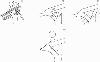
The tympanic part of the facial nerve, the greater superficial petrosal nerve, the cochlea and the vestibulum are all parallel structures (Figure 2a). This means that the tympanic segment of the facial nerve and the greater superficial petrosal nerve can be used as reference points to determine the most medial point of the cochlea and vestibulum. This study found that the distance from the tympanic segment of the facial nerve to the medial wall of the vestibulum was approximately 4 mm. The distance from the origin of the greater superficial petrosal nerve to the medial wall of the basal turn of the cochlea was found to be approximately 3 mm (Figure 1g). An imaginary line can be constructed parallel to the tympanic segment of the facial nerve, passing through Bill's crest. The present study found that the distance from this line to the medial wall of the vestibulum and cochlea was not more than 2 mm. This means that the surgeon can work safely from a point 2 mm medial to the vertical crest without risking damage to the cochlea or vestibulum (Figure 1f). Todd measured the distances from the centre of the superior portion of the basal turn of the cochlea to the geniculate ganglion and to the greater petrosal nerve, and found them to be 2.0–3.2 mm and 1.8–2.8 mm, respectively.Reference Todd28
Anatomical descriptions. See text for explanation of figure parts. TFN = tympanic facial nerve; VFT = vestibulofacial trigone; LFN = labyrinthine facial nerve; SVN = superior vestibular nerve; BTC = basal turn of cochlea; V = vestibulum

Rhoton and Tedeschi, in their study of 100 temporal bones, found that all or part of the geniculate ganglion was exposed in the floor of the middle fossa in 15 per cent of cases, and was covered completely in 15 per cent, but with no bone extending over the greater petrosal nerve. The greatest amount of coverage of the petrosal nerve was 6 mm.Reference Rhoton and Tedeschi27 Isaacson and Vrabec, using computed tomography examination, found the overall incidence of a dehiscent geniculate ganglion to be 14.5 per cent in the 365 sides reviewed.Reference Isaacson and Vrabec29 This means that, unless the ganglion is uncovered and located, the hiatus facialis is an unsafe surgical landmark. In solving this problem of location, the present study found the distance between the origin of the greater superficial petrosal nerve and the processus cochleariformis to be between 5.1 and 7.1 mm.
The anatomy of the bony area between the vertical crest and the tympanic segment of the facial nerve is not well defined in the literature. The vertical crest is only one corner of this vestibulofacial trigone (Figure 2b). This trigone has an anterior crest that separates the tympanic and labyrinthine segments of the facial nerve and a posterior corner between the tympanic portion of the facial nerve and the superior vestibular nerve. Thus, the vestibulofacial trigone is surrounded by the tympanic and labyrinthine segments of the facial nerve and the superior vestibular nerve. As this study shows, this trigone has approximately a 2–3 mm anterior–posterior and lateral–medial dimension. Deep to the vestibulofacial trigone are the end-points of the basal turn of the cochlea, anteriorly, and the vestibule, posteriorly (Figure 2c).
• This anatomical study used cadaveric temporal bones to define six reproducible measurements that relate the processus cochleariformis to inner-ear structures (and which are encountered during middle cranial fossa surgery), and to define 14 other measurements relating inner-ear structures to adjacent structures within the intact bone
• These measurements give important clues about the position of the cochlea, vestibulum, greater superficial petrosal nerve and labyrinthine segment of the facial nerve
• If classical landmarks are indiscernible during middle cranial fossa surgery, then the processus cochleariformis, with its intimate and constant relationship to inner-ear structures, is a safe and reproducible landmark
It is often suggested that the tympanic and labyrinthine segments run in straight lines, but this is not exactly true. The tympanic segment describes a curve with an anterolateral convexity between the processus cochleariformis and the origin of the greater superficial petrosal nerve. Similarly, the labyrinthine segment describes a curve with posterolateral convexity. It is important to be aware of these variants, in order to uncover the facial nerve without injury (Figure 2d). Another important point is that the most superior segment of the facial nerve is the labyrinthine segment. As it surrounds the cochlea it produces a bulge along the basal turn of the cochlea.
The superior semicircular canal is located a few millimetres posterior and parallel to the labyrinthine segment of the facial nerve.Reference Rhoton and Tedeschi27 In the present study, the mean angle of the labyrinthine segment was found to be 51° to the coronal plane, and the angle of the superior semicircular canal to the coronal plane was found to be 45°. The arcuate eminence may be readily apparent in some temporal bones but obscure in others. Kartush et al. have cautioned that the relationship between the arcuate eminence and the superior semicircular canal may be variable in some patients, but that the superior semicircular canal tends to be perpendicular to the petrous ridge. According to these authors, the apex of the semicircular canal lies 10 mm posterior and 5 mm medial to the origin of the greater superficial petrosal nerve.Reference Kartush and Brackmann30, Reference Kartush, Kemink and Graham31 Seo et al. found that the arcuate eminence corresponded exactly or well with the superior semicircular canal in only nine of 52 petrous bones examined.Reference Seo, Ito, Sasaki, Nakagawara and Nakamura32 Similarly, Bulsara et al. found the relationship between the arcuate eminence and the superior semicircular canal to be highly variable; the distance between the tips of the two structures was found to be between 2.7 and 10.4 mm.Reference Bulsara, Leveque, Gray, Fukushima, Friedman and Villavicencio33 The arcuate eminence has a variable position, and therefore should not be used as a substitute for the superior semicircular canal in topographical orientation of the internal auditory canal.Reference Fisch23 The present study gives an important clue about the position of the superior semicircular canal, showing, as it does, a 45° angle from the processus cochleariformis to the coronal plane.
Conclusion
The vertical crest lies at a 20° angle from the processus cochleariformis to the coronal plane, at a distance of 5 to 6 mm from the processus cochleariformis. The inexperienced surgeon should open the tegmen tympani and use the processus cochleariformis as a landmark if difficulties arise during middle cranial fossa surgery. The experienced surgeon can also use this structure as a landmark, especially if the classical landmarks are not discernible. With its intimate and constant relationship to inner-ear structures, the processus cochleariformis is a safe and dependable landmark.