Introduction
Keloids are scars that expand beyond the boundaries of the original injury as they heal. Keloids of the head and neck are very conspicuous, especially when occurring on the pinna, and are not easy for patients to cover. They cause significant morbidity as they are unsightly and uncomfortable, causing tightness, tenderness and itching; they thus impair the patient's quality of life and may cause psychological handicap.Reference Bock, Schmid-Ott, Malewski and Mrowietz1 Keloids often recur after various treatment attempts, and their management remains a challenging task. The underlying pathological cause is lack of the control mechanism that regulates wound healing and fibrous tissue production following skin trauma. Histologically, there is excessive accumulation of hyalinised collagen, which is thought to arise from intrinsically normal fibroblasts responding to an abnormal extracellular signal.
Keloids on the pinna can develop as a result of otoplasty, ear piercing or skin trauma.
The success rates of the different keloid treatment modalities vary markedly. Previous studies of keloid treatment have been limited by low patient numbers, short follow-up times and lack of objective assessment parameters.
Current keloid treatments include topical or intralesional steroids, cryotherapy, application of silicone gel, surgery followed by low dose radiotherapy, electron beam irradiation, pulsed dye lasers, high dose brachytherapy, intralesional bleomycin, intralesional 5-fluorouracil, and topical imiquimod. Interferon α-2b, topical tacrolimus, botulinum toxin and verapamil have also been tried.Reference Bailey, Waite, Clayton and Rustin2 Combination therapy is often used; however, no published keloid treatment has produced predictable results.Reference Al-Attar, Mess, Thomassen, Kauffman and Davison3
Mitomycin C is an alkylating agent which inhibits DNA synthesis by forming a cross-linkage of strands of the double helix, thus preventing neoplastic cell proliferation. It inhibits cell division, fibroblast proliferation, protein and collagen synthesis, and angioneogenesis.
At our centre, we conducted a prospective study on 20 patients (26 pinna lesions) in whom we used a new keloid treatment technique of surgical shave excision combined with topical application of mitomycin C and subsequent secondary healing.
Materials and methods
We selected for the study patients presenting to our out-patient department complaining of pinna swelling following ear piercing. All patients were women ranging in age from 18 to 50 years. A total of 20 patients with 26 pinna keloids (some bilateral) were included in the study.
Informed consent was obtained for surgery and mitomycin C application.
Pre- and post-operative photographs were taken to document patient progress.
All procedures were performed by the same surgeon, who used a similar technique in all cases.
Local anaesthetic was injected under and around the keloid. All of the keloid was removed by shaving to the level of the surrounding skin. Haemostasis was achieved with bipolar cautery. A mitomycin C solution of 1 mg/ml was prepared by adding 2 ml sterile water to powdered mitomycin C obtained from a 2 mg vial. Gauze cut to the shape of the scar and saturated with the mitomycin C solution was laid over the wound for 3 minutes and then removed. The wound was then patted dry and a dry dressing applied. Patients were instructed to re-dress the site as necessary and to keep it dry.
Three weeks later, any crust was removed and mitomycin C solution (1 mg/ml) was reapplied to the scar for 3 minutes as before.
Patients were reviewed at two, four and six months. At each visit, the scar and its symptoms were evaluated. Our criteria for successful treatment comprised patient satisfaction which was assessed using a linear analogue scale from 0 (disappointed) to 10 (delighted) and measurements of the thickness of any persisting keloid tissue. The aesthetic results of the keloid therapy were graded by a doctor uninvolved in the patient's treatment, as follows: 1 = excellent; 2 = good; 3 = acceptable; 4 = poor; 5 = bad; and 6 = unacceptable. Results of 3 or below were considered to represent success, whereas results of 4 or more were considered to represent treatment failure.
One representative case is presented here.
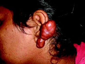
A 45-year-old woman presented to our out-patient department complaining of a swollen left pinna present for the last two years. She reported four previous complete excisions of similar swellings at the same site, at various hospitals. She had also received local steroid injections six months previously, with no benefit. On examination, there were two separate swellings over the left pinna, 5.4 × 4.3 cm and 1 × 1 cm in size (Figure 1). The swellings consisted of firm, nodular, non-encapsulated masses of hyperplastic scar tissue over the free posterior margin and adjoining medial and lateral surfaces of the pinna. The overlying skin was shiny and adherent to the underlying mass. The mass was tethered to the pinna perichondrium and cartilage. The mass was excised and managed according to the study protocol described above.
Pre-operative clinical photograph showing a large, lobulated keloid mass over the left pinna.

Results
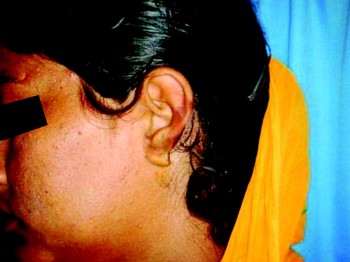
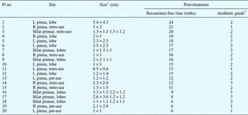
Patients were followed up for an average of one year (range six to 24 months). The combination of surgical excision and topical mitomycin C application was highly effective in treating pinna keloids. All patients were satisfied with the results. The wound healed well with normal skin (Figure 2), and no recurrences were noted in the follow-up period. None of the patients reported any adverse skin reactions or side effects that could be attributed to mitomycin C. The aesthetic outcome was good in all patients, although results were better for smaller keloids and those located in the preauricular region. The results are summarised in Table I.
Post-operative clinical photograph showing a well healed wound with normal skin and no sign of recurrence.

Patient keloid data

*Approximate in some cases. †See text for explanation. Pt no = patient number; mths = months; L = left; R = right; retro-aur = retro-auricular; pre-aur = pre-auricular
Discussion
Certain dark-skinned races are more prone to the development of keloids; in these populations, the incidence is between 4 and 16 per cent.Reference Stucker, Hoasjoe and Aarstad4 Keloids occur more often in certain sites, such as the ear lobe, upper chest, back and shoulder area. Keloids differ significantly from normal scars: the ratio of type I to type III collagen is elevatedReference Stucker and Goco5 and there is increased total collagen production (as much as 20-fold), with randomly orientated collagen fibres. There appears to be a genetic component to keloid formation, with certain associated human leukocyte antigen subtypes and familial inheritance patterns.Reference Botwood, Lewanski and Lowdell6
In general, nonessential cosmetic surgery should be avoided in predisposed racial groups, to avoid the risk of keloid formation. Surgical incisions should be made along skin tension lines without crossing them, and wounds should be closed without any tension. Subcuticular closure is preferable. Predisposing factors to the formation of keloid comprise: wounds in areas of high skin tension; wound healing by secondary intention; and chronic inflammation of wounds.
After simple excision of keloid, recurrence rates are as high as 50 to 60 per cent. There is a significant recurrence rate following the majority of treatment regimens, such as corticosteroid injection, cryotherapy, silicone contact therapy, pressure therapy and radiation therapy. Regardless of the technique employed, surgical removal of keloid causes further injury to the dermis, leading to fibroblast proliferation and extreme amounts of collagen formation, and thus keloid scar re-formation.
Mitomycin C is an anti-tumour antibiotic isolated from Streptomyces caespitosus. A study on endoscopic sinus surgery found that mitomycin C had an anti-fibroblastic effect without inhibiting epithelialisation.Reference Gupta and Motwani7 This drug has been shown to prevent scar tissue formation after glaucoma filtration surgery, subglottic surgery (in a canine model), tracheal stenosis repair, paediatric choanal atresia surgery and maxillary antrostomy. There have been no reports of adverse reactions to mitomycin C used in this context. In vitro studies found that fibroblasts treated with mitomycin C had decreased DNA synthesis and decreased density, compared with fibroblasts treated with buffered saline; three weeks after application, the cell count increased and DNA synthesis recovered in some of the treated fibroblasts.Reference Simman, Alani and Williams8 In the current study, topical mitomycin C was therefore reapplied three weeks after the initial application.
• This study investigated the role of mitomycin C in reducing keloid recurrence
• The authors used a surgical shave excision combined with topical application of mitomycin C and secondary wound healing, in 20 patients with keloid of the external ear
• No keloid recurrence was noted in a six to 24 month follow-up period
The English language literature contains four reports of the use of mitomycin C as a treatment for keloids. Two studies used keloid excision followed by topical application of plain cotton with 1 ml of mitomycin C solution (0.4 mg/ml) in eight and 10 patients, respectively, and found it effective in preventing recurrence.Reference Talmi, Orenstein, Wolf and Kronenberg9, Reference Stewart and Kim10 Another study, using the same concentration in a similar manner, concluded that the use of mitomycin C made no difference to the incidence of keloid recurrence.Reference Sanders, Gage-White and Stucker11 The most recent study involved 10 patients, and recommended using topical mitomycin C 1 mg/ml for 3 minutes after shave removal of keloid.Reference Bailey, Waite, Clayton and Rustin2 These authors further concluded that repeat application of mitomycin C after three weeks prevented keloid recurrence. The current study successfully followed the same technique and procedure in a larger group.
Topical application of mitomycin C has been shown in numerous studies to be safe and to have no side effects. Additionally, mitomycin C is inexpensive and readily available. As no effective treatment for keloid is currently available, utilisation of this therapeutic agent may improve treatment outcomes.
Conclusion
Successful treatment of keloids remains a challenge because this disease process has a high propensity for recurrence. Various therapies have previously been reported, and success rates are highly variable. We believe that shave excision followed by topical mitomycin C application is a promising treatment option for the management of pinna keloids.