Apersistent left-sided superior caval vein draining into the coronary sinus is a common finding occuring as an isolated lesion in 0.3–0.5% of the normal populationReference Biffi, Boriani, Frabetti, Bronzetti and Branzi 1 and is found in ~4% of the population having congenitally malformed hearts.Reference Parikh, Prasad, Iyer, Desai and Mohankrishna 2 The persisting left-sided caval vein most commonly drains into the right atrium through the coronary sinus, and hence is associated with normal haemodynamics. It is well recognised, nonetheless, that occasionally the caval vein drains directly to the roof of the left atrium, with so-called unroofing of its anticipated passage through the inferior left-sided atrioventricular groove.Reference Tuchman, Brown, Huston, Weinstein, Rowe and Crumpton 3 – Reference Davis, Jordaan and Snyman 5 In this setting, the mouth of the coronary sinus is usually large, serving as an interatrial communication permitting left-to-right shunting, with concomitant dilation of the right heart chambers. Occasionally, however, the orifice of the coronary sinus can be small and restrictive, or even atretic.Reference Adatia and Gittenberger-de Groot 6
The terminology left caval vein to roof of left atrium, although anatomically correct, fails to consider the coronary ostium. The coronary ostium, however, is crucial as it dictates not only the clinical presentation, but also the choice of intervention.
If intervention is indicated on clinical grounds, the left-sided caval vein can simply be occluded, either by surgeryReference de Leval, Ritter, McGoon and Danielson 7 – Reference Mutialu, Fajardo, Sullivan and Tsang 9 or by catheter-based interventions.Reference Troost, Gewillig and Budts 10 , Reference Geggel, Perry, Blume and Baker 11 Published literature to date has not adequately emphasised that the choice of intervention very much depends on the size of the opening of the coronary sinus to the right atrium. If the orifice is large, then a surgical approach is favoured, permitting ligation of the left-sided caval vein or redirection usually to the right atrial appendage together with closure of the mouth of the coronary sinus. If the orifice is small, then a catheter-based approach may be suitable after ensuring the presence of adequate decompressing channels.
In this study, we describe our recent experience with two patients, both having a left-sided superior caval vein draining to the left atrial roof with evidence of symptomatic systemic desaturation, but in whom catheter occlusion was possible because of a very small mouth of the coronary sinus. It is of interest that both were initially diagnosed with asthma, and the cardiac diagnosis was delayed for a number of years, suggesting that this condition does need to be considered in any patient with desaturation.
Persistent left superior caval vein to a de-roofed coronary sinus is at the extreme spectrum of coronary sinus de-roofing as first described by Kirklin in his classification of coronary sinus anomalies.
The coronary sinus can be partially or completely de-roofed. When completely de-roofed, it can be in the presence or absence of a left-sided caval vein. In addition, when completely de-roofed, it can be in the presence or absence of a patent coronary ostium, which when present can be restrictive, of normal size, or even larger than normal. This coronary ostium is not mentioned in the classification and not always considered by authors on this subject. In view of the clinical importance of the size of the coronary ostium, we suggest in this study an additional consideration in this commonly used classification system.
Cases
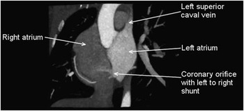
The first patient was a 32-year-old female referred to our service by the respiratory team with a 15-year history of exertional dyspnoea, escalating in severity over the last 2 years, on a background diagnosis of childhood asthma. She underwent a radionucleotide ventilation-perfusion scan, during which an injection of contrast into a vein in the left arm resulted in direct opacification of the left atrium. She suffered from a transient neurological deficit concomitant with the injection, suggesting the presence of a right-to-left shunt. This prompted a CT scan, which revealed the presence of a left-sided superior caval vein draining directly to the roof of the left atrium, with absence of the anticipated delineation between the coronary sinus in the left atrioventricular groove and the cavity of the left atrium. The right atrial mouth of the coronary sinus was shown to be markedly restrictive (Fig 1).
The CT image shows a left-sided superior caval vein draining directly to the roof of the left atrium, with failure of formation of the walls that would normally separate its ongoing passage into the left atrioventricular groove from the cavity of the left atrium. The right atrial mouth of the coronary sinus is restrictive.

The patient also had absence of the left thumb, and congenital scoliosis secondary to hemivertebrae. An echocardiogram showed the heart to be positioned in the right chest, with its apex pointing to the right, but was otherwise unremarkable, with all chambers being of the expected size. The persistent left-sided caval vein, however, was not visualised. A 6-minute walk test showed a reduction in baseline saturations from 96 to 89%.
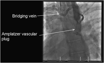
After discussion with the multidisciplinary team, the patient was scheduled for catheterisation in the first instance to confirm the anatomy and second to undertake test occlusion of the persistent left-sided superior caval vein. At catheterisation, angiography confirmed the CT findings. Following test occlusion of the caval vein with a sizing balloon of 18 mm for 10 minutes, angiography revealed the presence of a small bridging vein, which permitted adequate decompression of the left-sided venous structures to the right-sided superior caval vein (Fig 2). Mean pressures in the venous circuit remained unchanged following occlusion, at 13 mmHg, with the diameter of the occluded caval vein measuring 14 mm. The vein was therefore occluded using an 18 mm Amplatzer Vascular Plug Type II (St. Jude Medical, St. Paul, Minnesota, United States of America), which was inserted just cranial to the junction with the left atrium (Fig 2).
The angiogram shows the device in place, along with the decompressing bridging vein.

A CT scan performed 2 months after the procedure revealed the device to be properly seated, and demonstrated again the small and restrictive right atrial orifice of the coronary sinus, with no evidence of right-sided volume overload. Her 6-minute walk test was now normal, with saturations of 98% at rest and 97% at peak exercise. She was referred for genetic counselling in view of a possible diagnosis of Holt–Oram syndrome.
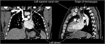
The second patient was a 43-year-old male, with dyspnoea at rest and on exertion, who was also referred to our service by the respiratory team. His resting oxygen saturations were 90%, falling to 67% on exercise. He had developed polycythaemia, with haemoglobin measured at 189 g/dl. Both his echocardiogram and pulmonary function tests were thought to be normal. Computed thoracic tomographic scanning, however, revealed a left-sided superior caval vein draining directly to the left atrium, with a very small right atrial mouth of the coronary sinus, and almost complete unroofing of the anticipated wall between the sinus and the left atrium (Fig 3).
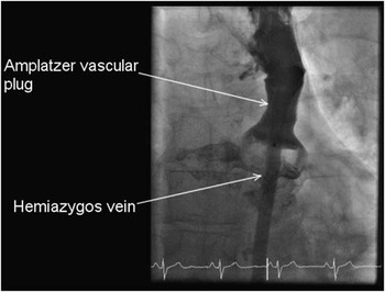
At cardiac catheterisation, angiography confirmed the CT findings. Test occlusion of the caval vein for 10 minutes induced a mild increase in his venous pressures, which rose from 8 to 10 mmHg with flow demonstrated through the hemiazygos vein into the inferior caval vein. We proceeded to occlude the vein with a 22 mm Amplatzer Vascular Plug Type II device, having measured the diameter of the caval vein at 18 mm. The device was placed cranial to the junction of the caval vein with the left atrium, but caudal to the origin of the hemiazygos vein (Fig 4). Subsequent to the procedure, the resting saturations increased to 98%.
At follow-up, he reported resolution of his symptoms, and was able to walk 540 m in a 6-minute walk test, with his saturations remaining at 96% throughout. This patient was also noted to have clinodactyly, and had been diagnosed previously with congenital anosmia.
Discussion
Persistence of drainage of a left-sided caval vein to the left atrial roof involves so-called “unroofing” of the anticipated passage of the coronary sinus through the left-sided inferior atrioventricular groove as well as loss of the division between the left caval vein wall and the left atrial wall. It can occur in isolation or in association with other CHDs; the associated right-to-left shunt presents clinically as desaturation with some patients occasionally presenting with signs of systemic embolisation. Previously reported genetic associations with persistent left-sided caval vein and a normal coronary sinus include the Holt–Oram and Coffin–Siris syndromes;Reference Thal, Boyella and Arsanjani 12 however, the associations with these two syndromes and a left caval vein to a de-roofed coronary sinus have not yet been reported.
Raghib et alReference Raghib, Ruttenberg, Andersen, Amplatz, Adams and Edwards 4 pointed to the frequent occurrence of an inferoposteriorly located interatrial communication, which in the setting of otherwise normal development would correspond with the right atrial mouth of the coronary sinus. Sometimes, the orifice of the coronary sinus in the setting of a persistent left-sided caval vein to an unroofed coronary sinus can also be atretic.Reference Adatia and Gittenberger-de Groot 6 The size of the right atrial mouth of the coronary sinus has received little previous attention when assessing management. Our experience shows that this feature is crucial both for presentation and decision making.
When the mouth of the sinus is large and unrestrictive, there will be a significant left-to-right shunt at the atrial level, producing the physiology anticipated for atrial septal defects, along with dilation of the right heart chambers. There will, of course, be a degree of desaturation produced by the presence of desaturated blood from the left superior caval vein entering the left atrium. The level of desaturation produced, however, will be negated by the increased pulmonary blood flow, and pulmonary venous return into the left atrium. In this setting, the management may need to involve surgical intervention, as not only can the caval vein be ligated or redirected to the right atrium but the mouth of the sinus can also be closed.
In contrast, when the right atrial mouth of the “unroofed” coronary sinus is restrictive or atretic, the clinical picture is primarily that of right-to-left shunting and desaturation, without right-sided volume loading. This situation, as seen in both our patients, is amenable to device occlusion of the caval vein, again having ascertained the presence of a decompressing vein or hemiazygous system for adequate run-off.
A previous classification dating to 1986 for this lesion identified subsets of so-called “complete unroofing” with or without a persistent left-sided superior caval vein, along with “partial unroofing” of either the midportion or the terminal portion of the presumed venous channel.Reference Kirklin and Barratt-Boyes 13 Although commonly quoted by other authors on this subject, the classification does not pay attention to the size of the right atrial mouth of the coronary sinus. We suggest, in view of its clinical significance, that this feature also needs to be considered when applying the classification below:
Type I – completely unroofed, with left superior caval vein.
Type II – completely unroofed, without left superior caval vein.
Type III – partially unroofed midportion.
Type IV – partially unroofed terminal portion.Reference Kirklin and Barratt-Boyes 13
The first type “completely unroofed, with LSVC” can occur with an atretic or restrictive coronary sinus orifice, which should then be thought of as a separate clinical entity to the “completely unroofed, with LSVC” with a normal/large coronary orifice.
Differentiation between these two subtypes of left-sided caval vein with complete unroofing, but with adequate as opposed to restrictive or atretic mouths of the coronary sinus, is important on clinical grounds as the former will result in milder desaturation and symptoms related to a left-to-right shunt at an atrial level as described above, whereas the latter variant will result in more significant desaturation alone as in our two patients. The degree of desaturation will vary depending on the size of the left compared with the right superior caval veins.
Careful assessment of the atrial septum is vital before any intervention. Those with a normal or larger-than-normal coronary sinus orifice should be evaluated further, and a decision for surgery versus catheter-based intervention should be made on the basis of the orifice size and the degree of right-sided volume loading using the same parameters as those used in the assessment of an atrial septal defect. Patients with a restrictive/atretic coronary sinus orifice should be considered for a catheter-based approach if otherwise suitable.
The CT images in frontal (left-hand panel) and lateral (right-hand panel) projections show a left-sided superior caval vein draining directly into the roof of the left atrium. The right atrial orifice of the coronary sinus, however, was poorly seen. Note the origin of the hemiazygos vein.

The Amplatzer device is shown in place, with decompression via the hemiazygos vein.

Acknowledgements
The authors thank their colleagues and their patients for their support.
Financial Support
This research received no specific grant from any funding agency or from commercial or not-for-profit sectors.
Conflicts of Interest
None.
Ethical Standards
The authors assert that all procedures contributing to this work comply with the ethical standards of the relevant national guidelines.