INTRODUCTION
When the Soviet Union existed, human visceral leishmaniasis (VL) was recorded as occurring widely in the towns and rural areas of the Central Asian Republics and the Caucasus, and its epidemiology suggested that it was a zoonosis possibly caused by Leishmania infantum (Ponirovskii et al. Reference Ponirovskii, Strelkova, Goncharov, Zhirenkina and Chernikova Iu2006). Owing to intensive control measures, its incidence decreased dramatically in Azerbaijan, Armenia, Georgia, Kazakhstan, Kyrgyzstan, Tajikistan, Turkmenistan and Uzbekistan (Ponirovskii et al. Reference Ponirovskii, Strelkova, Goncharov, Zhirenkina and Chernikova Iu2006). The disease has almost been forgotten by local doctors. For instance, the last reports of VL in Tajikistan dated back to the year 1958. However, the continuing existence of stable foci of VL in the Namangan and Fergana Regions of Uzbekistan has been documented in epidemiological studies (Finkelstein, Reference Finkelstein1956; Maroli et al. Reference Maroli, Krasnonos and Gafurov2001) In fact, the Namangan Region in the north-western part of the Fergana Valley is the only place in Uzbekistan where a surveillance system for VL exists, and from 2004 to 2007, 34 cases were registered there, with a peak of 15 cases in 2007. These numbers are probably an underestimate because of: (i) lack of appropriate diagnostic methods, (ii) non-specific clinical symptoms, (iii) lack of an efficient reporting system, and (iv) lack of awareness of the local medical personnel. From the few data available, there seems to be a reactivation of endemic foci, especially of those situated along the Fergana Valley.
Diagnosis of VL has always been done using clinical criteria, confirmed parasitologically by detecting amastigotes in smears of bone marrow aspirates stained with Giemsa's stain. Identification of the species of Leishmania has been impossible so far owing to the failure to culture strains for more than 2 passages after their isolation, even in media recommended for strains of L. infantum.
This study identified L. infantum as the causative agent of VL in Namangan region, Uzbekistan, by amplifying and sequencing the ribosomal internal transcribed spacer 1 (ITS1) using DNA extracted from 5 Giemsa-stained smears of human bone marrow aspirates. Further typing of 13 strains from Uzbekistan and Tajikistan by multilocus microsatellite typing (MLMT) revealed that parasites belong to zymodeme MON1 and that they form a distinct genetic group within the MON1 strains.
MATERIALS AND METHODS
Sampling and identification of the causative agent of VL
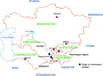
DNA samples were extracted from the amastigotes in the smears of bone marrow aspirates stained with Giemsa's stain of 13 patients from the Namangan and Jizzakh regions in Uzbekistan, and Penjikent region in Tajikistan which is located close to the border of Uzbekistan (Fig. 1). The leishmanial DNA samples were extracted from pre-existing bone marrow smears from cases of VL examined and treated during the period from 2006 to 2008 by the diagnostic unit of The Isaev Insitute in Samarkand, which offers its service to local physicians. Diagnosis of VL in Uzbekistan is by microscopical examination of bone marrow aspirate smears stained with Giemsa's stain for the presence of leishmanial amastigotes. Written consent is not required for this procedure in Uzbekistan. The cases are mostly children, who are accompanied by their parents, who give their verbal consent.
Map of Central Asia, showing the study area.

DNA was extracted from clinical materials on glass slides by using the phenol-chloroform method as described previously (el Tai et al. Reference el Tai, Osman, el Fari, Presber and Schönian2000; Schonian et al. Reference Schönian, Schweynoch, Zlateva, Oskam, Kroon, Graser and Presber1996), suspended in TE-buffer and stored at 4°C until use. Identification of the Leishmania parasites causing VL in Namangan region was done by using the ITS1-PCR described by Schonian et al. Reference Schönian, Nasereddin, Dinse, Schweynoch, Schallig, Presber and Jaffe2003). The ITS1 PCR products (~320 bp) of the 5 extracted DNA samples amplified were sequenced in the Department of Genetics of The Humboldt University of Berlin, employing the same primers used for the PCR. The sequences obtained were processed and aligned, using the multiple alignment programme BioEdit (Hall, Reference Hall1999) and edited manually.
Multilocus microsatellite typing (MLMT)
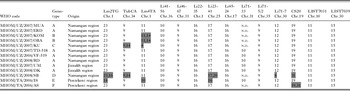
The 13 DNA samples were also analysed for variation in 14 multilocus microsatellite markers in the manner previously described (Kuhls et al. Reference Kuhls, Keilonat, Ochsenreither, Schaar, Schweynoch, Presber and Schönian2007; Ochsenreither et al. Reference Ochsenreither, Kuhls, Schaar, Presber and Schönian2006). Multilocus microsatellite profiles, based on the repeat numbers in 14 microsatellite markers, were assigned to each sample (Table 1) and compared to the MLMT profiles of 75 strains of L. infantum, some of which coincide with the enzyme profile of strains of L. infantum of the Montpellier zymodeme MON-1, further referred to as MON-1 strains, and other ones that do not (=non-MON-1), and 26 strains of L. donovani all from different places where VL is endemic (Kuhls et al. Reference Kuhls, Chicharro, Canavate, Cortes, Campino, Haralambous, Soteriadou, Pratlong, Dedet, Mauricio, Miles, Schaar, Ochsenreither, Radtke and Schönian2008; Seridi et al. Reference Seridi, Amro, Kuhls, Belkard, Zidane, Al-Jawabreh and Schönian2008; Alam et al. Reference Alam, Kuhls, Schweynoch, Sundar, Rijal, Shamsuzzaman, Raju, Salotra, Dujardin and Schonian2009; Chargui et al. Reference Chargui, Amro, Haouas, Schönian, Babba, Schmidt, Ravel, Lefebvre, Bastien, Chaker, Aoun, Zribi and Kuhls2009) (Supplementary Table 1, Online version only).
The multilocus microsatellite profiles of the 13 strains of Leishmania infantum isolated from Uzbeki and Tajiki VL cases
(UZ, Uzbekistan. TA, Tajikistan. Shading indicates different allele.)

n.d.=not done.
Microsatellite data analysis
MLMT data were processed by using STRUCTURE software (Pritchard et al. Reference Pritchard, Stephens and Donnelly2000) to investigate the population structure. This programme uses a model-based clustering method independent of a particular mutation model. A series of runs (10 runs for each K, a burn-in period of 20 000 iterations and a run of 200 000 iterations) was performed for each value of K (number of populations) between 1 and 10 to quantify the amount of variation of likelihood for each K. ΔK was calculated based on the rate of change in the log probability of data between successive K values (Evanno et al. Reference Evanno, Regnaut and Goudet2005). Microsatellite data were analysed with respect to diversity of alleles (A), mean number of alleles (MNA), expected heterozygosity (He) and observed heterozygosity, (Ho) using GDA software (http://lewis.eeb.uconn.edu/lewishome). Genetic differentiation was assessed by calculating Fst values and comparing p-values using the MSA software (Dieringer and Schlötterer, Reference Dieringer and Schlötterer2002).
RESULTS
ITS1 sequencing
To identify the Leishmania parasites causing VL in Namangan region at the species level, ITS1 PCR products from 5 different patients were sequenced. All sequences were submitted to GenBank (Accession nos FM164416, FM164417, FM164418, FM164419, FM164420s). The BLAST search (www.ncbi.nlm.nih.gov/BLAST/) showed 100% homology between these sequences and previously reported ITS1 sequences of L. infantum (GenBank Accession nos EU326227, AM502245, AJ634355, AJ634354).
Multilocus microsatellite typing
Six different MLMT profile variants were exposed among the 13 extracts of amastigote DNA, which differed from one another in 7 loci. Seven of the extracts had the same profile; another 2 presented with identical profiles, and the remaining 4 had their own profiles (Table 1).
MLMT profiles obtained for DNA extracts of Uzbeki and Tajiki patients were compared to that of strains of L. infantum and L. donovani of other geographical origins. The provenances of the DNA extracts studied and the strains of L. infantum and L. donovani from which they came as well as the MLMT microsatellite subpopulations to which they belong are given in the Supplementary Table 1 (Online version only).
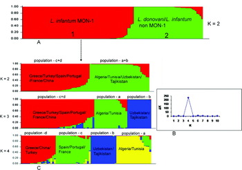
Bayesian model-based clustering approach, implemented in STRUCTURE confirmed the existence of 2 main groups (ΔK=2) in our sample set (Fig. 2): one comprising the DNA samples from strains of L. infantum falling in the Montpellier zymodeme MON-1 (MON-1 samples); the other of those from strains of both L. infantum not falling in the Montpellier zymodeme MON-1 (non-MON-1 samples) and L. donovani. The 13 Uzbeki and Tajiki DNA samples clustered in the group of MON-1 DNA samples, suggesting that the parasites that caused the Uzbek and Tajikistan cases of VL belong to the species L. infantum and are most closely related to strains of zymodeme MON-1 (Fig. 2 and Supplementary Table 1 in Online version only).
Population structure in the data set based on STRUCTURE analysis of 14 microsatellite markers. (A) General population structure of the 114 DNA samples from the strains of Leishmania infantum MON-1/non-MON-1 and L. donovani studied and including the 13 Uzbeki and Tajiki DNA extracts. Individuals are represented by a single vertical line divided into K colours, the length of the coloured segments indicating the individual's estimated proportion of membership of the strain in that population. (B) The derived graph for ΔK shows a peak at K=4, indicating the existence of 4 populations in the dataset. (C) Population substructure of the cluster comprising the DNA samples from strains of the L. infantum zymodeme MON-1, as shown by plots of clusters, where K is the number of populations assumed.

A second run of structure analysis was performed with only the DNA samples from strains of L. infantum of the MON-1 zymodeme, in which the first split, at K=2, separated the DNA samples of strains from Algeria, Tunisia and Uzbekistan from those from Greece, Turkey, Spain, Portugal, France and China. Successive splits were followed by increasing the number for K. Calculation of ΔK confirmed the existence of 4 clusters, which were well defined and stable at K=4: (a) Algeria+Tunisia; (b) Uzbekistan+Tajikistan; (c) Spain+Portugal+France; (d) Greece+Turkey+China (Fig. 2). Clusters a, c and d were exposed in earlier studies where more samples were included and the clusters were larger (Kuhls et al. Reference Kuhls, Chicharro, Canavate, Cortes, Campino, Haralambous, Soteriadou, Pratlong, Dedet, Mauricio, Miles, Schaar, Ochsenreither, Radtke and Schönian2008; Seridi et al. Reference Seridi, Amro, Kuhls, Belkard, Zidane, Al-Jawabreh and Schönian2008; Chargui et al. Reference Chargui, Amro, Haouas, Schönian, Babba, Schmidt, Ravel, Lefebvre, Bastien, Chaker, Aoun, Zribi and Kuhls2009).
Population characteristics
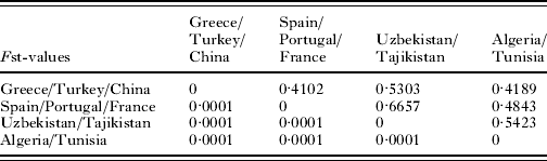
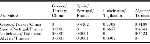
F statistics revealed a significant amount of population structure among the clusters in the L. infantum MON-1 group. All Fst values (Table 2) were significant and higher than 0·25 indicating very great genetic differentiation between the populations. The Uzbeki and Tajiki samples were found to be distinctly separate from the other DNA sample groups of the L. infantum zymodeme MON-1 studied here.
Fst values and corresponding P-values for the four populations of Leishmania infantum MON-1 as assumed by STRUCTURE
(Fst values are in the upper triangle; P-values in the lower triangle.)
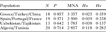
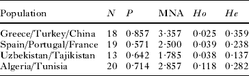
Calculations of mean number of alleles, proportion of polymorphic loci, expected and observed heterozygosity demonstrated that the Uzbeki and Tajiki samples were the least diverse (Table 3). The mean number of alleles (MNA), within populations, ranged between 1·78 for Uzbekistan/Tajikistan and 3·35 for Greece/Turkey/China. Also P, the proportion of polymorphic loci, was relatively lower for Uzbekistan/Tajikistan (0·64) and highest for Greece/Turkey/China (0·85). Homozygous allele combinations predominated in the small, geographically well-defined Uzbeki/Tajiki cluster, with an expected heterozygosity (He) of 0·13, whereas the other 3 populations presented greater heterozygosity.
Characterization of the populations found for strains of Leishmania infantum MON-1 of different geographical origin
(N, number of strains; P, proportion of polymorphic loci; MNA, mean number of alleles per locus; Ho, observed heterozygosity; He, expected heterozygosity.)
DISCUSSION
This molecular biological study showed that L. infantum is the causative agent of VL currently occurring in the Namangan region, Uzbekistan and confirms past suspicions owing to the zoonotic nature of the disease in Uzbekistan and neighbouring Turkmenistan (Ponirovskii et al. Reference Ponirovskii, Strelkova, Goncharov, Zhirenkina and Chernikova Iu2006). This was done without the need to isolate and culture the leishmanial parasites, a process that has always failed regarding the parasites causing VL in Uzbekistan. This identification fits with an entomological survey which showed that Ph. longiductus, a known vector of L. infantum, is present in the Namangan region (Maroli et al. Reference Maroli, Krasnonos and Gafurov2001). Since parasite strains could not be isolated and thus identified and since Ph. sergenti, a proven vector of L. tropica, was very prevalent in the study area, atypical visceralizing strains of L. tropica (Schnur et al. Reference Schnur, Chance, Ebert, Thomas and Peters1981) could not be excluded. Hence the need for species identification, which was achieved by applying DNA extraction and PCR-sequencing to the bone marrow aspirate smears stained with Giemsa's stain from human cases of VL.
This MLMT approach of applying 14 microsatellite markers, previously shown to be polymorphic for strains of the L. donovani complex, showed that the microsatellite profiles of the parasites causing human VL in the Namangan and Jizzakh regions in Uzbekistan, and Penjikent region in Tajikistan, basically coincide with those of strains of L. infantum that also have the enzyme profile correlated with the zymodeme MON-1. These findings suggest that they also probably belong to this zymodeme, which is the most prevalent zymodeme of L. infantum. Approximately, 70% of all characterized strains of L. infantum are strains belonging to the zymodeme MON-1. They are geographically widely distributed, occurring in more than 30 countries (Desjeux, Reference Desjeux1991; Gallego et al. Reference Gallego, Pratlong, Fisa, Riera, Rioux, Dedet and Portus2001). The advantage of microsatellite typing is that it can differentiate among strains within the zymodeme MON-1, discerning geographical and genetically differentiated populations and detect what might be hybrids and mosaic genotypes (Kuhls et al. Reference Kuhls, Chicharro, Canavate, Cortes, Campino, Haralambous, Soteriadou, Pratlong, Dedet, Mauricio, Miles, Schaar, Ochsenreither, Radtke and Schönian2008; Seridi et al. Reference Seridi, Amro, Kuhls, Belkard, Zidane, Al-Jawabreh and Schönian2008; Chargui et al. Reference Chargui, Amro, Haouas, Schönian, Babba, Schmidt, Ravel, Lefebvre, Bastien, Chaker, Aoun, Zribi and Kuhls2009). Strains of L. infantum of the zymodeme MON-1 from the Mediterranean Basin had individual MLMT profiles with a high degree of heterozygosity. By comparison, the MLMT profiles of the DNA samples obtained from 13 Uzbeki and Tajiki cases of VL assigned the parasites to a distinct cluster that is genetically clearly separate from the populations of strains of L. infantum of the zymodeme MON-1 from Europe, the Middle East and North Africa. This distinctiveness probably indicates that the parasites circulating in the Uzbeki and Tajiki foci studied have been restricted there for a long time rather than having been recently introduced from elsewhere by human or animal reservoir migration. A comprehensive study of the epidemiology of human VL in these foci, including investigations on reservoir hosts and insect vectors, is warranted.
This study was supported by INTAS, grant 05-1000008-8043. We would like to thank the Deutsche Akademische Austauschdienst (DAAD) for providing a Ph.D. Fellowship to M. Z. Alam.