INTRODUCTION
Polyclads are exclusively marine except for the genera Limnostylochus and Limnoplana (Faubel, Reference Faubel1983; Prudhoe, Reference Prudhoe1985), bilaterally symmetrical, unsegmented members of the phylum Platyhelminthes under the clade Rhabditophora (Ehlers, Reference Ehlers1986). In systematics, they are further classified into two sub-orders, Cotylea and Acotylea. The classification is primarily based on the presence or absence of a ventral sucker as well as on certain variations within the composition and arrangement of the reproductive system. Additionally, importance has been given to the external anatomical features, especially for cotyleans, such as number of eyespots, their position, shape of pseudotentacles, pharynx and body colour and pattern (Hyman, Reference Hyman1954; Newman & Canon, Reference Newman and Cannon1994, Reference Newman and Cannon1995). In a tropical and temperate marine environment, these worms are known to inhabit coral reefs and rocky intertidal zones; a few species also occur in mangroves and deep sea environments (Quiroga et al., Reference Quiroga, Bolaños and Litvaitis2006).
Laidlaw (Reference Laidlaw1902) studied five families, namely, Planoceridae, Leptoplanidae, Cestoplanidae, Pericelidae and Pseudocerotidae along with 10 species from the Laccadive Islands, which is the first and largest collection of polyclad flatworms from the Indian region ever made. A few recent studies documented the species mainly from coral reef islands of India, i.e. Lakshadweep (Prabhakaran, Reference Prabhakaran2008; Apte & Pitale, Reference Apte and Pitale2011) and Andaman and Nicobar (Sreeraj & Raghunathan, Reference Sreeraj and Raghunathan2011, Reference Sreeraj and Raghunathan2013). Besides these, three pseudocerotid worms from the Saurashtra coast (Bhadja, Reference Bhadja2010) have also been observed. Hence, collectively, only six families and 37 species of flatworms are known from Indian waters. This paper presents the first report of the family Prosthiostomidae with the species Prosthiostomum trilineatum from the west coast of India. Recently, occurrence of this species from the Andaman Islands has been mentioned by the Zoological Survey of India (ZSI-e-News, 2012). The family Prosthiostomidae was described by Lang (Reference Lang1884) and further assessed by Faubel (Reference Faubel1984). The genus Prosthiostomum is known to be peculiar to this family and encompasses 51 species distributed worldwide (Tyler, Reference Tyler2013). Laidlaw (Reference Laidlaw1902) recorded P. elegans and P. cooperi from the Maldives, which is the geographically closest report of this genus to the Indian west coast; he also hypothesized the abundance of Prosthiostomidae members within the Indian Ocean.
MATERIALS AND METHODS
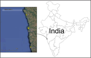
The Ratnagiri coast (16.98°N 73.3°E) from the Konkan region of Maharashtra State can be characterized by rocky cliffs, and narrow and wide shorelines with a steep and gentle continental slope. Heterogeneous habitat structure of rocky shores viz. pebbles, cobbles, boulders and rock pools of various sizes or depths are found at various zones of the littoral area. Present collections were carried out during low daytime tide by handpicking the worms with the help of a paintbrush, within the littoral areas of Kasheli (16.73705N 73.30627E), Undi (17.227299N 73.234972E) and Alawa (17.024326N 73.270228E) from the Ratnagiri district of Maharashtra State (Figure 1). Photographs were taken in the wild as well as ex situ to record true colour and pattern. Animals were then fixed in 10% frozen buffered formalin and 70% ethanol for long-term preservation (Quiroga et al., Reference Quiroga, Bolaños and Litvaitis2004). Studies on key anatomical features were carried out using a stereo microscope (Leica EZ4 D) in-built camera. The identification is based solely on the original description of Yeri & Kaburaki (Reference Yeri and Kaburaki1920) and the systematic classification system followed which was established by Faubel (Reference Faubel1984). Sagittal serial sections of reproductive system (6 µm) were obtained by specimen (Pclad-0103) embedding in paraplast and stained with haematoxilin and eosin. Remaining specimens are deposited in collections of the Bombay Natural History Society.
Geographical distribution of Prosthiostomum trilineatum from Ratnagiri, west coast of India (image courtesy of: Google Earth).

RESULTS
SYSTEMATICS
Order POLYCLADIDA Lang, Reference Lang1884
Suborder COTYLEA Lang, Reference Lang1884
Family PROSTHIOSTOMIDAE Lang, Reference Lang1884
Genus Prosthiostomum Quatrefages, 1845
Prosthiostomum trilineatum Yeri & Kaburaki, Reference Yeri and Kaburaki1920
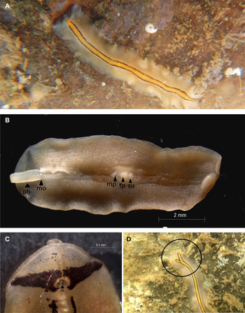
(Figure 2A)
Prosthiostomum trilineatum: (A) under pebble in shallow rockpool; (B) ventral surface showing pharynx (ph), mouth (mo), male gonopore (mp), female gonopore (fp), sucker (su); (C) anterior end with cerebral eyespots (ce), marginal eyespots (me); (D) caudal region showing extension from medial longitudinal stripe.

MATERIAL EXAMINED
One specimen (9.7 mm × 2.8 mm, preserved) found under rock pebble at Kasheli, Ratnagiri 11 May 2012, coll. V. Bhave (BNHS_Pclad-0044). One specimen (8.8 mm × 3.4 mm, preserved) found under rock pebble within rock pool at Undi, Ratnagiri 31 October 2012, coll. R. Pitale (BNHS_Pclad-0046). Two specimens (10.1 mm × 3 mm, used for histology; one preserved) found under rock pebble at Alawa, Ratnagiri, 7 February 2012, coll. Rajendra Pawar and Vishwas Shinde (BNHS_Pclad- 0103).
DIAGNOSIS
Elongated body; rounded anteriorly; tapers posteriorly, marginal eyespots confined to frontal end and not reaching to lateral margin, orange-yellow, black and white transverse band at frontal end and longitudinal orange-yellow stripe bordered with black runs medially; throughout the length, pair of free prostatic vesicles, penis with sclerotized stylet.
DESCRIPTION
The animal is broadly rounded anteriorly, elongated and tapering at the caudal end, which can only be noticed in a completely extended state. Dorsally, tentacles are absent and smooth dorsal surface is milky white, appeared buff in live specimens. At anterior extremity, marginal eyespots (26) are found in two irregular rows with sub-surface appearance for few. These eyespot rows are confined to anterior margin and do not reach to lateral margin. Immediately behind the rows of marginal eyespots, three distinct (orange-yellow, black and white respectively) arc-like transference bands are present. Black band lies exactly above the position of brain and found interrupted medially by a notch at its relative posterior margin. This colourless notch possesses two slightly oval cerebral eye clusters (13 each) (Figure 2C). Orange-yellow median stripe bordered by black band found originating from this notch and extends throughout the length (Figure 2A).
Semi-transparent ventral region possesses mouth, opening medially just behind the cerebral area, followed by pharynx, male gonopore, female gonopore and sucker respectively (Figure 2B). Pharynx plicatus is cylindrical and approximately 2.4 mm long and found extended during fixation. Male gonopore, female gonopore and sucker are close set, placed centrally and about 0.47 mm, and 0.34 mm distant from each other respectively.
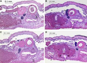
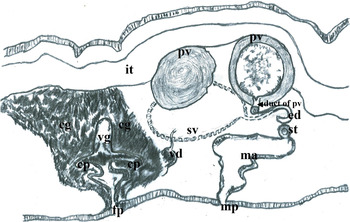
Single male copulatory system with numerous testes located dorsally. Pair of free prostatic vesicles (0.22 × 0.24 mm and 0.20 × 0.24 mm) is present dorsally beneath the intestine, spherical and muscularized (Figure 3A). Vasa differentia arranged lateral to the system and run posteriorly. Spermiducal bulbs are muscular and found organized adjacent to the prostatic vesicles (Figure 3B). Seminal vesicle is not visible within sections. Ejaculatory duct is found ventral to the prostatic vesicles (Figure 3A). Penis with sclerotized stylet found further down to the ejaculatory duct probably contracted or bent during fixation as visible only in the form of rounded structure (Figure 3C). Male antrum is not very deep (0.35 mm long) and approximately perpendicular to the body (Figure 4).
Sagittal sections of reproductive structures: (A–C) male reproductive structures of male antrum (ma), prostatic vesicle (pv), spermiducal bulbs (sb), ejaculatory duct (ed), stylet (st); (D) female reproductive structure of vagina (vg), cement pouch (cp), cement gland (cg), intestine (it).

Diagrammatic presentation of reproductive structures of male antrum (ma), seminal vesicle (sv), vas deference (vd), prostatic vesicle (pv), ejaculatory duct (ed), stylet (st); female reproductive structures of vagina (vg), cement pouch (cp), cement gland (cg), intestine (it).

Female reproductive system (Figure 3D) comprises the short vagina (0.12 mm) with lateral invagination of cement pouches (0.10 mm long). The cement glands are numerous found concentrated around the vagina and these even reach to the ventral margin of the intestine.
REMARKS
The family Prosthiostomidae was established by Lang (Reference Lang1884). Apart from external morphological features (rounded anteriorly, elongated body, mouth behind the brain, plicate pharynx, tentacles absent) he emphasized the presence of free prostatic vesicles. Faubel (Reference Faubel1984) in further assessment retaining the similar characters described by Lang revised the family and five genera were determined. He discriminated the genera mainly on the basis of male complex bound in common muscle bulb, presence of free pair prostatic vesicles, penis armed or unarmed, presence or absence of main frontal intestinal branch, presence of ductus genito-intestinalis in female reproductive system.
Prosthiostomum trilineatum was originally described from Hatake-jima, Kii Peninsula, Japan by Yeri & Kaburaki (Reference Yeri and Kaburaki1920). Species description was based on single immature specimen thus only external morphological characters were explained. This description refers the presence of two anterior transverse bands (yellow and black); however white band beneath the black is missing within text or even in the drawing presented. Our specimens fit with the characters mentioned earlier except for the presence of frontal white transverse band. Newman & Canon (Reference Newman and Cannon2003, Reference Newman and Cannon2005) noted P. trilineatum having similar colour pattern to that described here. Another colour morph of the species concerned was recognized by Newman & Canon (Reference Newman and Cannon2003, Reference Newman and Cannon2005). This colour morph exhibits yellow dorsal surface with frontal transverse orange band pointed anteriorly and two longitudinal orange stripes running on either side of median white stripe. Newman & Canon (Reference Newman and Cannon2005) mentioned the fact of existence of diverse colour-patterns for single species within the polyclad flatworms. Thus existence of three colour patterns (white band present, white band absent and only orange stripes-bands), can be considered as usual variation and forms the P. trilineatum species complex. This fact can be convincing when comparative analysis of the reproductive system histology and DNA barcodes reveals the similarity. Hence histological details and molecular data from these morphs are absolutely essential to resolve this question.
The specimen with voucher number BNHS-Pclad-0044 was found within a shallow rock pool of the high tide region, covered by Enteromorpha sp. under a rock, whereas another specimen with voucher number BNHS-Pclad-0046 was found under barren rock within a rock pool inhabited by zooanthus and algae. BNHS-Pclad-0044 has an extension from the median strip near the caudal region which might be the trace of regeneration (Figure 1D). Currently this species is known from Japan, Micronesia, Papua New Guinea, the Red Sea, Singapore (Newman & Canon, Reference Newman and Cannon2005) and recently mentioned from Andaman Island, India (ZSI-e-News, 2012), and with this report extends to the northern west coast of India.
DISCUSSION
The present paper increases the species diversity of polyclads from the Indian coast to 38 and is the first description of a polyclad from Ratnagiri, west coast of India. The Konkan coast of Maharashtra can be characterized by the presence of a heterogeneous habitat structure on rocky shores, which in turn supports diverse faunal and floral assemblages. Patchy coral reefs are known from this coast, which is also the preferred habitat of this group of animals. However, a literature review clearly indicates the gaps in the polyclad studies in this region that have resulted in the lack of baseline data. In another context, the Konkan coast is being targeted for a large number of coastal infrastructure projects including power plants, ports, shipyards, mines and related development activities. The cumulative effect of these activities can be adverse on benthic communities due to presumable anomalies in temperature and sedimentation rate. These fluctuations will definitely alter the composition of present benthic assemblages. Apart from the impending habitat loss, the possibility of them being temperature sensitive (Rawlinson, Reference Rawlinson2008) and due to their dependency on sessile invertebrates for food, polyclads will be susceptible to probable adverse environmental conditions. Hence, in this frame of reference, the creation of baseline data about the diversity and ecology of these worms from the region concerned has extreme conservation importance.
ACKNOWLEDGEMENTS
The authors are grateful to Dr Marcela Bolaños for providing valuable guidance while identifying histological characters of this species. Special thanks to Ms Eriko Kudo from Kyushu University Library, Japan for making available required references and to Ashok Bhagat for expertly providing histological sections. We also thank Dr Vinay Deshmukh from the Central Marine Fishery Research Institute for permitting work in his laboratory and Mr Bala Mhadgut for his help during microscopic imaging. We greatly appreciate Ms Pooja Nagale for providing the field image, Mr Rajendra Pawar and Mr Vishwas Shinde for collection and assistance during fieldwork, and Gopi Naidu for some technical help.