Introduction
The CDT is a simple and valid screening tool for dementia (Shulman, Reference Shulman2000; Pinto and Peters, Reference Pinto and Peters2009), correlating highly with global cognitive function (Wolf-Klein et al., Reference Wolf-Klein, Silverstone, Levy and Brod1989; Brodaty and Moore, Reference Brodaty and Moore1997). A wide range of cognitive functions is assessed by the CDT, including orientation, selective and sustained attention, auditory comprehension, verbal working memory, numerical knowledge, visual memory and reconstruction, visuospatial organization, motor execution (praxis), and executive function (Royall et al., Reference Royall, Cordes and Polk1998; Shulman, Reference Shulman2000). While the literature on the use of the CDT in screening for the syndrome of dementia is extensive (Sunderland et al., Reference Sunderland1989; Wolf-Klein et al., Reference Wolf-Klein, Silverstone, Levy and Brod1989; Mendez et al., Reference Mendez, Ala and Underwood1992; Rouleau et al., Reference Rouleau, Salmon, Butters, Kennedy and McGuire1992; Shulman, Reference Shulman2000), it is not clear whether the CDT is valid for discriminating between the subtypes of dementias.
Due to the variety of skills required for successful completion of the CDT, specific errors have been linked to different types of dementias (Eknoyan et al., Reference Eknoyan, Hurley and Taber2012). Hence, it would be reasonable to assume that scores and the pattern of errors on the CDT would differ between different dementia and hence the CDT may aid in the differential diagnosis of dementia. The type of clock-drawing errors can contribute to the clinical evaluation of patients with suspected neuropsychiatric disorders and syndromes as these errors likely represent different neuropsychological functions (Eknoyan et al., Reference Eknoyan, Hurley and Taber2012). There are two general scoring approaches – qualitative (Rouleau et al., Reference Rouleau, Salmon, Butters, Kennedy and McGuire1992; Kozora and Cullum, Reference Kozora and Cullum1994; Libon et al., Reference Libon, Malamut, Swenson, Sands and Cloud1996) and quantitative (Sunderland et al., Reference Sunderland1989; Mendez et al., Reference Mendez, Ala and Underwood1992; Shulman et al., Reference Shulman, Gold, Cohen and Zucchero1993; Watson et al., Reference Watson, Arfken and Birge1993). There have been at least 19 CDT scoring systems described since its use in the 1960s (Mainland et al., Reference Mainland, Amodeo and Shulman2014). Rouleau et al. and Libon et al. were among the first to devise qualitative scoring systems for the CDT (Rouleau et al., Reference Rouleau, Salmon, Butters, Kennedy and McGuire1992; Libon et al., Reference Libon, Malamut, Swenson, Sands and Cloud1996). They found that patients with AD demonstrated different qualitative errors compared to patients with subcortical deficits, such as Huntington's disease and ischemic VaD. The types of qualitative errors that can be found also depend on the method used for clock drawing, e.g. whether or not a pre-drawn circle is used (Rouleau et al., Reference Rouleau, Salmon, Butters, Kennedy and McGuire1992; Libon et al., Reference Libon, Malamut, Swenson, Sands and Cloud1996). In one of the most commonly used systems of evaluating qualitative clock-drawing errors (Rouleau et al., Reference Rouleau, Salmon, Butters, Kennedy and McGuire1992), errors include: (i) graphical difficulties, in which lines are not precise resulting in distortions of the clock face or numbers that are difficult to read; (ii) stimulus-bound responses, where there is the tendency of the drawing to be dominated or guided by a single stimulus e.g. the hands are set at 10–11 instead of 10 after 11; (iii) conceptual deficits, comprised of deficits in accessing the knowledge of the attributes, features, and meaning of a clock, resulting in a drawing that does not look like a clock or drawing with hands that do not communicate a time; (iv) spatial/planning deficits, which are errors in the layout of the numbers of the clock; and finally (v) perseveration errors, which are the continuation or recurrence of activity without appropriate stimulus (e.g. presence of more than two hands or writing of numbers beyond 12 or inappropriate recurrence of numbers). Rouleau et al. (Reference Rouleau, Salmon, Butters, Kennedy and McGuire1992) found that patients with AD were more likely to draw larger clocks than patients with Huntington's disease, which may be a result of poor visuospatial planning due to impairment of executive and visuospatial functioning. Another finding was that graphical errors were more common in Huntington's disease patients than AD patients, probably as a result of secondary disruption of frontostriatal circuits essential for the functions of fine motor control and planning (Rouleau et al., Reference Rouleau, Salmon, Butters, Kennedy and McGuire1992).
Currently, the literature reveals conflicting data regarding the ability of the CDT to distinguish patients with various types of dementia. The present qualitative review aims to determine the usefulness of the CDT in differentiating AD from the other subtypes of dementia, including VaD, DLB, PDD, and FTD.
Method
Study design
A literature search of electronic databases, MEDLINE, PSYCINFO, and EMBASE was conducted. Searches included the following keywords – clock drawing or CLOX and dementia or Parkinson's Disease or AD or DLB or VaD. The search was carried out in November 2013 and no limitation on publication date was used. Citations from the reference lists of previously gathered articles were also manually searched to ensure that significant work would not be missed. To determine the usefulness of the CDT in differentiating AD from the other subtypes of dementia and since AD is the most common form of dementia, studies which specifically compared performance on the CDT between patients with AD and other dementias were selected. Studies were assessed for inclusion based on the following criteria: (1) studies that used the CDT, independent of the scoring system applied; (2) studies that compared AD patients with patients of other types of dementias; (3) diagnosis of dementia in patients were made based on clearly defined criteria and/or autopsy; (4) studies published in English.
The combined searches yielded 866 potentially relevant references. Where a title or an abstract appeared to describe a study that included the comparisons of cognitive testing in people with AD and other types of dementias, the full article was retrieved and examined for relevance. Fifty-three articles were retrieved and reviewed for final inclusion; 33 of these had to be excluded because they did not fulfill the selection criteria. Twenty publications met the inclusion criteria. Given the small and heterogeneous studies identified, a quantitative review was not performed.
Results
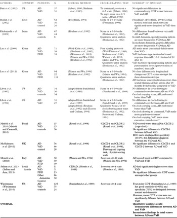
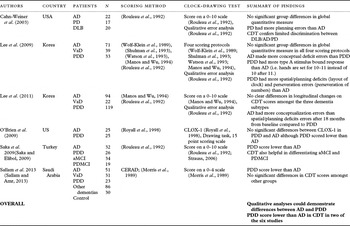
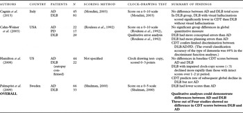
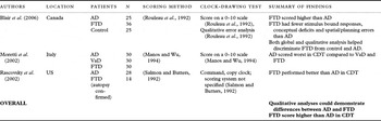
Papers were published between 1992 and 2013 and were mostly conducted in the USA (Barr et al., Reference Barr, Benedict, Tune and Brandt1992; Libon et al., Reference Libon, Swenson, Barnoski and Sands1993; Reference Libon, Malamut, Swenson, Sands and Cloud1996; Rascovsky et al., Reference Rascovsky2002; Cahn-Weiner et al., Reference Cahn-Weiner, Williams, Grace, Tremont, Westervelt and Stern2003; Hamilton et al., Reference Hamilton2008; O’Brien et al., Reference O’Brien, Wadley, Nicholas, Stover, Watts and Griffith2009; Wiechmann et al., Reference Wiechmann, Hall and O’Bryant2010) (n = 8). Two studies were from Italy (Moretti et al., Reference Moretti, Torre, Antonello, Cazzato and Bava2002; Cagnin et al., Reference Cagnin2013) and two studies were from Korea (Lee et al., Reference Lee, Kim, Choi and Sohn2009; Reference Lee, Oh, Jeong, Sohn, Lee and Lee2011). The remainder came from Israel (Heinik et al., Reference Heinik, Solomesh, Raikher and Lin2002), Japan (Kitabayashi et al., Reference Kitabayashi, Ueda, Narumoto, Nakamura, Kita and Fukui2001), UK (McGuinness et al., Reference McGuinness, Barrett, Craig, Lawson and Passmore2010), Brazil (Matioli and Caramelli, Reference Matioli and Caramelli2010), Saudi Arabia (Sallam and Amr, Reference Sallam and Amr2013), Turkey (Saka and Elibol, Reference Saka and Elibol2009), Sweden (Palmqvist et al., Reference Palmqvist, Hansson, Minthon and Londos2009), and Canada (Blair et al., Reference Blair, Kertesz, McMonagle, Davidson and Bodi2006). Eleven studies (Barr et al., Reference Barr, Benedict, Tune and Brandt1992; Libon et al., Reference Libon, Swenson, Barnoski and Sands1993; Reference Libon, Malamut, Swenson, Sands and Cloud1996; Kitabayashi et al., Reference Kitabayashi, Ueda, Narumoto, Nakamura, Kita and Fukui2001; Heinik et al., Reference Heinik, Solomesh, Raikher and Lin2002; Moretti et al., Reference Moretti, Torre, Antonello, Cazzato and Bava2002; Lee et al., Reference Lee, Kim, Choi and Sohn2009; Matioli and Caramelli, Reference Matioli and Caramelli2010; McGuinness et al., Reference McGuinness, Barrett, Craig, Lawson and Passmore2010; Wiechmann et al., Reference Wiechmann, Hall and O’Bryant2010; Lee et al., Reference Lee, Oh, Jeong, Sohn, Lee and Lee2011; Sallam and Amr, Reference Sallam and Amr2013) compared CDT scores between AD and VaD patients (Table 1). Six studies (Cahn-Weiner et al., Reference Cahn-Weiner, Williams, Grace, Tremont, Westervelt and Stern2003; Lee et al., Reference Lee, Kim, Choi and Sohn2009; O’Brien et al., Reference O’Brien, Wadley, Nicholas, Stover, Watts and Griffith2009; Saka and Elibol, Reference Saka and Elibol2009; Lee et al., Reference Lee, Oh, Jeong, Sohn, Lee and Lee2011; Sallam and Amr, Reference Sallam and Amr2013) were identified which compared CDT scores between AD and PDD patients (Table 2). Four studies (Cahn-Weiner et al., Reference Cahn-Weiner, Williams, Grace, Tremont, Westervelt and Stern2003; Hamilton et al., Reference Hamilton2008; Palmqvist et al., Reference Palmqvist, Hansson, Minthon and Londos2009; Cagnin et al., Reference Cagnin2013) compared CDT scores between AD and DLB patients (Table 3); and three studies (Moretti et al., Reference Moretti, Torre, Antonello, Cazzato and Bava2002; Rascovsky et al., Reference Rascovsky2002; Blair et al., Reference Blair, Kertesz, McMonagle, Davidson and Bodi2006) compared CDT scores between AD and FTD patients (Table 4). Five of the studies (Moretti et al., Reference Moretti, Torre, Antonello, Cazzato and Bava2002; Cahn-Weiner et al., Reference Cahn-Weiner, Williams, Grace, Tremont, Westervelt and Stern2003; Lee et al., Reference Lee, Kim, Choi and Sohn2009; Reference Lee, Oh, Jeong, Sohn, Lee and Lee2011; Sallam and Amr, Reference Sallam and Amr2013) compared CDT scores between AD patients and more than one type of dementia. Patients were recruited from memory clinics, geriatric assessment clinics, neurology clinics, and research centers.
Comparison of clock-drawing test between Alzheimer's dementia and patients with vascular dementia

*Includes clock size and shape of clock errors.
**Includes turning of paper when writing numbers, numbers written in counterclockwise order regardless of deficits in spatial layout or location within the clock and number perseveration errors.
Comparison of clock-drawing test results between Alzheimer's dementia and Parkinson's disease dementia patients

Notes: aMCI, amnestic mild cognitive impairment; PDMCI, mild cognitive impairment associated with Parkinson's disease.
Comparison of clock-drawing test results between Alzheimer's dementia and patients with Lewy bodies dementia

Comparison of clock-drawing test results between Alzheimer's dementia and frontotemporal dementia patients

Methodological characteristics of the studies
There are considerable methodological variations in the studies included. For example, sample characteristics differ widely across studies. The sample sizes varied from 50 to 235 patients, with samples consisting of patients with differing severity of dementia. Diagnosis of AD was mostly based on the NINCDS-ADRDA (McKhann et al., Reference McKhann, Drachman, Folstein, Katzman, Price and Stadlan1984). DSM-IV (Diagnostic and statistical manual of mental disorders (4th ed., text rev.), 2000) was used in one study (Heinik et al., Reference Heinik, Solomesh, Raikher and Lin2002) and two studies (Rascovsky et al., Reference Rascovsky2002; Hamilton et al., Reference Hamilton2008) had autopsy-confirmed diagnoses. The diagnosis of probable VaD was made using the NINDS-AIREN (Roman et al., Reference Roman1993) criteria in most studies, except one (Barr et al., Reference Barr, Benedict, Tune and Brandt1992) that used the DSM-III-R (Diagnostic and statistical manual of mental disorders (3rd ed., revised.), 1987) criteria, two (Heinik et al., Reference Heinik, Solomesh, Raikher and Lin2002; Matioli and Caramelli, Reference Matioli and Caramelli2010) that used the DSM-IV(Diagnostic and statistical manual of mental disorders (4th ed., text rev.), 2000) criteria and one study (Libon et al., Reference Libon, Malamut, Swenson, Sands and Cloud1996) used the California Criteria of Chui et al. (Reference Chui, Victoroff, Margolin, Jagust, Shankle and Katzman1992). The diagnosis of DLB was made according to the consensus criteria recommended by the Consortium of DLB (McKeith et al., Reference McKeith1996) in all the studies except one study had autopsy-confirmed diagnosis of DLB (Hamilton et al., Reference Hamilton2008). PDD was diagnosed when the Parkinson's disease patients met the DSM-IV (Diagnostic and statistical manual of mental disorders (4th ed., text rev.), 2000) criteria for dementia with the clinical diagnosis of the movement disorder at least 1 year prior to the onset of the dementia or using the clinical diagnostic criteria for dementia associated with Parkinson's disease (Emre et al., Reference Emre2007). FTD was diagnosed using the Neary et al. (Reference Neary1998) and McKhann et al. (Reference McKhann, Drachman, Folstein, Katzman, Price and Stadlan1984) criteria in one study (Blair et al., Reference Blair, Kertesz, McMonagle, Davidson and Bodi2006), the Lund and Manchester (Clinical and neuropathological criteria for frontotemporal dementia. The Lund and Manchester Groups, 1994) guidelines in another study (Moretti et al., Reference Moretti, Torre, Antonello, Cazzato and Bava2002), and was autopsy confirmed in the third study (Rascovsky et al., Reference Rascovsky2002).
The versions of CDT used in the studies also differed in the methods of administration and scoring. The studies included used either a blank sheet of paper with instructions to draw a clock face or a pre-drawn circle for the CDT. Most patients were instructed to set the clock hands at 10 past 11 (11:10), although other times such as 2:45 and 1:45 were also used less frequently in the studies that utilized the Sunderland et al. (Reference Sunderland1989) CDT, and the CLOX (Royall et al., Reference Royall, Cordes and Polk1998) methods. Some studies used a clock-copying task such as simple instructions to copy a clock shown or the CLOX (Royall et al., Reference Royall, Cordes and Polk1998) test. Scoring systems varied from a simple three-point system (Hamilton et al., Reference Hamilton2008) to a 15-point scoring system (Freedman, Reference Freedman, Leach, Kaplan, Wnocur, Shulman and Delis1994; Royall et al., Reference Royall, Cordes and Polk1998). Qualitative error analyses, when included, followed the Rouleau et al. system (Rouleau et al., Reference Rouleau, Salmon, Butters, Kennedy and McGuire1992).
Differentiating AD from VaD
In general, quantitative scores evaluating performance on the CDT were not shown to be useful in differentiating AD from VaD (Table 1). The findings were inconsistent across studies and although AD patients were found to perform better than VaD patients in clock copying scores in three studies (Libon et al., Reference Libon, Swenson, Barnoski and Sands1993; Reference Libon, Malamut, Swenson, Sands and Cloud1996; Matioli and Caramelli, Reference Matioli and Caramelli2010), the majority (Barr et al., Reference Barr, Benedict, Tune and Brandt1992; Libon et al., Reference Libon, Swenson, Barnoski and Sands1993; Reference Libon, Malamut, Swenson, Sands and Cloud1996; Kitabayashi et al., Reference Kitabayashi, Ueda, Narumoto, Nakamura, Kita and Fukui2001; McGuinness et al., Reference McGuinness, Barrett, Craig, Lawson and Passmore2010; Wiechmann et al., Reference Wiechmann, Hall and O’Bryant2010) found no significant differences in clock drawing to command scores between AD and VaD patients. One study (Moretti et al., Reference Moretti, Torre, Antonello, Cazzato and Bava2002) found that AD patients scored lower than VaD patients and two studies (Heinik et al., Reference Heinik, Solomesh, Raikher and Lin2002; Sallam and Amr, Reference Sallam and Amr2013) found that VaD patients scored lower than AD patients in the CDT. However, for the four studies (Libon et al., Reference Libon, Malamut, Swenson, Sands and Cloud1996; Kitabayashi et al., Reference Kitabayashi, Ueda, Narumoto, Nakamura, Kita and Fukui2001; Lee et al., Reference Lee, Kim, Choi and Sohn2009; Reference Lee, Oh, Jeong, Sohn, Lee and Lee2011) that included qualitative error analyses, significant differences could be identified between AD and VaD patients. VaD patients appeared to demonstrate more spatial/planning deficits (Kitabayashi et al., Reference Kitabayashi, Ueda, Narumoto, Nakamura, Kita and Fukui2001; Lee et al., Reference Lee, Kim, Choi and Sohn2009), graphical difficulties (Libon et al., Reference Libon, Malamut, Swenson, Sands and Cloud1996; Kitabayashi et al., Reference Kitabayashi, Ueda, Narumoto, Nakamura, Kita and Fukui2001), and perseveration errors (Lee et al., Reference Lee, Kim, Choi and Sohn2009) than AD patients. When evaluated over a period of 18 months, AD patients tend to show more conceptual errors than VaD patients compared to baseline (Lee et al., Reference Lee, Oh, Jeong, Sohn, Lee and Lee2011).
Differentiating AD from PDD
Patients with PDD scored lower than AD patients in three of the six studies which compared CDT in PDD and AD patients (O’Brien et al., Reference O’Brien, Wadley, Nicholas, Stover, Watts and Griffith2009; Saka and Elibol, Reference Saka and Elibol2009; Sallam and Amr, Reference Sallam and Amr2013), although in one study, this was not statistically significant (O’Brien et al., Reference O’Brien, Wadley, Nicholas, Stover, Watts and Griffith2009) (Table 2). In the other three studies, no significant differences were found in CDT scores between PDD and AD patients (Cahn-Weiner et al., Reference Cahn-Weiner, Williams, Grace, Tremont, Westervelt and Stern2003; Lee et al., Reference Lee, Kim, Choi and Sohn2009; Reference Lee, Oh, Jeong, Sohn, Lee and Lee2011). Qualitative analyses of the type of errors seem to suggest a difference between AD and PDD patients. In PDD patients, more spatial/planning errors were observed compared to AD patients (Cahn-Weiner et al., Reference Cahn-Weiner, Williams, Grace, Tremont, Westervelt and Stern2003; Lee et al., Reference Lee, Kim, Choi and Sohn2009). AD patients made more conceptual deficit errors than PDD patients (Lee et al., Reference Lee, Kim, Choi and Sohn2009; Reference Lee, Oh, Jeong, Sohn, Lee and Lee2011).
Differentiating AD from DLB
In patients with DLB, no significant differences were found between total CDT scores in three of the four studies included (Table 3) (Cahn-Weiner et al., Reference Cahn-Weiner, Williams, Grace, Tremont, Westervelt and Stern2003; Hamilton et al., Reference Hamilton2008; Cagnin et al., Reference Cagnin2013). The fourth study however, found that DLB patients had lower scores than AD patients (Palmqvist et al., Reference Palmqvist, Hansson, Minthon and Londos2009). In the only study examining qualitative differences between these groups, DLB patients were found to have more conceptual and planning errors than AD patients (Cahn-Weiner et al., Reference Cahn-Weiner, Williams, Grace, Tremont, Westervelt and Stern2003). Interestingly, it was also found that DLB patients with visual hallucinations scored lower than DB patients without visual hallucinations in one study (Cagnin et al., Reference Cagnin2013), and another study showed that the clock-drawing score predicted the rate of subsequent global decline in DLB but not AD patients (Hamilton et al., Reference Hamilton2008).
Differentiating AD from FTD
In FTD patients, all three studies (Moretti et al., Reference Moretti, Torre, Antonello, Cazzato and Bava2002; Rascovsky et al., Reference Rascovsky2002; Blair et al., Reference Blair, Kertesz, McMonagle, Davidson and Bodi2006) found that FTD patients scored higher than AD patients (Table 4). In the only study that examined the clock-drawing errors qualitatively, FTD patients had fewer stimulus-bound responses, conceptual deficits, and spatial/planning errors than AD patients (Blair et al., Reference Blair, Kertesz, McMonagle, Davidson and Bodi2006).
Discussion
This review suggests that qualitative analyses of the CDT may be helpful for the differential diagnosis of AD from other dementias such as VaD, PDD, DLB, and FTD. Furthermore, CDT scores may also help differentiate AD from FTD. However, quantitative scores in general, regardless of the scoring system used, were not useful for differentiating AD from VaD, PDD, and DLB. This is likely due to the intrinsic nature of the CDT assessing several cognitive skills at the same time for its completion. Hence although a single overall score is able to demonstrate the presence of cognitive impairment, it is limited in delineating specific domains of cognitive impairment. An examination of the type of CDT errors may be more useful in localizing the domain of cognitive dysfunction and assist in the diagnosis of the type of dementia.
Royall et al. hypothesized that the clock drawing to command (CLOX1) is more indicative of executive impairment than clock copying (CLOX2) (Royall et al., Reference Royall, Cordes and Polk1998), which focuses more on visuoconstructional abilities. Whilst there are some studies that support this theory with lower scores in CLOX1 than CLOX2 in VaD patients (You et al., Reference You, Chen, Zhang, Zhou, Cai and Li2011), there are also studies that conclude that CLOX2 is a more sensitive measure of executive deficits (Libon et al., Reference Libon, Swenson, Barnoski and Sands1993; Cosentino et al., Reference Cosentino, Jefferson, Chute, Kaplan and Libon2004). This may be explained by AD patients being more impaired in semantic knowledge, resulting in poorer CLOX1 scores than VaD patients. This would account for our finding that the total score of the CDT does not appear to be a sensitive measure to differentiate between AD and VaD. However, based on the four studies in this review that included qualitative analysis of the CDT between AD and VaD patients, there is a suggestion that the type of CDT errors may help to distinguish between the two. Most studies agree that frontal executive dysfunction is most characteristic of VaD (Roh and Lee, Reference Roh and Lee2014), and subcortical VaD differentially affects the frontosubcortical circuits that mediate fine motor control and planning. The spatial and planning deficits of the CDT seen more commonly in patients with VaD than AD are likely to be due to subcortical involvement in VaD (Looi and Sachdev, Reference Looi and Sachdev1999).
Patients with PDD or DLB generally have greater visuospatial, attention, and executive function impairment than AD patients (Ferman et al., Reference Ferman2006; Salmon and Bondi, Reference Salmon and Bondi2009). In this review, spatial/planning deficits on the CDT were more common in PDD or DLB patients than AD patients (Cahn-Weiner et al., Reference Cahn-Weiner, Williams, Grace, Tremont, Westervelt and Stern2003; Lee et al., Reference Lee, Kim, Choi and Sohn2009; Reference Lee, Oh, Jeong, Sohn, Lee and Lee2011). As is the case with VaD, this is likely to be due to subcortical involvement present in PDD and DLB. Planning errors on the CDT have been shown to be more common in patients with greater subcortical involvement (e.g. Huntington's disease), than those with predominantly cortical involvement (Rouleau et al., Reference Rouleau, Salmon, Butters, Kennedy and McGuire1992). Conceptual errors were found to be more common in AD than PDD patients in two of the four CDT studies with qualitative analyses (Lee et al., Reference Lee, Kim, Choi and Sohn2009; Reference Lee, Oh, Jeong, Sohn, Lee and Lee2011). This may be explained by the earlier impairment of semantic memory in AD patients (Salmon and Bondi, Reference Salmon and Bondi2009) compared to patients with PDD. In the only study, that included a qualitative analysis of the CDT comparing AD and DLB patients, conceptual errors were unexpectedly found to be greater in DLB patients than AD patients (Cahn-Weiner et al., Reference Cahn-Weiner, Williams, Grace, Tremont, Westervelt and Stern2003). The authors explain that picture comprehension is affected in DLB patients due to a combination of semantic and visuoperceptual impairments and other studies have also found that DLB patients demonstrate similar semantic deficits compared to AD patients (Calderon et al., Reference Calderon, Perry, Erzinclioglu, Berrios, Dening and Hodges2001; Lambon Ralph et al., Reference Lambon Ralph, Powell, Howard, Whitworth, Garrard and Hodges2001). Given the difficulty in clinically differentiating PDD and DLB from AD, patterns of deficits in the CDT may have some clinical utility in distinguishing between AD and PDD/DLB patients, although quantitative scores per se are not likely to be useful.
All three studies that administered the CDT to AD and FTD patients found that FTD patients consistently scored higher than AD patients (Moretti et al., Reference Moretti, Torre, Antonello, Cazzato and Bava2002; Rascovsky et al., Reference Rascovsky2002; Blair et al., Reference Blair, Kertesz, McMonagle, Davidson and Bodi2006). This finding is supported by several studies which suggest that the impairment in FTD is largely restricted to executive function rather than other cognitive functions (Forstl et al., Reference Forstl, Besthorn, Hentschel, Geiger-Kabisch, Sattel and Schreiter-Gasser1996; Rascovsky et al., Reference Rascovsky2002). Another study has also found that FTD patients perform better on tests of visuospatial abilities sensitive to the dysfunction of medial temporal structures compared to AD patients (Perri et al., Reference Perri2005). In the only study that included a qualitative analysis of the CDT comparing FTD and AD patients, FTD patients had fewer stimulus-bound responses, conceptual deficits, and spatial/planning errors than AD. This is also in keeping with another study showing that behavioral variant FTD patients often perform in the normal range on traditional executive function tests measuring working memory, planning, mental flexibility, response inhibition, and concept formation, at initial evaluation (Gregory and Hodges, Reference Gregory and Hodges1996; Perri et al., Reference Perri2005).
The comparability of the studies reviewed is limited due to the substantial methodological heterogeneity including the selection of the sample, the modality of CDT being administered, and the scoring system utilized. Despite these differences, it still appears that the CDT score itself confers limited discrimination between AD and VaD, PDD and DLB, and should not be used in isolation. Quantitative scores however, do appear to discriminate AD and FTD. Notably, the studies that included a qualitative analysis of the CDT found several significant differences between the type of errors made by patients with AD compared to patients with VaD, PDD, DLB, and FTD. Future studies should continue to evaluate the qualitative aspects rather than the quantitative aspects of the CDT. Qualitative analysis of the CDT may aid as an adjunct tool in the differential diagnosis of dementia.
Funding
None.
Conflict of interest disclosure
None.
Description of authors’ roles
K. Shulman and L. Tan formulated the research question. N. Herrmann and K. Shulman supervised the data collection. L. Tan collected, analysed the data and wrote the paper. N. Herrmann, B. Mainland, and K. Shulman assisted with writing the article.
Acknowledgments
The authors would like to acknowledge Mr Henry Lam for assisting with the library resources at the Sunnybrook Health Sciences Centre in obtaining the list of articles for this review.