Introduction
The transcervical approach to Zenker's diverticulum for inversion and cricopharyngeal myotomy remains a viable treatment option in today's endoscopic-dominant world. Zenker's diverticulum is a condition that was initially treated via a transcervical approach to the pulsion sac. Advances in technology have brought about changes in the approach to the pouch. Today, it is most commonly treated endoscopically via a transoral route. Indeed, by 2004, 83 per cent of otolaryngologists in the UK favoured the endoscopic route to the sac.Reference Siddiq and Sood1 However, the transcervical approach to the pharyngeal pouch (i.e. sac excision, suspension or inversion) still has proponents today.Reference Mantsopoulos, Psychogios, Kunzel, Zenk, Iro and Koch2 The interesting history of Zenker's diverticulum and its treatment has been collated and described by Simic et al. in detail.Reference Simić, Gurski and Pesko3
A recent systematic review of the literature suggests that the two approaches, transcervical and endoscopic, provide comparable overall outcomes, albeit with a difference in the pattern of complications.Reference Verdonck and Morton4 It should be noted that the various transcervical approaches are often reported en bloc, where excision accounts for the vast majority (84 per cent) of open procedures.Reference Verdonck and Morton4 However, of the various external approach options, the inversion technique is under-represented in the literature, with only 76 patients from 4 studies, compared with 1990 cases of sac excision.Reference Verdonck and Morton4
Inverting the sac is our favoured approach for Zenker's diverticulum; it does not involve a breach in the oesophageal mucosa (unlike transcervical excision, endoscopic laser or endoscopic stapling). In our experience, this technique is effective, and it carries a low risk of complications and recurrence. Because reports of inversion in the literature comprise only small numbers of cases, we decided to review and report our own recent experience with this technique.
Materials and methods
Approval for this study was obtained from the Regional Ethical Committee and the Institutional Clinical Board. A retrospective audit was performed of patients who underwent transcervical treatment for Zenker's diverticulum performed by two surgeons from 2003 to 2013.
Data on demographics, surgery, length of hospital stay, recurrence and other complications were extracted from the clinical records, and the results were compared to those in the literature. Statistical analysis was performed using IBM SPSS® Statistics software, version 19.0. Cross-tabulation analysis was performed on categorical variables and independent samples t-tests were run on the continuous variables to determine whether the groups (inversion vs cricopharyngeal myotomy only) were significantly different.
Surgical technique
Preliminary oesophagoscopy allows inspection of the sac, which is then packed with ribbon gauze. An anaesthetic-type tube is inserted into the oesophagus. The proximal ends of the gauze and the tube remain accessible under the drapes to enable removal by the anaesthetist when required by the surgeon. An incision is made on the anterior margin of the sternocleidomastoid and the diverticulum is approached via careful dissection. Once identified, the sac is grasped using Babcock's forceps, and the ribbon gauze is then withdrawn by the anaesthetist. The cricopharyngeus is divided in the midline along at least 3 cm using a scalpel and/or scissors, to which the endotracheal tube in the oesophagus provides useful counter pressure. The sac, once dissected free, has a purse-string suture (using 3-0 Vicryl®) placed around its neck. The tube prevents inadvertent over-closure which can lead to stenosis. Once the tube is withdrawn, the sac is inverted. A suction drain is routinely left in situ.
Patients can immediately resume fluids and eat breakfast the following day, and the drain is removed if this is tolerated. The majority of patients are discharged having resumed a normal diet after a single night in hospital. Follow-up barium swallow tests are not routinely performed.
Results
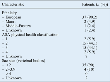
All 41 patients underwent myotomy of the cricopharyngeus muscle. The patients’ characteristics are shown in Table I. Mean patient age was 75.8 years (standard deviation (SD) = 11.4), with a near equal distribution of males and females (21 males and 20 females). The ethnic make-up was predominantly European. American Society of Anaesthesiologists' classifications of physical health were most commonly 2 or 3 (mild or moderate systemic disease) reflecting the sample age group.
Patients’ characteristics

ASA = American Society of Anaesthesiologists
Sac size details were available for 39 patients, 35 of whom had a sac size of less than 2 vertebral bodies. Twelve patients had a sac that was shallow and did not require ‘inversion’.
Average operating time was 78.6 minutes (SD = 18.0 minutes). The distribution of length of hospital stay was non-parametric (Figure 1), with the median stay being 1 night but with the average stay (of 2.46 nights) being skewed by a few outliers with a stay of over 6 nights (range, 1–15 nights).
Length of hospital stay.
The surgical complication rate was 9.8 per cent (three perforations and one dental trauma). Those patients who experienced surgical complications stayed in hospital for 1, 5, 7 and 15 nights. Only one of the perforation cases developed surgical emphysema; none developed mediastinitis. One patient was transferred to a medical ward after developing cardiac issues that required closer monitoring. The non-surgical complication rate was 4.9 per cent (two cases of urinary retention). Three patients (7.1 per cent) required a second operation, although only one required a true revision (repeat cricopharyngeal myotomy) while the remaining two had their initial procedures abandoned (dental trauma and stiff neck restricting access).
Cross-tabulation and independent samples t-test analyses did not demonstrate any statistically significant difference between the inversion and cricopharyngeal myotomy groups for any of the independent variables. However, with a more complete dataset of sac sizes, we would expect the inversion group to show at least a trend toward larger pouches.
Discussion
This series compares well to reported cases of endoscopic stapling. A recent systematic review on the treatment of Zenker's diverticulum provides an ideal resource for comparison.Reference Verdonck and Morton4 The average length of hospital stay in this current series (2.46 nights) compares favourably with the reported mean length of hospital stay for endoscopic stapling (3.2 days).Reference Verdonck and Morton4 Our complication rate of 9.8 per cent is slightly higher but not dissimilar to the mean pooled complication rate for endoscopic stapling (7.4 per cent), and is slightly lower than the average complication rate for all transcervical approaches (11 per cent).Reference Verdonck and Morton4 There were no deaths in our case series. In terms of failure to resolve symptoms, there were only two cases abandoned (4.9 per cent; dental trauma and difficult access), compared to the average failure rate of 18.9 per cent in endoscopic stapling.Reference Verdonck and Morton4 A single case had a recurrence of symptoms requiring further intervention (2.4 per cent), compared to the reported average recurrence rate for endoscopic stapling of 18.4 per cent.Reference Verdonck and Morton4
The vast majority of sacs encountered (85 per cent) were less than two vertebral bodies in size. We find the vertebral body classification useful, though no classification system has been universally adopted. Moreover, sac sizes are not routinely reported in the literature. From a practical standpoint, we believe that small to medium sized sacs (less than two vertebral bodies) can be inverted without risk of oesophageal obstruction, though larger sacs may be more amenable to suspension. Small sacs can prevent the insertion of an endoscopic stapler for release of the cricopharyngeal bar. The transcervical approach can be applied effectively, irrespective of sac size.
A retrospective intra-unit comparison of transcervical techniques was published by Mantsopoulos et al. in 2012.Reference Mantsopoulos, Psychogios, Kunzel, Zenk, Iro and Koch2 Cricopharyngeal myotomy alone, cricopharyngeal myotomy with inversion, and cricopharyngeal myotomy with diverticulectomy were compared. In the 11 cases of inversion, a single complication (recurrent laryngeal nerve paralysis) was reported, while diverticulectomy (37 cases) was associated with 14 significant complications.Reference Mantsopoulos, Psychogios, Kunzel, Zenk, Iro and Koch2 Mantsopoulos and colleagues tended to treat Zenker's diverticulum classified as Brombart II–III sacs with inversion, while larger sacs classified as Brombart III–IV were excised. Length of hospital stay was longer than in our study. Inversion resulted in earlier discharge than diverticulectomy (8.9 vs 11 days; p = 0.024).Reference Mantsopoulos, Psychogios, Kunzel, Zenk, Iro and Koch2 A single recurrence was reported. Their report is in keeping with our own beliefs that the transcervical approach results in fewer recurrences and that preserving the oesophageal mucosa results in fewer complications.
A more recent study by Leibowitz et al. which examined a single unit's experiences with both endoscopic and transcervical techniques found no significant difference in length of hospital stay, or rates of complications or recurrences.Reference Leibowitz, Fundakowski, Abouyared, Rivera, Rudman and Lo5 Endoscopic carbon dioxide laser, endoscopic stapler and transcervical diverticulectomy techniques (68, 69 and 27 cases, respectively) were compared.Reference Leibowitz, Fundakowski, Abouyared, Rivera, Rudman and Lo5 Length of hospital stay was greater in all groups (3.87, 4.32 and 6.5 days, respectively) than in our transcervical inversion group (2.46 nights).Reference Leibowitz, Fundakowski, Abouyared, Rivera, Rudman and Lo5 Our complication rate (9.8 per cent) was higher than the average complication rate reported across the three techniques (5.77 per cent).Reference Leibowitz, Fundakowski, Abouyared, Rivera, Rudman and Lo5
The transcervical approach to Zenker's diverticulum is a technically demanding procedure. However, it allows both physical manifestations of the problem to be addressed: the cricopharyngeal bar and the pulsion sac. Inspection of the cricopharyngeus muscle allows accurate and complete myotomy to be performed, while the sac is directly visualised for consideration of inversion. Careful surgical technique avoids a breach of the oesophageal mucosa necessitated by endoscopic stapling and diverticulectomy. Though an easier procedure to perform, endoscopic stapling limits access to both the bar and the sac for inspection, palpation and treatment. This may explain the high rate of recurrence reported in case series (18.4 per cent).Reference Verdonck and Morton4
• This audit increases the number of reported cases of transcervical inversion of Zenker's diverticulum from 76 to 105
• Transcervical cricopharyngeal myotomy with or without inversion of Zenker's diverticulum results in far lower rates of recurrence than endoscopic stapling
• Transcervical cricopharyngeal myotomy also results in a shorter hospital stay, with a comparable complication rate
Despite the trend toward minimally invasive surgical techniques, the transcervical approach to Zenker's diverticulum should not be discounted. The advantages of direct visualisation and handling of the pouch outweigh the technical challenges in our opinion. In situations where a prolonged anaesthetic may be deleterious to a patient (e.g. because of co-morbidities), an endoscopic approach offers relief of symptoms, but it carries a higher rate of recurrence.
Conclusion
Transcervical cricopharyngeal myotomy with or without inversion of Zenker's diverticulum is effective, with low morbidity when performed by surgeons well versed in its technique. The approach has a complication rate comparable to endoscopic stapling, and it is superior in terms of length of hospital stay and rate of recurrence in our experience.