Introduction
Filarial infections still remain a major cause of morbidity and mortality in humans and livestock, despite current eradication efforts (Perumal et al., Reference Perumal, Gunawardene and Dassanayake2016). Setaria digitata is an important filarial nematode living in the peritoneal cavity of cattle and buffaloes, and usually causes mild disease (Perumal et al., Reference Perumal, Gunawardene and Dassanayake2016). Infection of abnormal hosts, such as goats, sheep and horses, by this nematode can cause cerebrospinal nematodiasis (CSN) that leads to lumbar paralysis and the eventual death of the animals (Voronin et al., Reference Voronin, Abeykoon, Gunawardene and Dassanayake2015). This disease inflicts significant economic losses on livestock farmers. Due to the mobility of the mosquito vector and the risk of development of resistance to drugs used for treatment, CSN is difficult to control (Yatawara et al., Reference Yatawara, Wickramasinghe, Nagataki, Rajapakse and Agatsuma2007).
The accurate identification and differentiation of filarial nematodes has important implications for the study of their epidemiology, population biology and systematics. Identification and differentiation of S. digitata and its congeners (e.g. S. labiatopapillosa and S. cervi) have traditionally been based on morphological features (Perumal et al., Reference Perumal, Gunawardene and Dassanayake2016). However, these criteria are often insufficient for specific identification and differentiation, particularly at the larval and/or egg stages (Gasser, Reference Gasser2006). Molecular techniques, such as analysis of mitochondrial (mt) genomes, have been widely used as genetic markers for the identification and differentiation of organisms, particularly for population genetic and systematic investigations among nematode species (Gao et al., Reference Gao, Zhao, Liu, Zhang, Zhang, Wang, Chang, Wang and Zhu2014; Liu et al., Reference Liu, Zhao, Song, Zhao, Cai, Zhao and Zhu2014; Xu et al., Reference Xu, Qiu, Liu, Zhang, Liu, Duan, Yue, Chang, Wang and Zhao2015; Wang et al., Reference Wang, Gu, Yang, Wang, Lai, Zhong and Liu2016; Zhao et al., Reference Zhao, Wang, Jia, Zhao, Hu, Yu and Liu2016). Recently, the mt genome of S. digitata from cattle in Sri Lanka has been sequenced (Yatawara et al., Reference Yatawara, Wickramasinghe, Rajapakse and Agatsuma2010), but S. digitata isolated from different hosts and geographic origins may have some differences because a previous study detected intra-specific variation in the mt cytochrome c oxidase subunit 1 gene (cox1) in Sri Lanka (Alasaad et al., Reference Alasaad, Pascucci, Jowers, Soriguer, Zhu and Rossi2012). Moreover, a recent study of S. digitata also detected sequence variation in the second internal transcribed spacers (ITS-2) of nuclear ribosomal DNA (rDNA) among individuals of mithun (Bos frontalis) in both Arunachal Pradesh and Nagaland (Chamuah et al., Reference Chamuah, Sakhrie, Lama, Chandra, Chigure, Bauri and Jacob2015).
The objectives of the present study were to characterize the mt genome of S. digitata from buffalo in China and to compare this mt genome with that of S. digitata from cattle in Sri Lanka.
Materials and methods
Parasites and DNA extraction
An adult nematode representing S. digitata was obtained from the peritoneal cavity of an infected buffalo at a slaughterhouse in Guangdong province, China. The nematode was washed in physiological saline, identified preliminarily to species based on host preference, morphological characters and predilection sites (Perumal et al., Reference Perumal, Gunawardene and Dassanayake2016), fixed in 70% (v/v) ethanol and stored at −20°C until use. Total genomic DNA was isolated from this filarial worm using sodium dodecyl sulphate/proteinase K treatment, followed by spin-column purification (Wizard® SV Genomic DNA Purification System, Promega, Madison, Wisconsin, USA).
Long PCR amplification and sequencing
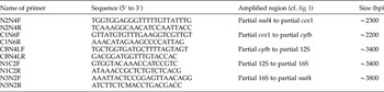
The primers (table 1) were designed in the conserved regions to amplify the complete mt genome of S. digitata (Sri Lanka isolate; NC_014282). Long polymerase chain reactions (PCR) were performed (total volume 25 μl, comprising 2 mm MgCl2, 0.2 mm of each deoxynucleoside triphosphate (dNTP), 2.5 μl 10 × rTaq buffer, 2.5 μm of each primer, 1.25 U rTaq polymerase (Takara, Dalian, China) and 1 μl of DNA sample) in a thermocycler (Biometra, Göttingen, Germany) under the following conditions: 92°C for 2 min (initial denaturation); then 92°C for 10 s (denaturation), 50°C for 30 s (annealing) and 60°C for 8 min (extension) for 10 cycles; followed by 92°C for 10 s, 50°C for 30 s and 60°C for 8 min for 20 cycles, with a cycle elongation of 10 s for each cycle; and a final extension at 60°C for 10 min. Samples not containing DNA (no-DNA controls) were included in each amplification run, and in neither case were amplicons detected in the no-DNA controls (data not shown). Each amplicon (5 μl) was examined by agarose (1%) gel electrophoresis, stained with ethidium bromide and photographed using a gel documentation system (UVItec, Cambridge, UK). PCR products were sent to Sangon Biotech (Shanghai, China) for sequencing from both directions using a primer walking strategy.
Sequences of primers used to amplify PCR fragments from Setaria digitata.

Sequence analyses
Sequences were assembled and aligned against the complete mt genome sequence of S. digitata (Sri Lanka isolate) using the computer program Clustal X 1.83 (Thompson et al., Reference Thompson, Gibson, Plewniak, Jeanmougin and Higgins1997) to identify gene boundaries by comparison with that of S. digitata (Sri Lanka isolate). The open reading frames and codon usage profiles of protein-coding genes were analysed by the Open Reading Frame Finder (http://www.ncbi.nlm.nih.gov/gorf/gorf.html) using the invertebrate mt code. Gene annotation, translation initiation, translation termination codons and the boundaries between protein-coding genes of the mt genome sequence of S. digitata (China isolate) were identified based on comparison with the mt genome sequences of S. digitata (Sri Lanka isolate) and other filarial nematodes reported previously (Yatawara et al., Reference Yatawara, Wickramasinghe, Rajapakse and Agatsuma2010; McNulty et al., Reference McNulty, Mullin, Vaughan, Tkach, Weil and Fischer2012). For analysis of tRNA genes, putative secondary structures of 22 tRNA genes were identified using tRNAscan-SE (Lowe & Eddy, Reference Lowe and Eddy1997), or by recognizing potential secondary structures and anticodon sequences by eye, and two rRNA genes were predicted by comparison with those reported previously (Yatawara et al., Reference Yatawara, Wickramasinghe, Rajapakse and Agatsuma2010).
Results and discussion
Mitochondrial genome organization
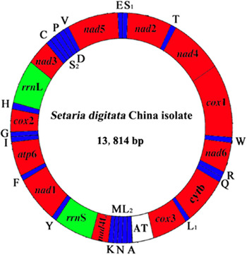
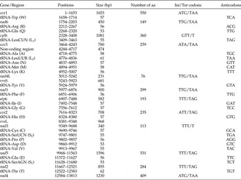
The complete mt genome sequence of S. digitata (China isolate) was 13,814 bp in size (fig. 1). It has been deposited in GenBank under the accession number KY284626. The mt genome of S. digitata (China isolate) contains 12 protein-coding genes (cox1–3, nad1–6, nad4L, atp6 and cytb), 22 transfer RNA genes, two ribosomal RNA genes and one non-coding region (table 2). All genes are transcribed in the same direction as other infraorder Spiruromorpha species sequenced to date (Keddie et al., Reference Keddie, Higazi and Unnasch1998; Hu et al., Reference Hu, Gasser, Abs El-Osta and Chilton2003; Liu et al., Reference Liu, Gasser, Otranto, Xu, Shen, Mohandas, Zhou and Zhu2013, 2015). The mt genome sequence of S. digitata (China isolate) has high T content and low C content (T = 55.1%, A = 19.7%, G = 18.4%, C = 6.8%), and its A + T content was 74.8%, in accordance with mt genomes of other infraorder Spiruromorpha species sequenced to date (Keddie et al., Reference Keddie, Higazi and Unnasch1998; Hu et al., Reference Hu, Gasser, Abs El-Osta and Chilton2003; Liu et al., Reference Liu, Gasser, Otranto, Xu, Shen, Mohandas, Zhou and Zhu2013, 2015).
Arrangement of the mitochondrial genome of Setaria digitata (China isolate). Gene scaling is only approximate. All genes are coded by the same DNA strand and are transcribed clockwise. All genes have standard nomenclature except for the 22 tRNA genes, which are designated by the one-letter code for the corresponding amino acid, with numerals differentiating each of the two leucine- and serine-specifying tRNAs (L1 and L2 for codon families CUN and UUR, respectively; S1 and S2 for codon families AGN and UCN, respectively). ‘AT’ refers to a non-coding region.

Mitochondrial genome organization of Setaria digitata from China.

aa, amino acids; Ini/Ter, initial/terminal.
A total of 3458 amino acids are encoded in the mt genome of S. digitata (China isolate). All protein-coding genes use TTT, ATT, TTG, ATG, GTT and ATA as their initiation codon. Four protein-coding genes use TAG as a stop codon whereas six genes use TAA and two genes use T as a termination codon. These results were consistent with that of S. digitata Sri Lanka isolate (Yatawara et al., Reference Yatawara, Wickramasinghe, Rajapakse and Agatsuma2010). The sizes of 22 tRNA genes identified in the mt genome of S. digitata (China isolate) ranged from 53 to 62 nucleotides (nt). Their putative secondary structures (not shown) are similar to those of other infraorder Spiruromorpha species (Keddie et al., Reference Keddie, Higazi and Unnasch1998; Hu et al., Reference Hu, Gasser, Abs El-Osta and Chilton2003; Liu et al., Reference Liu, Gasser, Otranto, Xu, Shen, Mohandas, Zhou and Zhu2013, Reference Liu, Jia, Wang, Zhao and Zhu2015). The rrnL is located between RNA-His and nad3, and rrnS is located between nad4L and tRNA-Tyr. The sizes of the rrnL and rrnS genes are 968 and 681 bp, respectively, for both S. digitata (China isolate) and S. digitata (Sri Lanka isolate) (table 2). The majority of nematode mtDNA sequences usually contain two non-coding regions with a significant size difference (Liu et al., Reference Liu, Gasser, Su, Nejsum, Peng, Lin, Li, Xu and Zhu2012; Jabbar et al., Reference Jabbar, Littlewood, Mohandas, Briscoe, Foster, Müller, von Samson-Himmelstjerna, Jex and Gasser2014; Yong et al., Reference Yong, Song, Eamsobhana, Goh and Lim2015), but there is only one non-coding region (AT-rich region) in the mt genome of S. digitata, which is located between cox3 and tRNA-Ala (fig. 1 and table 2), with 84.6% of A + T content. Furthermore, this region is devoid of consecutive sequences of [A] and [T], and there are no AT dinucleotide repeat sequences; these repeat regions have been reported in the mt genomes of Thelazia callipaeda (Liu et al., Reference Liu, Gasser, Otranto, Xu, Shen, Mohandas, Zhou and Zhu2013) and Gongylonema pulchrum (Liu et al., Reference Liu, Jia, Wang, Zhao and Zhu2015).
Comparative analyses between the China and Sri Lanka mt genome isolates
The S. digitata (China isolate) mt genome sequence was 25 bp shorter than that of S. digitata (Sri Lanka isolate). The identity of the nucleotide sequences between the entire mt genomes of S. digitata (China isolate) and S. digitata (Sri Lanka isolate) is 98.3%. For the 12 protein genes, this comparison revealed sequence differences at both the nucleotide (1.4%) and amino acid (2.2%) levels. The magnitude of nucleotide sequence variation in each gene between S. digitata (China isolate) and S. digitata (Sri Lanka isolate) ranged from 0.6 (nad1) to 2.7% (nad2). Amino acid sequences inferred from individual mt protein genes of S. digitata (China isolate) were compared with those of S. digitata (Sri Lanka isolate). The amino acid sequence differences ranged from 0.2 to 6.2%, with COX1 being the most conserved protein and NAD3 the least conserved.
Significance and implications
Although much attention has been paid to soil-transmitted nematodes as pathogens because of their major socio-economic impact on human populations, parasitic veterinary nematodes, such as members of the genus Setaria, have been seriously neglected. Setaria digitata can cause fatal cerebrospinal nematodiasis in goats, horses and sheep in many countries, including China (Khedri et al., Reference Khedri, Radfar, Borji and Azizzadeh2014). In the present study, the characterization of the mt genome of S. digitata (China isolate) provides a foundation for the improved diagnosis of cerebrospinal nematodiasis using molecular methods, as well as future, detailed studies of the population genetics and epidemiology/ecology of this parasite in China. As adult and larval stages of S. digitata cannot be identified reliably to species by morphology, molecular tools, using genetic markers in the mt cox1 gene, have been used for identification and differentiation of two Setaria species (Alasaad et al., Reference Alasaad, Pascucci, Jowers, Soriguer, Zhu and Rossi2012). The availability of the complete mt genome of the S. digitata China isolate will be useful in molecular epidemiological investigations of S. digitata.
In conclusion, this study determined the complete mtDNA sequence of S. digitata from China, which is slightly shorter (25 bp) than that of S. digitata from Sri Lanka. Nucleotide sequences between both mt genomes differed by 1.7% only. These mtDNA sequences should provide novel genetic data for addressing further questions regarding the population genetics of these and other related infraorder Spiruromorpha of socio-economic significance.
Financial support
This project was supported by the Scientific Research Fund of Hunan Provincial Education Department (16A102).
Conflict of interest
None.
Ethical standards
The performance of this study was strictly according to the recommendations of the Guide for the Care and Use of Laboratory Animals of the Ministry of Health, China, and our protocol was reviewed and approved by the Research Ethics Committee of Hunan Agricultural University.