Introduction
Primary melanocytic lesions of the central nervous system (CNS) are rare. They include diffuse leptomeningeal melanocytosis, melanocytoma and primary malignant melanoma.Reference Louis, Ohgaki, Wiestler and Cavenee1 The reported prevalence of primary CNS melanoma is 0.005 cases per 100 000 population.Reference Louis, Ohgaki, Wiestler and Cavenee1 A primary CNS melanoma located in the cerebellopontine angle region is even rarer, with only eight cases reported in the English language literature.Reference Liubinas, Maartens and Drummond2
We present a patient with a primary malignant melanoma of the cochlea extending into the internal auditory canal and cerebellopontine angle, in whom further investigation failed to identify any extracranial primary lesion.
Case report
A 71-year-old man with an unremarkable past medical history presented with sudden hearing loss in his left ear, in the absence of any other focal neurological symptoms.
Audiometric testing demonstrated total hearing loss in the left ear.
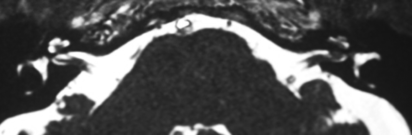
Magnetic resonance imaging (MRI) (T2-weighted sequence) demonstrated an isointense, 3 × 3 mm lesion within the left internal auditory canal, with cochlear enhancement (Figure 1). The images showed homogeneous enhancement with gadolinium (Figure 2).
Axial, T2-weighted magnetic resonance imaging scan of the internal auditory canals, showing a rounded, isointense nodule at the fundus of the left internal auditory canal, with cochlear extension.

Axial, T1-weighted, post-contrast, fat-saturated magnetic resonance imaging scan showing enhancement at the fundus of the left internal auditory canal. A = anterior; R = right; L = left; P = posterior
Given the location of the lesion, a presumptive diagnosis of an intracochlear schwannoma was made, and routine monitoring was advised.
Six months later, the patient suffered an acute vestibular disturbance with sudden onset of dizziness, nausea, vomiting and left frontal headache. Right-beating nystagmus on neutral, upward and horizontal eye movement was observed, with a positive head impulse test. The left-sided hearing loss was unchanged. Repeated MRI showed the previously observed lesion to have increased to a size of 9 × 3 mm.
The patient underwent a translabyrinthine and transcochlear removal of the tumour, together with placement of an osseointegrated screw fixture for a bone-anchored hearing aid. Intra-operatively, a dark-brown tumour involving the cochlear and facial nerves was noted to extend through the modiolus to fill the cochlea, and also to extend along the vestibulocochlear nerve into the cerebellopontine angle (Figure 3). Frozen section analysis was equivocal, indicating an epithelioid, pigmented lesion associated with haemorrhage. Although gross total resection was performed, a fragment of tumour was noted to be adherent to the facial nerve medial to the internal auditory meatus.
Intra-operative, operating microscope view of the tumour within the left internal auditory canal, following a translabyrinthine approach.
Histopathological analysis revealed a densely hypercellular tumour containing spindle and epithelioid cells with a fascicular and diffuse sheeted arrangement. The epithelioid tumour cells had round and oval nuclei, with prominent amphophilic nucleoli and a variable amount of pale cytoplasm. The cytoplasm of many tumour cells contained globular, finely granular, black-brown pigment. Moderate numbers of mitotic figures were identified (5 in 10 high power fields of 2 mm2; ×400 magnification). On immunohistochemical analysis, the tumour cells exhibited strong immunostaining for the melanocyte-associated markers Melan-A and S-100 peptide. No staining for glial fibrillary acidic protein was noted in the tumour cells. Consequently, a diagnosis of malignant melanoma was made.
Subsequent dermatological examination revealed a pigmented lesion on the right side of the neck and another on the right posterior pinna. Excisional biopsies and histopathological analysis confirmed the diagnosis of a dysplastic naevus and a morphoeic basal cell carcinoma, respectively.
The patient had an uncomplicated post-operative course. Staging investigations conducted five months after surgery, including whole-body positron emission tomography, and computed tomography of the chest, abdomen and pelvis, showed no evidence of metastatic disease. The patient underwent stereotactic radiotherapy using a 6 MV Novalis linear accelerator (Brainlab, Feldkirkchen, Germany); a dose of 55.56 Gy in 20 fractions was applied to the isocentre, with a covering isodose of 90 per cent, equivalent to 50 Gy, covering the target volume. The target volume was defined as the internal auditory canal, cochlea, tympanic cavity and petrous bone to below the stylomastoid foramen, for a final planned target volume of 12.7 cm3 (Figure 4).
Stereotactic radiotherapy treatment model marking the target volume, which included the left internal auditory canal, cochlea, tympanic cavity and petrous bone to below the stylomastoid foramen.
A routine MRI brain scan conducted approximately one year following the operation unfortunately showed multiple left cerebellar metastases and two adjacent enhancing nodules on the surface of the left medulla. To treat this, the patient received a course of whole-brain radiotherapy with a stereotactic boost to target the metastatic disease in the posterior fossa.
Three months following radiotherapy, the patient was admitted to hospital with increasing thoracic radicular pain, ataxia and a sensory level at the 10th thoracic vertebra. Spinal imaging confirmed diffuse leptomeningeal metastases involving the entire spinal cord.
Given the patient's limited prognosis and treatment options, he and his family decided upon conservative management, and he was discharged home with community palliative care services follow up.
Discussion
Tumours of the region encompassing the internal auditory canal and cerebellopontine angle make up 5–10 per cent of all intracranial tumours; fewer than 1 per cent of these are malignant.Reference Shramek, Gray, Wilkins and Rengachary3 Acoustic neuromas are by far the most common type reported (85 per cent), followed by meningiomas (5–10 per cent) and epidermoid tumours (6–7 per cent).Reference Shramek, Gray, Wilkins and Rengachary3 Primary CNS melanomas located in the internal auditory canal and cerebellopontine angle region are exceptionally rare; Liubinas and colleagues' review described only eight previously reported cases.Reference Liubinas, Maartens and Drummond2
Primary melanocytic lesions of the CNS are derived from the leptomeningeal melanocytes that arise from the neural crest during embryogenesis.Reference Louis, Ohgaki, Wiestler and Cavenee1 These have been reported in patients ranging from 17 to 71 years of age, with a peak incidence in the fifth decade. They can occur throughout the neuroaxis, with a slight predilection for the spinal cord and posterior fossa.
Clinical presentation varies depending on the location of the tumour. In the case of tumours in the internal auditory canal and cerebellopontine angle region, hearing loss, vertigo and/or facial nerve palsy have been described.Reference Brackmann and Doherty4
• An extremely rare case of primary melanoma in the cerebellopontine angle is presented
• Recognition of characteristic findings on presentation and imaging aids differentiation of cerebellopontine angle melanoma from acoustic neuroma
• Characteristic magnetic resonance imaging features include hyperintense T1 images with no fat suppression, hypointense T2 images, and hyperintense fluid-attenuated images with contrast
• Total surgical resection plus radiotherapy is the accepted treatment
On MRI scanning, melanocytic neoplasms of the CNS typically appear as a hyperintense image on T1-weighted sequences, which does not decrease in signal with fat-suppression sequences, and as a hypointense image on T2-weighted sequences; this is thought to be due to the paramagnetic properties of melanin. Melanocytic CNS neoplasms also exhibit hyperintensity on fluid-attenuated inversion recovery sequences and homogeneous enhancement with gadolinium, consistent with the vascular nature of melanomas.Reference Liubinas, Maartens and Drummond2, Reference Brackmann and Doherty4 In our case, we observed the typical MRI findings of a melanocytic lesion, including hyperintensity on T1-weighted sequences and contrast enhancement. However, when arriving at the initial, provisional diagnosis of vestibular schwannoma, the location of the tumour took precedence.
Macroscopically, melanocytic lesions in the CNS may appear black, red-brown, blue or non-pigmented.Reference Louis, Ohgaki, Wiestler and Cavenee1 The differential diagnosis of a pigmented lesion in the cerebellopontine angle includes primary and metastatic lesions. Malignant melanomas are the third most common tumours to metastasise to the CNS.Reference Louis, Ohgaki, Wiestler and Cavenee1 However, isolated metastasis to the cerebellopontine angle is rare. Pigmented primary lesions involving the cerebellopontine angle include primary melanocytic tumours, pigmented meningiomas and pigmented schwannomas.Reference Louis, Ohgaki, Wiestler and Cavenee1 Histopathological examination and immunohistochemical analysis are therefore vital in confirming the diagnosis. Features commonly associated with malignant melanoma include anaplastic epithelioid cells arranged in loose nests, fascicles or sheets, and displaying variable amounts of melanin, together with increased cellular density, pleomorphism and atypia. In our case, immunohistochemical analysis confirmed the diagnosis, with positive results for anti-melanosomal antibody MART-1 (Melan-A) and S-100 protein, and a negative result for glial fibrillary acidic protein; these findings are consistent with a diagnosis of meningeal melanoma.
Primary CNS melanoma is an aggressive tumour. The prognosis and clinical course correlates with the histological grade, completeness of resection and presence of distant metastases.Reference Louis, Ohgaki, Wiestler and Cavenee1 Total resection supplemented with whole-brain irradiation has been shown to have significantly better outcomes and is recommended in all cases.Reference Rodriguez, Gaetani, Danova, Bosi and Zappoli5 Post-operative surveillance imaging is also recommended at routine intervals.
Conclusion
Given the extreme rarity of malignant cerebellopontine angle tumours, coupled with their variation in clinical presentation and imaging appearance, clinical and neuroradiological findings are often inadequate to distinguish between a malignant lesion and a vestibular schwannoma. The diagnosis of an internal auditory canal or cerebellopontine angle melanoma should be considered in patients with: a history of previous cutaneous or other melanoma, MRI findings atypical for common tumours of this location, and accelerated audiovestibular symptoms and/or facial nerve palsy. Despite the presented patient's outcome, total surgical resection with post-operative radiotherapy remains the recommended treatment.