The ductus arteriosus is necessary in foetal life, allowing blood to bypass the lungs while maintaining systemic blood flow. In term infants, the ductus typically constricts within hours after birth; however, in preterm infants, it may remain patent. Persistent patent ductus arteriosus is associated with earlier gestational age and lower birth weight.Reference Koch, Hensley, Roy, Brown, Ramaciotti and Rosenfeld 1 , Reference Fanaroff, Stoll and Wright 2 Rates of patent ductus arteriosus in preterm infants range from 29 to 80%, depending on the population studied.Reference Fanaroff, Stoll and Wright 2 – Reference Shah, Sankaran and Aziz 6 Persistent patent ductus arteriosus can lead to increased pulmonary blood flow and inadequate systemic blood flowReference Moore and Brook 7 and is associated with complications including congestive heart failure,Reference Little, Pratt, Blalock, Krauss, Cooney and Custer 8 acute renal failure,Reference Vida, Lago and Salvatori 9 necrotising enterocolitis,Reference Cassady, Crouse and Kirklin 3 , Reference Dollberg, Lusky and Reichman 10 intraventricular haemorrhage,Reference Evans and Kluckow 11 pulmonary haemorrhage,Reference Finlay and Subhedar 12 bronchopulmonary dysplasia,Reference Koch, Hensley, Roy, Brown, Ramaciotti and Rosenfeld 1 and mortality.Reference Brooks, Travadi, Patole, Doherty and Simmer 13 , Reference Noori, McCoy and Friedlich 14
There is significant controversy about patent ductus arteriosus treatment in preterm neonates.Reference Benitz 15 – Reference Clyman, Couto and Murphy 17 Surgical ligation is often used for haemodynamically significant patent ductus arteriosus when medical treatment has failed, in unstable patients, or in those with contraindications to medical treatment, including necrotising enterocolitis, intraventricular haemorrhage, and acute renal failure. The definition of haemodynamically significant patent ductus arteriosus itself is disputed.Reference Sehgal and McNamara 18 Although in some cases surgical ligation is complicated by a period of postoperative haemodynamic instability,Reference El-Khuffash, Jain, Weisz, Mertens and McNamara 19 , Reference El-Khuffash, Jain and McNamara 20 surgical ligation has been shown to be safe and effectiveReference Little, Pratt, Blalock, Krauss, Cooney and Custer 8 , Reference Vida, Lago and Salvatori 9 and may decrease mortality.Reference Madan, Kendrick, Hagadorn and Frantz 21 On the other hand, acute complications have been reported, including vocal cord paralysis,Reference Jaillard, Larrue, Rakza, Magnenant, Warembourg and Storme 22 diaphragmatic paralysis,Reference Yon, Amka, Pildes and Tatooles 23 pneumothorax,Reference Jaillard, Larrue, Rakza, Magnenant, Warembourg and Storme 22 , Reference Gould, Montenegro and Gaynor 24 and chylothorax.Reference Gould, Montenegro and Gaynor 24 Recent studies have raised concerns about the long-term risks associated with patent ductus arteriosus ligation, including worse neurodevelopmental outcomes and increased retinopathy of prematurity and bronchopulmonary dysplasia;Reference Madan, Kendrick, Hagadorn and Frantz 21 , Reference Kabra, Schmidt, Roberts, Doyle, Papile and Fanaroff 25 , Reference Clyman, Cassady, Kirklin, Collins and Philips 26 however, determinations of causality are troubled by many confounding factors.Reference Weisz and McNamara 27 The rate of spontaneous patent ductus arteriosus closure in preterm infants may be higher than that previously thought,Reference Koch, Hensley, Roy, Brown, Ramaciotti and Rosenfeld 1 , Reference Nemerofsky, Parravicini, Bateman, Kleinman, Polin and Lorenz 28 , Reference Herrman, Bose, Lewis and Laughon 29 raising concerns about early ligation, or whether ligation is required. No consensus exists on the most appropriate selection criteria for surgical ligation or optimal timing of the procedure.Reference Cassady, Crouse and Kirklin 3 , Reference Vida, Lago and Salvatori 9 , Reference Gersony, Peckham, Ellison, Miettinen and Nadas 30 , Reference Cotton, Stahlman, Bender, Graham, Catterton and Kovar 31
Given this lack of consensus, we sought to describe patent ductus arteriosus ligation practices in a broad sample, using multiple years of a nationally representative data set to characterise patients and hospitals, explore changes over time, and identify predictors of ligation.
Materials and methods
Data source
Data were obtained from the Kids’ Inpatient Database, an all-payer, hospital administrative database of paediatric inpatient care in the United States. Released every 3 years, the Kids’ Inpatient Database is a stratified sample of discharges from all community, non-rehabilitation hospitals in states participating in the Agency for Healthcare Research and Quality’s Healthcare Cost and Utilization Project. It contains a nationally representative sample, including 80% of paediatric discharges and complicated births and 10% of uncomplicated births. The Kids’ Inpatient Database contains information from discharge abstracts, including diagnoses, procedures, demographics, and hospital characteristics, with safeguards to protect the privacy of the hospitals, physicians, and patients. The Kids’ Inpatient Database for 2009 included 4121 hospitals – 80% of all American Hospital Association-designated hospitals – from 44 states. The Kids’ Inpatient Database for 2006 included 3739 hospitals (73%) from 38 states, and for 2003 it included 3438 hospitals (71%) from 36 states. 32
Data in the Kids’ Inpatient Database are stratified by region, location, teaching status, hospital size, ownership, and whether the hospital is a freestanding children’s hospital. Discharges are also stratified by uncomplicated in-hospital births, complicated in-hospital births, and non-newborn paediatric discharges. Each discharge is weighted in proportion to the number of known American Hospital Association discharges nationally. Using discharge weights, individual discharges are extrapolated to produce national estimates. All the data are presented as weighted national estimates, not actual observations. 33 Cells based on 10 or fewer observations were suppressed due to the Healthcare Cost and Utilization Project data use agreement to prevent individual patient identification, but the values were included in the analysis. 34
Search strategy
We queried the 2003, 2006, and 2009 Kids’ Inpatient Database for International Classification of Diseases, Ninth Revision diagnosis codes associated with premature births, stratified by gestational age from <24 weeks to 32 weeks. Codes for prematurity based on gestational age were not used in the Kids’ Inpatient Database before 2003. Discharges were examined for an International Classification of Diseases code for patent ductus arteriosus (747.0). 35 We excluded patients with other congenital cardiac defects likely to require surgery in the 1st year of life, in whom patent ductus arteriosus ligation might be carried out during other cardiac surgical procedures. Entries were examined for International Classification of Diseases procedure codes for surgical patent ductus arteriosus ligation (38.85) and for patient characteristics such as gender, race, payer, and associated diagnoses as well as hospital characteristics such as type, teaching status, hospital size, location, state, and region.
Some preterm infants require transfer between hospitals for surgical ligation;Reference Gould, Montenegro and Gaynor 24 however, data in the Kids’ Inpatient Database are limited to a single hospitalisation, and thus we were unable to track patients transferred to another institution. To avoid counting these patients more than once in our analysis, we excluded patients admitted after 1 day of age. To determine the effect of this exclusion on our results, we attempted to identify the proportion that may have been transferred to another hospital for ligation by examining the disposition of each patient who did not undergo ligation. Moreover, seven states do not report age at admission in days, thus we excluded all discharges from these states.
Owing to an expected association between gestational age, patent ductus arteriosus diagnosis, and ligation, we examined the rate of patent ductus arteriosus diagnosis by gestational age and observed a drop-off in the rate of diagnosis in infants born after 28 weeks of gestation. In addition, a high rate of spontaneous closure of patent ductus arteriosus after 28 weeks of gestation has been reported.Reference Koch, Hensley, Roy, Brown, Ramaciotti and Rosenfeld 1 , Reference Nemerofsky, Parravicini, Bateman, Kleinman, Polin and Lorenz 28 Thus, we dichotomised gestational age into two categories – that is, ⩽28 weeks and 29–32 weeks – for all the analyses. As changes in survival in preterm infants over time could affect both the rate of diagnosis and the rate of ligation of patent ductus arteriosus, we examined length of stay as a crude surrogate for survival and calculated the length of stay by year and gestational age group.
Definitions
Payer was defined as “Medicaid”, “Private”, or “Other”, which included “Medicare”, “Self-pay”, and “No charge”. “Associated diagnoses” included acute renal failure, intraventricular haemorrhage, necrotising enterocolitis – including spontaneous intestinal perforation – congestive heart failure, and pulmonary haemorrhage.
Hospital type was based on the information provided by the National Association of Children’s Hospitals and Related Institutions. We evaluated three categories of the National Association of Children’s Hospitals and Related Institutions hospitals: “Not a Children’s Hospital”, “Children’s Unit in a General Hospital”, and “Children’s Hospital”. Hospital size was defined as “Small”, “Medium”, or “Large” by hospital bed number, based on stratified groups by hospital region, urban or rural location, and teaching status. Teaching status was defined based on the American Hospital Association Annual Survey Database. Hospital location was defined as rural or urban based on United States Census Bureau data.
In the analysis of data by state, patent ductus arteriosus diagnosis rate was defined as the number of discharges associated with the International Classification of Diseases codes for patent ductus arteriosus per preterm infant born at ⩽32 weeks of gestation in that state. Ligation rate was defined as the number of discharges associated with patent ductus arteriosus ligation per preterm infant born at ⩽32 weeks of gestation with patent ductus arteriosus in that state. Owing to an expected association between patency of the ductus arteriosus and altitude,Reference Bialkowski 36 we evaluated the correlation between patent ductus arteriosus diagnosis rate and average altitude of each state, using data obtained from the U.S. Census Bureau. 37
Statistical analysis
Survey analysis methods were used for stratified, 2-stage cluster samples. Associations between categorical variables were tested using the Rao–Scott χ2 tests. Logistic regression was used for multivariable modelling. The results of the logistic regression analysis are reported as odds ratios with 95% confidence intervals.
To identify predictors of patent ductus arteriosus ligation in preterm infants, univariate analysis was performed using the following variables: year, gender, gestational age, payer, associated diagnoses, hospital type, teaching status, size, rural or urban location, and region. Owing to the small numbers of ligations performed in several states and the exclusion of data from seven states due to lack of admission data in days, state was not examined in our univariate analysis. Race is not reported in the Kids’ Inpatient Database by several states and was missing for 24% of all discharges examined; therefore, race was excluded from the univariate analysis.
We created a multivariable model for the prediction of patent ductus arteriosus ligation for variables that showed significance at a p-value of <0.05 in the univariate analysis. Year was included as an ordinal variable for multivariable modelling, but for all other variables all the years included in the Kids’ Inpatient Database were examined together. Records with missing data were excluded from the multivariable analyses. To examine the effect of excluding patients admitted after 1 day of age on our results, we performed these analyses again using the entire data set, including those discharges with admission age >1 day. An Archer–Lemeshow–Hosmer F-adjusted mean residual goodness-of-fit test was performed to evaluate the multivariable models.Reference Archer, Lemeshow and Hosmer 38
All the calculations were performed using SAS for Windows 9.3 (SAS Institute Inc., Cary, North Carolina, United States of America).
Results
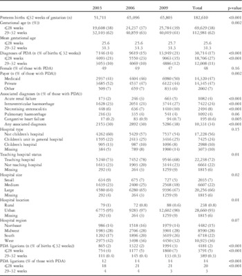
Of 19,440,796 paediatric discharges, we identified 283,377 discharges of preterm infants born at ⩽32 weeks of gestation. Of these discharges, 68,409 (24%) were excluded due to missing age at admission, 25,296 (9%) were excluded due to age >1 day at admission, and 7061 (2%) were excluded due to the presence of CHD. Of the remaining 182,610 discharges of preterm infants born at ⩽32 weeks of gestation, 69,629 (38%) were ⩽28 weeks of gestation. Length of stay increased over time for each gestational age category (p=0.03). Overall, 30,714 discharges (17%) included a diagnosis of patent ductus arteriosus. The number of patent ductus arteriosus diagnoses increased from 7146 in 2003 to 13,949 in 2009, representing an increase in the percentage of preterm infants with patent ductus arteriosus from 14% in 2003 to 21% in 2009 (p<0.001). Both gestational age subgroups (⩽28 weeks and 29–32 weeks) showed an increase in the rates of diagnosis of patent ductus arteriosus over the study period (p<0.001). A graph displaying the rate of patent ductus arteriosus diagnosis by gestational age and year is shown in Supplementary figure 1a. A graph displaying the rate of patent ductus arteriosus ligation by gestational age and year is shown in Supplementary figure 1b. The diagnosis rate was higher in the ⩽28-week gestational age group (27%) compared with the 29- to 32-week gestational age group (11%) for all years combined. Table 1 provides the number of patent ductus arteriosus diagnoses and ligations per year, as well as associated patient and hospital data.
Patient and hospital data, national estimated totals.

PDA=patent ductus arteriosus
A total of 4181 patent ductus arteriosus ligations were performed, with an overall rate of 14% of preterm infants ⩽32 weeks of gestation with a diagnosis of patent ductus arteriosus, 2% of all preterm infants ⩽32 weeks of gestation. The overall rate of patent ductus arteriosus ligation increased from 12% in 2003 to 14% in 2009 (p<0.001).
Additionally, 16% of discharges associated with patent ductus arteriosus without ligation ended with transfer to another hospital, and the majority of these discharges (73%) were from a non-children’s hospital.
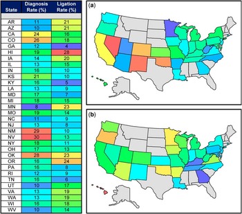
Significant variation was noted in patent ductus arteriosus diagnosis rate and ligation rate by state. Patent ductus arteriosus diagnosis rate, per number of preterm infants in that state, ranged from 8 to 30% (Fig 1). Ligation rate ranged from 4 to 28% of preterm infants with patent ductus arteriosus. No correlation was found between patent ductus arteriosus diagnosis rate and ligation rate by state (slope coefficient 0.009±0.20; Supplementary figure 2). With the known association between patent ductus arteriosus and altitude,Reference Bialkowski 36 we analysed rate of diagnosis by mean altitude in each state, 37 and found a significant correlation, as shown in Supplementary figure 3.
Variability of PDA diagnosis and ligation rate by state. ( a ) Diagnosis rate for each state determined by the number of PDA diagnoses per preterm infant in that state. ( b ) Ligation rate for each state determined by the number of PDA ligations per preterm infant with a PDA in that state. Data were excluded if cells contained <10 occurrences by data use agreement. PDA=patent ductus arteriosus.

In the univariate analysis, lower gestational age, presence of an associated diagnosis, discharge from a “Children’s Hospital” or “Children’s Unit in a General Hospital”, teaching hospital, and hospital region in the West or Midwest were associated with higher ligation rate (Supplementary table 1). When evaluated individually, each associated diagnosis was also associated with a higher rate of ligation (all p<0.001). There was no significant difference in the ligation rate by year, gender, payer, hospital size, or location.
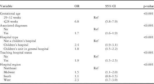
In the multivariable model for predictors of ligation (Table 2), lower gestational age, associated diagnoses, discharge from a “Children’s Hospital” or “Children’s Unit in a General Hospital”, and teaching hospital predicted increased likelihood of patent ductus arteriosus ligation (p<0.001). Compared with discharges from the Northeast, ligation was more likely in the Midwest and West (p<0.001). When length of stay was included in the multivariable model, we found that an additional day of length of stay had a statistically significant but minor impact on the probability of ligation (odds ratio 1.023, 95% confidence interval 1.022–1.025), but did not change the effect of the other variables. Goodness-of-fit testing did not indicate any overall departure of the multivariable model from the observed data (p=0.39).
Multivariable model of predictors of ligation.

OR=odds ratio; 95% CI=95% confidence interval
Archer–Lemeshow–Hosmer F-adjusted mean residual goodness-of-fit: p=0.39
As part of a sensitivity analysis, we examined the number of discharges, patent ductus arteriosus diagnoses, and ligations using the entire data set, without excluding those patients admitted after 1 day of age. Patient data are shown in Supplementary table 2. Similar to our initial analysis, the rates of patent ductus arteriosus diagnosis and ligation increased over time (p<0.001). The multivariable model revealed the same factors associated with ligation, except that ligation was also more likely to occur in the South than in the Northeast (Supplementary table 3).
Discussion
Our study represents a large examination of patent ductus arteriosus ligation in the United States. These results, based on weighted national estimates from a validated inpatient database, provide a real-world snapshot of ligations from 2003 to 2009.
We found that the rate of patent ductus arteriosus diagnosis has increased, both overall and in gestational age subgroups, consistent with previous studies. Shah reported an increase in patent ductus arteriosus in 3763 infants born at <29 weeks of gestation in Canada between two eras – 1996–1997 and 2006–2007. They suggested that this increase may have resulted from improved echocardiography technology, and thus improved detection.Reference Shah, Sankaran and Aziz 6 Advances in diagnostic tools are not likely to explain the increase seen in our study, which was conducted during a period of echocardiographic technology more than adequate to diagnose patent ductus arteriosus. Improvements in electronic medical records and other clinical documentation initiatives may contribute to increased recognition of patent ductus arteriosus in the discharge abstract. As we did find that an increase in length of stay by year paralleled the increase in diagnosis rate, this may be another explanation for increased diagnosis rate, as longer survival provides a greater opportunity to make a diagnosis of patent ductus arteriosus. Further investigation is required to determine whether our increased rate of diagnosis is due to increased detection or a true increase in the incidence of patent ductus arteriosus.
In addition, there is a known association between patency of the ductus arteriosus and altitude,Reference Bialkowski 36 and our analysis confirmed this positive correlation.
A striking finding of our study is the significant geographic variation in patent ductus arteriosus ligation practice. Regional variation in specific neonatal procedures has been described previously, including placement of intracranial pressure monitors, tracheotomy, and lumbar puncture in early onset neonatal sepsis.Reference Odetola, Tilford and Davis 39 – Reference Patrick, Schumacher and Davis 41 Such regional differences in our study may reflect variation in neonatology training, access to cardiology or surgical services, or the perception of risk of persistent patent ductus arteriosus.
We also found significant differences by hospital characteristics, including a significantly higher rate of patent ductus arteriosus ligation in teaching hospitals and children’s hospitals. This may reflect that infants born at earlier gestational age or with complex disease are frequently admitted to teaching and children’s hospitals due to regionalisation of care, or may reflect different practice patterns at such tertiary-care hospitals. The apparent lower rates of ligation at non-teaching hospitals and non-children’s hospitals may also be due to the need to transfer infants to another institution for the primary purpose of patent ductus arteriosus ligation at a centre with neonatal surgical expertise.
Our overall ligation rate, 14%, is lower than the rates of 19–36% reported previously, depending on the population examined, possibly due to differences in selection criteria.Reference Fanaroff, Stoll and Wright 2 , Reference Little, Pratt, Blalock, Krauss, Cooney and Custer 8 , Reference Vida, Lago and Salvatori 9 , Reference Kabra, Schmidt, Roberts, Doyle, Papile and Fanaroff 25 Without excluding infants admitted after 1 day of age, the overall ligation rate was 17%. We found that the most premature infants had a significantly higher likelihood of patent ductus arteriosus ligation, consistent with previous studies, which have shown a higher rate of failure of medical therapies with decreasing birth weight and gestational age.Reference Fanaroff, Stoll and Wright 2 , Reference Little, Pratt, Blalock, Krauss, Cooney and Custer 8 , Reference Vida, Lago and Salvatori 9 Lower gestational age infants are at higher risk for co-morbidities, such as necrotising enterocolitis, which are contraindications for medical treatment.Reference Dollberg, Lusky and Reichman 10 We found that the presence of such associated diagnoses correlated with a higher likelihood of ligation. Furthermore, these diagnoses may be a marker of increased complexity and illness severity, identifying patients less likely to tolerate waiting for a patent ductus arteriosus to close spontaneously.
Study limitations
Administrative, population-based databases allow the inclusion of large numbers of patients and provide a cross-section of national data; however, they are limited by the lack of clinical detail and missing data and are susceptible to coding errors. As the Kids’ Inpatient Database does not include medications, we were unable to compare surgical ligation with other therapies such as cyclooxygenase inhibitors. We were also unable to determine the methods of diagnosis, clinical significance of patent ductus arteriosus, or temporal sequence of diagnoses and procedures. The lack of available clinical and temporal data makes it difficult to discern specific causes of morbidity or mortality in this medically complex and fragile patient population, and thus we chose to focus our investigation on patient and hospital variables associated with ligation.
The diagnoses included in the Kids’ Inpatient Database are drawn from the discharge abstract for each hospitalisation and include a limited number deemed to be most important. This may underestimate the rate of patent ductus arteriosus diagnoses and highlight the more severe patent ductus arteriosus cases, which may be more likely to undergo ligation.
Conclusion
Patent ductus arteriosus diagnosis and ligation rates have increased in recent years. Significant geographic variation exists in the patent ductus arteriosus ligation rate in the United States, which persists after adjustment for potential confounding factors such as gestational age. This highlights the lack of consensus about this commonly performed procedure. Both patient and hospital factors influence the likelihood of ligation. Future studies examining the effects of practice variation on outcomes may suggest opportunities for improvement in the care of these infants.
Acknowledgement
None.
Financial Support
This research received no specific grant from any funding agency, commercial, or not-for-profit sectors.
Conflicts of Interest
The views expressed are those of the authors and do not necessarily reflect official National Heart, Lung, and Blood Institute positions.
Supplementary Material
For supplementary material referred to in this article, please visit http://dx.doi.org/10.1017/S1047951115001869