Breath-holding spells, a stereotyped sequence of clinical events, are not an uncommon clinical presentation and are a frightening experience for parents. During the spell, the child cries excessively because of frustration, pain, or both. At the end of prolonged expiration, the child becomes apnoeic for several seconds and then turns either blue (cyanotic spell) or pale (pallid spell), with some children experiencing both. Occasionally, tonic spasms occur, and the children may develop clonic movements.Reference DiMario 1 In general, breath-holding spells are benign disorders that often resolve with time. However, in the complex case with severe and frequent spells that are unresponsive to medication and are associated with seizures, life-threatening bradycardia, or asystole, aggressive intervention should be considered.Reference Kelly, Porter, McGoon, Espinosa, Osborn and Hayes 2 Neurologic and cardiologic evaluations are necessary in doubtful cases. The presence of cardiac pacing in breath-holding cases reported in international publications has demonstrated the importance of detailed investigation into possible changes in heart rhythm in these patients.Reference Kelly, Porter, McGoon, Espinosa, Osborn and Hayes 2 – Reference Vinik and Ziegler 7 Previous studies have shown that the underlying pathophysiologic mechanism in children with breath holding may be generalised autonomic dysregulation.Reference Anil, Nedunchezian, Jayanthini and Pathmanabhan 8 – Reference DiMario, Bauer, Volpe and Baxter 12 The pathophysiology of pallid breath holding is different from cyanotic breath holding, with loss of consciousness being preceded by 1 to several seconds of asystole.Reference Evans 13 , Reference Lombroso and Lerman 14 These children have been shown to have an exaggerated oculocardiac reflex, resulting in asystole in 61% of the children with pallid breath-holding spells and in only 25% of the children with cyanotic breath-holding spells.Reference Lombroso and Lerman 14 Thus, we performed cardiac rhythm and heart rate variability analyses using 24-hour Holter monitoring to evaluate the cardiac effects of autonomic dysregulation in children with breath-holding spells. Results in previous studies have been obtained from short-term electrocardiogram recordings, not from 24-hour Holter monitor recordings. Thus, the effect of night–day intervals on heart rate variability has not been described.
Heart rate variability, or beat-to-beat variation, provides information about autonomic nervous system activity – consisting of the sympathetic and parasympathetic system – in the sinoatrial node. Heart rate variability is usually analysed dynamically using 24-hour Holter monitoring or in 0.5–5-minute periods.Reference Malik 5 Time-domain and frequency-domain analyses are used in heart rate variability evaluation.
The time-domain method is the simplest measure of heart rate variability and estimates heart rate variability in 5-minute cycles.Reference Hartikainen, Tahvannainen and Kuusela 15 The time-domain method is limited, however, in its ability to discriminate between sympathetic and parasympathetic regulation of heart rate variability, and it can be affected by artefacts and extreme values.Reference Rajendra Acharya, Paul Joseph, Kannathal, Lim and Suri 16 Recent studies have demonstrated that frequency-domain methods are the only non-invasive measures that provide accurate information on autonomic nervous system activity by distinguishing between sympathetic and parasympathetic regulation of heart rate variability.Reference Malik 5 An increase in high frequency reflects a parasympathetic effect, and an increase in low frequency is more commonly associated with a sympathetic effect.Reference Malik 5 , Reference Rajendra Acharya, Paul Joseph, Kannathal, Lim and Suri 16 , Reference Sztajzel 17 The total power reflects the activation of the entire autonomous nervous system. There is a strong relationship between time-domain heart rate variability parameters and frequency-domain parameters, and one measurement is not superior to the other; the information each provides is complementary.Reference Malik 5 , Reference Kayıkçıoğlu and Payzın 18
Methods
In all, 68 children with breath-holding spells – diagnosed via history from parents – who were assessed in the Pediatric Cardiology Department of Erzurum Regional Education and Research Hospital between May, 2011 and June, 2013 were included in this study. Cases included in this study consisted of breath-holding children who had severe, recurrent (>10/month) spells. The control group consisted of 39 healthy children without cardiac problems who were referred to Pediatric Cardiology for murmur assessment. Control subjects were chosen to match the age and sex distributions of the breath-holding children.
Age, sex, spell type (cyanotic or pallid), spell level (each had multiple severe spells – more than 3 – with loss of consciousness within the 3 months prior), and spell frequency (>10/month) of breath-holding children were determined.Reference Anil, Nedunchezian, Jayanthini and Pathmanabhan 8 The spell types, according to descriptions provided by the child’s family, were divided into two groups (blue or pale) based on the colour change during the spell.
The left ventricular function of all cases in the study group was evaluated for valvular abnormalities and additional abnormalities using echocardiography. Patients with haemodynamically and clinically significant cardiac lesions or patients with epileptic activity upon electroencephalography were excluded.
Rhythm, maximum and minimum heart rates, mean heart rates, QTc intervals, and heart rate variability of breath-holding children and controls were investigated using 24-hour Holter monitoring with a three-channel electrocardiography recording instrument (Holter for Windows, H4W, version 10.00d, Rozinn Electronics Inc., Glendale, NY, United States of America). Heart rate variability analyses were performed using a Holter for Windows (H4W), version 10.00d, Rozinn Electronics Inc. analyser system. The heart rate variability variables, calculated automatically by the device after manual cleaning of artefacts, were used.
Time-domain and frequency-domain analyses were used for heart rate variability assessment. The standard deviation of all NN intervals, standard deviation of all the 5-minute NN intervals, mean of the standard deviations of all NN intervals for all 5-minute segments, percentage of differences between adjacent RR intervals >50 ms, and square root of the mean of the sum of squares of the differences between adjacent NN intervals were assessed in the time-domain analyses. Total power (<0.4 Hz), high frequency: 0.15–0.4 Hz, low frequency: 0.003–0.04 Hz, and low frequency/high frequency ratio were evaluated in the frequency-domain analyses. Age (months), maximum and minimum heart rate, mean heart rate, systolic blood pressure, systolic ejection fraction, QTc intervals, and heart rate variability parameters of breath-holding children were statistically compared with those of the control group, as well as between the pallid and cyanotic groups.
Statistical analysis
The Statistical Package for Social Sciences for Windows 11.5 (SPSS, SPSS Inc., Chicago, Illinois, United States of America) was used for statistical analyses. Descriptive statistics are expressed as the mean±standard deviation, with percentages for categorical variables. The Kolmogorov–Smirnov test was used to determine the suitability of sample values to a normal distribution. Normally distributed group values were compared using a Student’s t-test, whereas those not normally distributed were compared using the Mann–Whitney U-test. Fisher’s exact test and Yates Continuity Correction binary test (Yates-corrected χ 2) were used to compare qualitative data. All quantitative results are reported with 95% confidence intervals, and a p-value <0.05 was considered significant.
Results
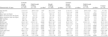
In all, 68 breath-holding children including 56 with cyanotic spells (39 boys, 17 girls; 5–48 months) and 12 with pallid spells (six girls, six boys; 8–44 months), and 39 healthy controls (21 boys, 17 girls; 4–46 months) were evaluated. There were no significant differences in mean age, mean heart rate, maximum heart rate, minimum heart rate, systolic ejection fraction, systolic blood pressure, QTc intervals, and heart rate variability parameters between the breath-holding children and controls. When clinical and heart rate variability results for each spell type were compared with those for the controls, significant differences were detected in standard deviation of all NN intervals, mean of the standard deviations of all NN intervals for all 5-minute segments, percentage of differences between adjacent RR intervals >50 ms, and square root of the mean of the sum of squares of the differences between adjacent NN intervals values between the pallid and cyanotic groups. Between the pallid and control group, there were significant differences in mean heart rate, mean minimum heart rate, standard deviation of all NN intervals, mean of the standard deviations of all NN intervals for all 5-minute segments, percentage of differences between adjacent RR intervals >50 ms, square root of the mean of the sum of squares of the differences between adjacent NN intervals, total power, high frequency, and low frequency/high frequency values (p<0.05). However, no significant differences were observed between the cyanotic group and controls (Table 1).
Demographic and clinical features, and heart rate variability results of breath-holding children and control group.

Ef=ejection fraction; HF=high frequency; LF=low frequency; pNN50=percentage of differences between adjacent RR intervals >50 ms; rMSDD=square root of the mean of the sum of squares of the differences between adjacent NN intervals; SBP=systolic blood pressure; SDANN=standard deviation of all the 5-minute NN intervals; SDNN=standard deviation of all NN intervals; SDNNi=mean of the standard deviations of all NN intervals for all 5-minute segments
The first two columns (*) compare the data for the two breath-holding groups, and the other columns (+) compare these groups with controls
Significant values are in bold
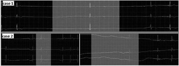
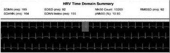
When 24–48-hour Holter findings for children with pallid spells were reviewed separately, sinus pauses lasting at least 2.5 seconds but <5.6 seconds were detected in two cases (Fig 1). Long QTc (520 ms) accompanied with high heart rate variability was also noted in another case (Fig 2).
Hypervagotonic sinus dysregulation seen in Holter electrocardiography in two cases with pallid breath holding. Upper panel: Holter electrocardiography shows two sinus pauses at 3.12 and 2.6 seconds (first case). Lower panel: Holter electrocardiography shows four sinus pauses at 2.08, 2.16, 5.6, and 2.52 seconds (second case).

Long QTc (520 ms) accompanied by high heart rate variability observed in Holter electrocardiography in a pallid breath-holding patient.

Discussion
Children with breath holding are classified into two groups –cyanotic and pallid – according to their skin colour during a spell.Reference DiMario 1 The cyanotic type constitutes 60% of cases, whereas the pallid type occurs in 20–30% of cases. Both types may also occur in some patients.Reference DiMario 1 , Reference DiMario 19 Our clinical differentiation between types in this study revealed that 82.4% of breath-holding cases had cyanotic spells, and 17.6% had pallid spells.
Many factors may play a role in the aetiopathogenesis of breath holding.Reference Holmes 20 Various studies have shown that the underlying pathophysiologic mechanism in breath-holding children may be generalised autonomic dysregulation.Reference Anil, Nedunchezian, Jayanthini and Pathmanabhan 8 – Reference DiMario, Bauer, Volpe and Baxter 12 Heart rate response to deep inspiration, heart rate response to standing position, Valsalva manoeuvre, diastolic blood pressure response to continuous isometric contraction, and response to the tilt-table test may be used to clinically evaluate autonomic dysregulation.Reference Vinik and Ziegler 7 , Reference Boysen, Lewin, Hecker, Leichter and Uhlemann 21 , Reference Ewing and Clarke 22 However, heart rate variability assessment is a more practical approach compared with these methods, which are hard to apply in early childhood, when breath-holding spells often occur.
Heart rate variability is a non-invasive and repeatable electrocardiographic method that measures the efficacy of sympathetic and parasympathetic components of the autonomic nervous system on the sinoatrial node. Its importance and usefulness is accepted for the evaluation of autonomic dysregulation.Reference Malik 5 , Reference Lahiri, Kannankeril and Goldberger 23
In non-invasive studies performed by DiMario et al,Reference DiMario, Bauer and Baxter 9 – Reference DiMario, Bauer, Volpe and Baxter 12 alteration of autonomic dysregulation was emphasised for reducing the pathophysiologic mechanism in breath-holding spells. Although parasympathetic exaggeration is seen in pallid breath holding, it does not occur in cyanotics. Using standard electrocardiography records, Kolkiran et alReference Kolkiran, Tutar, Atalay, Deda and Cin 24 found that asystole frequency was higher during high heart rate variability in pallid breath-holding cases. Previous studies have demonstrated the effects of autonomic dysregulation on heart rate variability with short-time standard electrocardiography recording in children with breath holding. Although short-time electrocardiography recording is also a method used for heart rate variability analyses, it would be difficult for infants to remain calm during the recording. Therefore, we decided the results would not be accurate. In addition, it was shown that heart rate variability may be affected differently depending on the time of day (day–night).Reference Noritake, Takase, Kudoh, Kugai, Kurita and Nagata 25 – Reference Boudreau, Yeh, Dumont and Boivin 27 Thus, in the present study, we analysed the results from 24-hour electrocardiography records. In our study, standard deviation of all NN intervals, mean of the standard deviations of all NN intervals for all 5-minute segments, percentage of differences between adjacent RR intervals >50 ms, and square root of the mean of the sum of squares of the differences between adjacent NN interval values in children with pallid breath holding were significantly higher than in those with cyanotic breath holding. Standard deviation of all NN intervals, mean of the standard deviations of all NN intervals for all 5-minute segments, percentage of differences between adjacent RR intervals >50 ms, square root of the mean of the sum of squares of the differences between adjacent NN intervals, total power, and high frequency values were also significantly higher in children with pallid breath holding than those in the control group. The low frequency/high frequency ratio, mean heart rate, and mean minimum heart rate in children with pallid breath holding were significantly lower than the values in controls. Diurnal influence was observed in standard deviation of all NN intervals, standard deviation of all the 5-minute NN intervals, mean of the standard deviations of all NN intervals for all 5-minute segment parameters calculated directly from the NN interval – interval between two consecutive normal stimuli originating from the sino-atrial node – and there were few effects on heart rate of short-time alterations due to respiration. Indexes – square root of the mean of the sum of squares of the differences between adjacent NN intervals, percentage of differences between adjacent RR intervals >50 ms – calculated from differences between NN intervals are short duration measurements and reflect high-frequency variations in heart rate. These are free from diurnal effects and reflect changes on autonomic tonus controlled via the vagal nerve.Reference Malik 5 , Reference Kayıkçıoğlu and Payzın 18 Increased high frequency reflects parasympathetic effect and low frequency/high frequency ratio reflects the relationship between the sympathetic and parasympathetic systems – increased vagal tonus compared with sympathetic tonus.Reference Malik 5 , Reference Kayıkçıoğlu and Payzın 18 , Reference Gula, Krahn and Skanes 28 Researchers have determined that changes in vagal tonus related to respiration rate and variability may also affect the heart rate because of the vagal efferent signal to the heart.Reference Grossman, Karemaker and Wieling 29 , Reference Katona and Jih 30 From our data, although diurnal influence was higher in children with pallid breath holding than in cyanotics, both diurnal influence and autonomic tonus changes controlled via the vagus nerve were higher in children with pallid breath holding compared with controls. The increase in total power and high frequency values and the decrease in mean average heart rate, mean minimum heart rate, and low frequency/high frequency ratio in cases with pallid breath holding may originate from cardioinhibition caused by vagal tonus and vigorous parasympathetic stimulus.
The presence of cardiac pacing applied to children with breath holding in international publications has shown the importance of detailed investigation of possible changes in heart rhythm in these patients. In the pathophysiology of pallid breath holding, asystole lasting 1 to several seconds was reported, along with loss of consciousness.Reference Evans 13 , Reference Lombroso and Lerman 14 Kelly et alReference Kelly, Porter, McGoon, Espinosa, Osborn and Hayes 2 applied permanent pacemaker implantation to 10 patients with pallid breath holding along with bradicardia due to asystole during 1985–1995 and reported that this treatment may be successful and reliable. Another study reported improvement in an infant with frequent pallid breath-holding spells accompanied by loss of consciousness with sustained asystole lasting 26 seconds on Holter monitoring via permanent pacemaker treatment.Reference Di Pino, Calabrò, Gitto, Bianca and Oreto 3 The association of sudden infant death due to reversible cardiac asystole caused by vigorous vagal activity was studied as a risk factor in children with pallid breath holding.Reference Lucet, de Bethmann and Denjoy 31 , Reference Lucet, Le Gall and Shojaeï 32 When the Holter findings of children with pallid breath holding were separately reviewed, sinus pauses lasting 2.5–5.6 seconds were detected in two cases, and long QTc accompanied with high heart rate variability was noted in another case.
Conclusions
We would like to explain that autonomic dysfunction involves excessive autonomic breath-holding activity (parasympathetic), not autonomic nervous system failure, in some children with breath holding. Holter monitoring (24 hours) and heart rate variability parameters, particularly in children with pallid spells, are crucial for evaluating cardiac rhythm changes. However, more cases of pallid breath holding are required for 24-hour Holter monitoring and heart rate variability analyses.
Acknowledgement
None.
Conflicts of Interest
None.