

In the healthcare field, technological innovation takes multiple, hardly predictable forms. High-cost devices installed in a hospital for a given use are sometimes used for new emerging purposes without prior evaluation.
This continuous implementation process is a real challenge for economic evaluation. Ideally, any innovative use of a given device should prompt full economic evaluation. This, however, requires early knowledge of the health impact of the innovation. This condition is not met in most cases, because health outcomes are only known in the medium- or long-term. However, in the absence a full economic assessment, and even if the conclusions are not definitive, we believe that the evaluation process is necessary and that it should take place as early as possible, with an evaluation of both the resources used and the results obtained.
Positron emission tomography (PET), a functional imaging technique recently introduced in nuclear medicine, provides a good example of this situation. The technique, which is widely used in oncology, makes use of the metabolic properties of malignant cells, such as excessive consumption of glucose most commonly determined by means of a radioactive analog of glucose, FDG (2-[18F]fluoro-2-deoxy-D-glucose). Unlike conventional imaging techniques such as computed tomography (CT) and magnetic resonance imaging (MRI), which provide precise anatomical images but cannot distinguish between cancerous and noncancerous tissues, PET can be used to visualize isolated metastases, or to verify whether treated tumor sites still contain cancer cells.
It has been demonstrated that PET is effective for the initial diagnosis of both non-small-cell lung cancer (NSCLC) and Hodgkin's disease and/or the assessment of disease extent (Reference Drummond, Sculpher, Torrance, O'Brien and Stoddart7;8). In these applications, several cost-effectiveness studies (Reference Alzahouri, Lejeune, Woronoff-Lemsi, Arveux and Guillemin1–Reference Bradbury, Facey, Laking and Sharp3;Reference Chuck, Jacobs and Logus4;Reference Gambhir, Hoh, Phelps, Madar and Maddahi9;Reference Gugiatti, Grimaldi and Rossetti11;Reference Juweid and Cheson13–Reference Kosuda, Ichilara, Watanabe, Kobayashi and Kusano17;Reference Nguyen, Peloquin and Lacasse19–Reference Von Schulthess, Steinert, Dummer and Weder24) have suggested that PET could save time and money by better guiding diagnostic procedures and reducing unnecessary and inappropriate treatments.
The use of PET could also provide a valuable aid to treatment decision making in radiotherapy, particularly for these two diseases. Thus, combined with CT scan, which is the gold standard imaging modality, PET would provide a better definition of the target volume and the dose to be delivered, or even lead to a change in the indication for radiotherapy. However, there has been little evaluation of this use of PET imaging (Reference Gregoire, Haustermans, Geets, Roels and Lonneux10), and, to our knowledge, no assessment of effects and costs has been performed.
A prospective, multicenter study developed as part of a French national program was conducted in eight hospitals throughout the country; each of them with only one PET equipment unit. The objective of the study was to evaluate the benefit of using PET for decision making in radiotherapy, as a complement to the standard strategy, planning CT. More specifically, it involved assessing the impact of using PET on both the choice of treatment and the resources used (costs incurred and costs avoided) for patients with nonoperable NSCLC and Hodgkin's disease.
METHODS
Most economic evaluation studies focus on a comparison of mutually exclusive options, and generally conclude that one option is cost-effective, compared with a differential cost-effectiveness acceptability threshold that is assumed to be known beforehand (6;Reference Drummond, Sculpher, Torrance, O'Brien and Stoddart7). On the contrary, the present study focuses on two nonexclusive strategies involving two different diseases. Each strategy must, therefore, be compared with the standard. In the context of a full economic evaluation taking the form of a cost-effectiveness analysis, a prioritization scheme could be developed, based on one of the following three conclusions: (i) neither of the innovative strategies proposed is interesting to implement because differential cost-effectiveness ratios are too high, (ii) both are interesting, or (iii) one is interesting but the other is not.
For the reasons mentioned above, we were not able to conduct a full economic evaluation that would have required long-term monitoring of patients. However, even though we were conducting a short-term assessment, we adopted the methodologically relevant approach by comparing the innovative strategy with the reference strategy for each disease.
Patient Enrolment
All patients with unresectable NSCLC or Hodgkin's disease considered for radiotherapy in the one of the nine participating hospitals between 1 January 2004 and 31 December 2005 were recruited. More specifically, inclusion criteria were as follows:
NSCLC. To be eligible, patients must have stage I, II, IIIA, or IIIB disease, a WHO performance score lower than or equal to 1, be age 75 years or less and able to receive exclusive conformal radiotherapy with curative intent. Stage IV patients were not selected because of the high probability of metastatic disease at presentation, in which case radiotherapy is not an option.
Hodgkin's Disease. Only patients with stages I and II were included. Stages III and IV were not selected because of the high degree of treatment heterogeneity across centers, and the fact that radiotherapy is not always an option.
Study Design
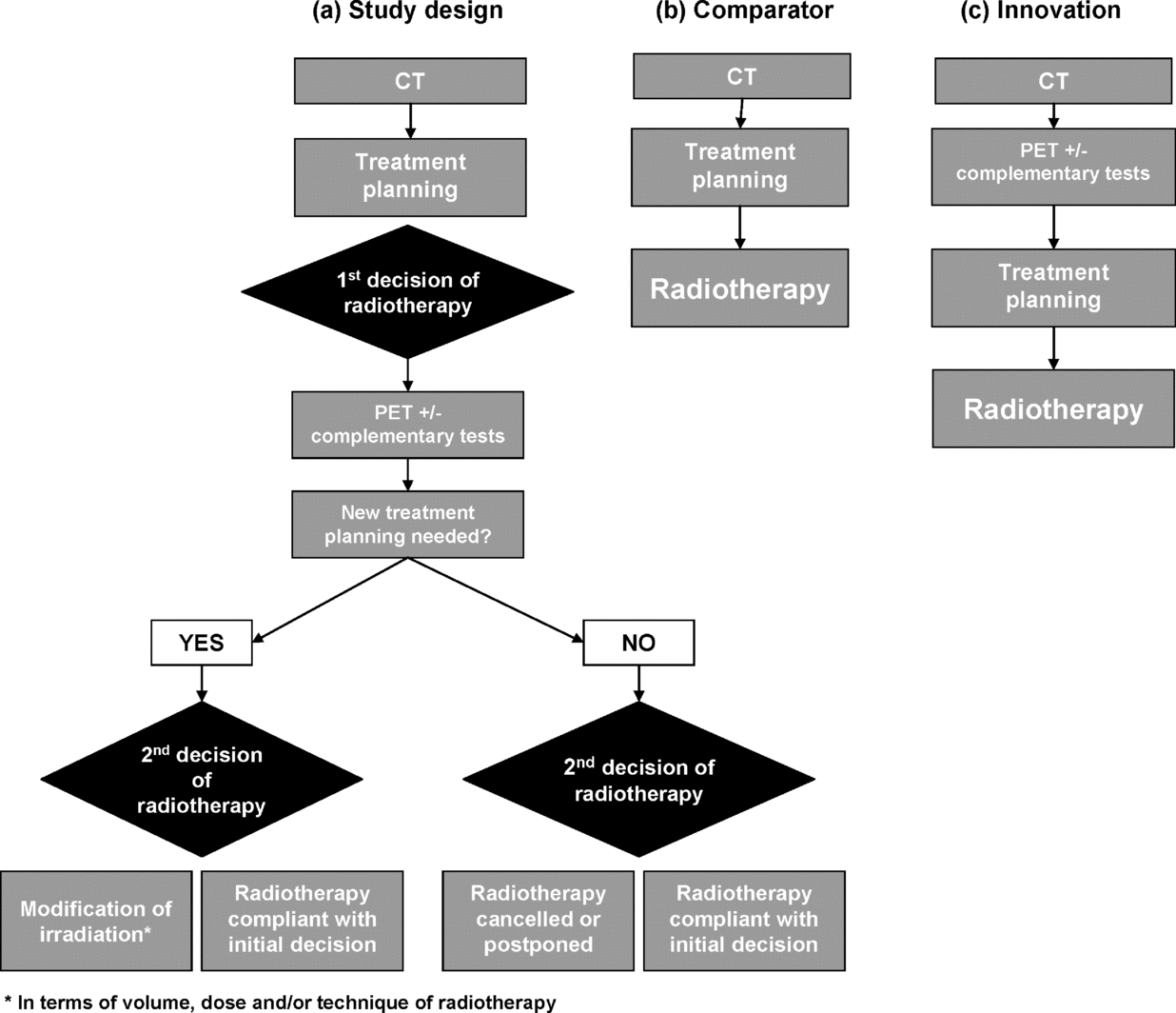
This prospective study included all patients who met the inclusion criteria and used a design where each subject was his/her own control. The protocol was as follows (Figure 1a): (i) In accordance with standard clinical practices, patients had a CT scan that was used for initial radiation treatment planning: definition of target volume, dose, and number of fractions. (ii) A PET scan was then performed. (Two types of PET were available, depending on the center: either a dedicated PET scanner [two centers] or a hybrid PET–CT system [seven centers]). Dedicated machines are progressively replaced by latest generation hybrid systems, which provide better localization of FDG binding sites by simultaneous acquisition of anatomical images by means of a CT, using the same patient positioning.) To facilitate the interpretation of results, additional tests could be prescribed.
Study protocol: standard strategy and innovative strategy. CT, computed tomography; PET, positron emission tomography.

Two alternatives were then possible: (i) new treatment planning was not necessary and a second decision regarding radiotherapy was made: either the initial decision was confirmed, or the treatment was cancelled, or it was postponed until after chemotherapy. Or, (ii) a new treatment plan was made, with either confirmation of the first decision or change of treatment: changes could involve the dose delivered (secondarily affecting the number of treatment fractions), or the area irradiated, or the type of radiotherapy (for example, conformal radiotherapy replaced by conventional radiotherapy).
Based on the information collected under the protocol, it was possible to reconstruct, for each patient, both the standard management strategy, that is, the definition of radiotherapy treatment based on CT alone without use of PET (Figure 1b), and the innovative strategy, that is, the definition of radiotherapy treatment (or its cancellation) by use of PET in complement to CT and the possible use of additional tests (Figure 1c). The mid- to long-term consequences of changes in radiotherapy treatments, either on health outcomes or on subsequent healthcare costs, were not studied.
Outcome Evaluation
In the absence of information about how radiation treatment changes impacted patients' health in the medium to long-term, the outcome criterion selected was the contribution of PET to decision making, evaluated by means of the changes observed. Because each patient served as his or her own control, two outcomes were possible: either PET had no influence on the decision, or the decision was changed, with modifications of the dose and/or the volume and/or the radiotherapy technique, or postponement or even cancellation of treatment.
Cost Evaluation
The direct costs incurred or saved by using PET for radiotherapy treatment decision making, as compared to the standard strategy (CT alone), were evaluated from a societal point of view. Only changes induced in the short-term, whether in tests (PET and additional tests) or in radiation treatment, were taken into account; treatment changes that might be introduced later were not considered.
In the context of a comparative approach, the costs common to both strategies (with or without use of PET) had to be identified, then eliminated from consideration: this applies to initial treatment planning, which is done in both cases. A second treatment planning could be necessary in the context of our protocol but, as only one planning is performed in usual practice (Figure 1), either with or without PET, the second session was not taken into account.
Thus, we studied (i) the costs of the PET scan itself, (ii) the costs of additional tests intended for the interpretation of the PET scan, and (iii) the costs incurred or saved by changes in radiation treatment dictated by using PET. We also evaluated the transportation costs incurred or saved, whether attributable to examinations or to treatment.
To assess the costs of the PET examination, we favored a microcosting approach, which is particularly well-suited to evaluating the cost of an emerging technique (Reference Valk, Pounds, Tesar, Hopkins and Haseman22;Reference Verboom, van Tinteren and Hoekstra23). Two questionnaires were developed: the “patient questionnaire,” successively completed by the departments of nuclear medicine and of radiotherapy for each patient, and the “center questionnaire” completed only once by the hospital. Both were used to collect information on physical quantities and unit costs (questionnaires are available from the authors upon request). Unit costs were expressed in euros of year 2005.
Staff hours were collected for each patient, and their cost was calculated based on average salary costs of staff with 10 years of experience. Staff included operators, who were mainly involved in the preparation and injection of FDG, and in the acquisition and reconstruction of images, and nuclear medicine doctors, mainly involved in the interpretation of images. Radiotherapists could also be involved in the interpretation of PET images and in ensuing consultations.
Equipment included the PET scanner itself, related nuclear medical equipment (computer consoles and software, flat panel, alignment lasers, and specific phantoms) and equipment dedicated to the preparation and injection of FDG (lead chambers, dose preparation systems, syringe racks, and so on). To better reflect market trends and realities, we recorded costs of acquisition rather than catalogue prices. We calculated the mean cost of performing a PET examination using the equipment described, with no distinction as to the type of device (hybrid PET–CT or dedicated PET). These costs were amortized over the lifespan of the equipment. The usual lifespan of the PET equipment is 7 years, as was the case in six of the eight centers. In the other two centers, upgrading the machine extended its lifespan to 10 years. The lifespan of associated equipment was also 10 years.
In accordance with the French Guidelines for the Economic Evaluation of Health Care Technologies (5), the interest rate used in calculating the annuity for PET and associated equipments was 3 percent. Costs of maintenance and quality controls of the PET were estimated annually.
Preparatory work before the installation of the PET scanner incurred other equipment-related expenses (modifications to bring preexisting facilities up to specifications, construction of additional space dedicated to PET and injection of FDG) with an assumed lifespan of 25 years. Again, the corresponding annuity was calculated using an interest rate of 3 percent. All annual costs or annuities were divided by the number of examinations performed annually.
FDG was purchased externally from a radiopharmaceutical supplier. The cost of FDG per patient resulted from adding the purchase price of one dose with the cost of delivery, which varied with the distance to the center. We also took into account other operating costs such as the price of image printing.
Additional tests to assist in the interpretation of PET were valued based on the reimbursement rates for the procedures performed (Common Classification of Medical Procedures); these rates are determined based on estimated production costs.
Changes in radiotherapy did not systematically induce a variation in the cost of treatment, such as when the number of fields to be irradiated varied but the dose or the technique was constant. On the other hand, cost was sensitive to (i) changes in the technique used, (ii) variations in doses, because the number of sessions was correspondingly changed (at a mean rate of two grays per session, an increase (or decrease) in the delivered dose is systematically associated with an increase (or decrease) in the number of sessions.), and (iii) of course, cancellation of treatment. The impact of treatment changes on costs was evaluated as a function of variables like the number of radiotherapy sessions or the reimbursement rates per session for each type of radiotherapy (French DRG).
Patient transportation to the hospital was generally provided by a medical vehicle. Costs were calculated on the basis of the rate per kilometer determined by the French Social Fund, multiplied by the distance between home and hospital, which reflects the true traveling cost.
Statistical Analysis
Statistical analysis was performed using SAS software version 8.2 (SAS Institute, Cary, NC). Depending on the source of the data (“center” questionnaire or “patient” questionnaire), the unit of observation was either the hospital or the patient.
We will successively present the costs (i) of performing PET scans, (ii) incurred by additional tests, and (iii) incurred or saved by changes in radiotherapy treatment. Mean costs per patient (in 2005 euros) were stratified according to the disease (Hodgkin's disease vs. NSCLC), except the costs of performing PET scan, which do not vary with the disease.
RESULTS
Two hundred nine patients from eight participating hospitals were included in the analysis. The diseases treated were distributed as follows: 112 NSCLC (53.6 percent) and 97 Hodgkin's diseases (46.4 percent).
Changes in Radiotherapy Treatment Decisions
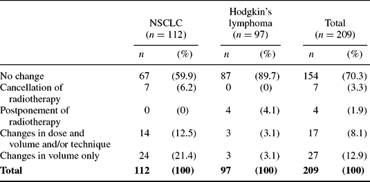
In accordance with the study protocol, taking PET into account for radiation treatment decision could require performing a second dosimetry (Figure 1). This was the case for forty-four patients with NSCLC (39.3 percent) and six patients with Hodgkin's disease (6.2 percent). Table 1 shows changes in the therapeutic schedule following the use of PET, with wide variations between diseases.
Modifications of Treatment Strategy following PET Scan According to Type of Disease

PET, positron emission tomography; NSCLC, non-small-cell lung cancer
The therapeutic strategy was modified in 40.1 percent of patients with NSCLC, with either changes of radiation treatment (33.9 percent) or, to a lesser extent, cancellation of radiotherapy (6.2 percent). Only 10.3 percent of patients with Hodgkin's disease underwent treatment changes, with either postponement of treatment after chemotherapy (4.1 percent) or changes in the treatment regimen (6.2 percent).
Changes in Costs
Cost of the PET Equipment. The average acquisition price of the six PET machines with a lifespan of 7 years was €2,037,000 (SD €449,000); it was close to €2,830,000 for the two machines with a lifespan of 10 years. The mean annual cost for maintenance and quality control was €143,645 (SD 28,120). The average cost of associated equipment was €85,106 (SD €51,835).
Renovation expenses to meet PET specifications varied widely between centers, from €178,748 up to €1,922,618 when complete refitting of the building was required, for an assumed lifespan of 25 years. There were a mean of 2,135 (SD 624) examinations per year and per center. The mean distance between hospital and home, 40.6 km, was relatively large due to the rarity of equipment.
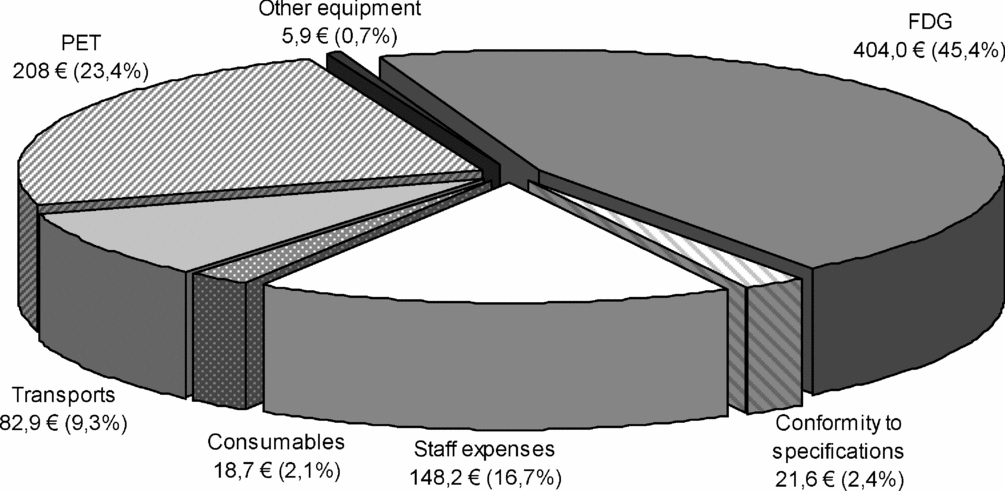
Figure 2 shows the mean cost per patient of performing a PET scan, as well as the distribution of expenses. The total cost per patient was €889.3 (€806.4 when excluding transport). FDG was the largest cost item (45.4 percent). Other expenditures were, in descending order, the PET scanner itself (23.4 percent), staff expenses (16.7 percent), patient transport (9.3 percent), renovation expenses to meet specifications (2.4 percent), consumables (2.1 percent), and other equipment (0.7 percent).
Distribution of mean costs of performing a positron emission tomography (PET) scan (in 2005 euros). FDG, 2-[18F]fluoro-2-deoxy-D-glucose.

Cost of Additional Tests Intended for Helping the Interpretation of PET. Additional tests, including ultrasonography, CT scan, colonoscopy, or cervical dissection and analysis, were performed for nine (8 percent) patients with NSCLC and four (4.1 percent) patients with Hodgkin's disease. For patients undergoing additional examinations, the mean cost of tests per patient was €659.6 for NSCLC and €580.2 for Hodgkin's disease, with, respectively, €48 and €123 for extra transport. Averaged over the total patient population, the costs of additional examinations were €53 and €23.9 for NSCLC and Hodgkin's disease, whereas related transport costs were €3.9 and €5.1, respectively.
Costs Incurred or Saved by Changes in Radiotherapy Treatment. Changes were either a reduction or an increase in dose, a change in volume, in the number of radiation fields and/or in the type of radiotherapy, or the cancellation or postponement of treatment (Table 1).
For NSCLC, performing a PET scan resulted in a mean savings of €367.9 calculated on all patients. Treatment was modified in 45 patients. There was no cost impact for twenty-four patients for whom only the volume to be irradiated was changed (Table 1). Cost variations were reported for the other twenty-one patients: cancellation of treatment (seven patients) resulted in a total savings of €37,282, dose increase (six patients) resulted in an additional cost of €4,032, and dose reduction (eight patients, including one switch from conformal radiotherapy to conventional radiotherapy) resulted in a savings of €7,950.
For Hodgkin's disease, changes in treatment following PET produced a mean increase of €10 calculated on all patients. Changes were made in ten patients, with no change in cost for seven of them: modification of the radiation volume (three patients) and postponement of treatment (four patients) (Table 1). Cost variations were reported for three patients: two received a higher dose than initially planned (including one switch from conventional radiotherapy to conformal radiotherapy) for a total additional cost of €1,398, and one received a smaller dose corresponding to a savings of €426. Transport expenses related to treatment changes resulted in a savings of €152.7 per patient with NSCLC and an extra cost of €2.9 per patient with Hodgkin's disease.
Summary of Findings
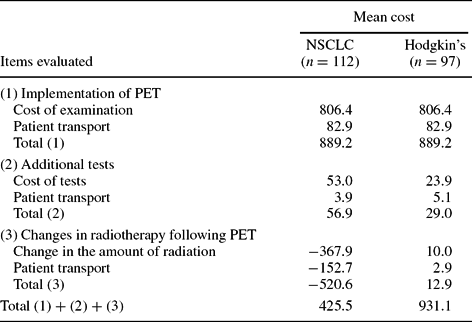
Table 2 shows all mean costs per patient, incurred or avoided by using PET, calculated on the total population of patients enrolled. With a mean cost per patient of €889 for performing PET, the extra cost incurred by additional tests was comparatively low for both diseases, because less than 10 percent of the patients were concerned. As for treatment changes, they were far more frequent in the NSCLC group than in the Hodgkin's group, 40 percent versus 10 percent (Table 1), bringing about a strong cost decrease in the NSCLC group and a minor increase in the Hodgkin's group. As a result, the consequence of using PET was an overall increase in resource consumption of €425 for NSCLC and €931 for Hodgkin's disease.
Costs Incurred or Avoided by Performing PET (Mean per Patient Enrolled) (2005 €)

PET, positron emission tomography; NSCLC, non-small-cell lung cancer.
DISCUSSION
The present study evaluated the interest of using PET as an aid in radiotherapy treatment decision making. At this early stage, it was not possible to evaluate the mid- to long-term effects of the innovation, either on health outcomes or on the resources used. However, in the absence of a full economic assessment, we believe that some type of evaluation is necessary and that it should be performed as early as possible. This is why we sought to evaluate the impact of using PET in the short-term, on both the choice of treatment and the resources used (costs incurred and costs avoided).
Although the study involved all French centers that were willing to participate, throughout the country, and enrolled all eligible patients over the 2 years of enrolment, our quest for exhaustiveness does not guarantee that results can be generalized to other sites. Highly contrasting results were obtained, demonstrating the interest of using PET for NSCLC, whereas it is far less clear for Hodgkin's disease. PET significantly affected the therapeutic decision for 40 percent of the patients with NSCLC. As compared to CT alone, changes induced by PET generated a monetary savings of over €500 per enrolled patient, bringing down the mean net cost of using PET to €425 per patient.
On the contrary, using PET led to a change in radiotherapy treatment in only 10 percent of stage I and II Hodgkin's patients, bringing up the mean total cost of using PET to €931 per enrolled patient. It cannot be ruled out that PET might be more beneficial for stages III and IV, because the disease is more progressive and therapeutic uncertainties may have more serious consequences than in stages I and II. Unfortunately, these stages were not included in the study due to the heterogeneity of practices among the participating centers.
Some of the items that make up the cost of the PET examination itself, estimated at €800 per patient, are likely to change over time, as the technology becomes more widespread. This could be the case for (i) the price of FDG, which represents half the cost of the examination, and (ii) the cost of equipment, which accounts for 25 percent.
The cost of FDG is highly dependent on conditions of production and the lifetime of the product, which is unfortunately very short. A cyclotron is required to produce fluorine-18, a radioisotope with a half-life of less than 2 hours (110 min), which makes storage impossible. In 2005, there were only three industrial producers of FDG in France, with eleven production sites that were sometimes several hundred kilometers away from the center requiring the product. This resulted in high delivery costs (transport expenses and product loss). The other radioactive markers potentially used for PET in oncology (11C-methionine and 18F-fluoromisonidazole) have an even shorter half-life and are, therefore, more costly. Consequently, the cost of FDG is unlikely to decrease in the short-term.
As for equipment costs, the medical imaging market, especially the sector of PET, is oligopolistic, with only three industrial producers represented in France. Given this low competition, a reduction in the cost of equipment is unlikely in the short-term. Increasing the number of examinations would systematically reduce the cost of equipment per patient, but the capacity to perform more examinations with the current level of material and staff resources in the centers is highly questionable.
In the longer term, however, the number of PET machines is due to increase as a result of the government's healthcare policy (18). Producers may, therefore, segment the market, as was the case for CT, by offering more affordable entry-level PET machines. The demand for FDG will probably increase accordingly, but the possible emergence of new producers and/or production sites should promote competition, with a likely price-lowering effect.
We recall that our results are limited to the short-term effects of using PET. Subsequent effects in terms of health outcomes or resources consumption were not taken into account. When radiotherapy treatment is maintained, PET makes it possible to better identify tumor tissues, and, therefore, to better define target volumes and deliver higher doses to these areas while sparing nearby healthy tissue. This should induce a reduction in relapse rates and/or toxicity, with favorable consequences on both patients' health and costs. The same goes for postponement of treatment in Hodgkin's disease: the administration of chemotherapy can avoid relapses and heavy related treatments (dose intensification, bone marrow transplantation).
CONCLUSIONS
The impact of using PET on radiotherapy treatment decisions seems to be particularly high in NSCLC, and much lower in Hodgkin's disease. In addition, the short-term impact on costs is more favorable in NSCLC. Based on these two criteria, the advantages of this new use of PET seem to be higher for NSCLC than for Hodgkin's disease.
However, if PET were to become a standard for determining the adequacy of radiotherapy treatment in NSCLC, which is a particularly common disease (12), it would not be feasible given the currently available resources and would need additional funding. Whether this use of PET should be prioritized as a standard at this early stage of innovation remains a matter of debate.
CONTACT INFORMATION
Raphaël Remonnay, MSc (remonnay@lyon.fnclcc.fr), PhD Student, GATE, GATE-CNRS UMR 5824; Project Manager, GATE-GRESACE, Centre Léon Bérard, Magali Morelle, MSc (carrere@lyon.fnclcc.fr), GATE, GATE-CNRS UMR 5824; GATE-GRESAC, Centre Léon Bérard, Pascal Pommier, MD, PhD (pommier@lyon.fnclcc.fr), Radiotherapist, Radiation Oncology Department, Regional Cancer Center Léon Bérard, 28 rue Laennec, Lyon, France 69008
Francesco Giammarile, MD, PhD (francesco.giammarile@chu-lyon.fr), Professor, Faculté de médecine Lyon sud, University of Lyon, 43, boulevard du 11 novembre 1918, Villeurbanne, France 69622; Chief, Nuclear Medicine Department, Centre Hospitalier Lyon-Sud, Pierre Bénite, 69495
Marie-Odile Carrère, PhD (carrere@lyon.fnclcc.fr), Professor, GATE, University of Lyon; Coordinator, GATE, GATE-CNRS UMR 5824, 28 rue Laennec, Lyon, France 69008