Introduction
The cell cycle is controlled by a series of protein complexes with kinase activity, composed of a cyclin protein subunit and an accompanying cyclin-dependent kinase subunit (Cdk). During the G1 phase in animal cells, the active complex is formed by any of three cyclin Ds and Cdk4 or Cdk6, which phosphorylates and inhibits the antiproliferative protein pRB (or retinoblastoma protein), thus initiating a positive loop towards establishment of the S phase (Pines, Reference Pines1995). Activity of G1 cyclin/Cdks is regulated in several ways, including the addition of inhibitory phosphorylations by specific kinases, and the association with different proteins, the role of which is to inhibit their kinase activity. There are two main classes of inhibitory proteins: the Cip/Kip family and the Ink4 family (Sherr and Roberts, Reference Sherr and Roberts1999; Rank et al., Reference Rank, Evans and Sharma2000). The former contains three members: p21Cip1, p27Kip1 and p57Kip2. The Ink4 family contains four members: p15Ink4, p16Ink4, p18Ink4 and p19Ink4. These proteins associate with either the kinase or the cyclin moieties of the protein complex and inhibit its catalytic activity (Pavletich, Reference Pavletich1999).
Plant cells have conserved most of the basic mechanisms for cell cycle control, including the presence of cyclin/Cdk complexes, although some peculiarities exist. Whereas three main types of D cyclins have been described for animal cells, Arabidopsis cells contain 10 different types of D cyclins (Renaudin et al., Reference Renaudin, Doonan, Freeman, Hashimoto, Hirt, Inzé, Jacobs, Kouchi, Rouzé, Sauter, Savouré, Sorrell, Sundaresan and Murray1996; Vandepoele et al., Reference Vandepoele, Raes, De Veylder, Rouzé, Rombauts and Inzé2002; Wang et al., Reference Wang, Zhou, Gilmer, Cleary, John, Whitwill and Fowke2003). Also, only one type of Cdk appears to associate with plant D cyclins (Cdk-A), while two different Cdks associate with animal D cyclins (Cdk4 or 6) (Pines, Reference Pines1995).
The search for cyclin/Cdk inhibitors in plants has revealed a family of proteins related to the animal p27Kip1 protein that have been named Kip-related proteins (KRPs) (De Veylder et al., Reference De Veylder, Beeckman, Beemster, Krols, Terras, Landrieu, Van der Schueren, Maes, Naudts and Inzé2001; Verkest et al., Reference Verkest, Weinl, Inzé, De Veylder and Schnittger2005; Weinl et al., Reference Weinl, Marquardt, Kuijt, Nowack, Jakoby, Hülskamp and Schnittger2005). There are seven of these in Arabidopsis (KRP1 to 7) (Jasinski et al., Reference Jasinski, Riou-Khamlichi, Roche, Perennes, Bergounioux and Glab2002; Zhou et al., Reference Zhou, Fowke and Wang2002). The similarity is based on the amino terminal of p27Kip1, which corresponds to the carboxy end of the plant KRPs (about 20 amino acids). These proteins have been classified in two groups: group A proteins interact with Cdk-A and cyclins D1, 2 and 3, while group B proteins interact with cyclin Ds, but not with Cdk-A (Jasinski et al., Reference Jasinski, Riou-Khamlichi, Roche, Perennes, Bergounioux and Glab2002; Wang et al., Reference Wang, Kong, Sun, Zhang, Zhang, Altman, de Pamphilis and Ma2004), suggesting distinct functions for each type. It is also suggested that their main action occurs during the G1 phase (Wang et al., Reference Wang, Qi, Schorr, Cutler, Crosby and Fowke1998; Coelho et al., Reference Coelho, Dante, Sabelli, Sun, Dilkes, Gordon-Kamm and Larkins2005; Nakai et al., Reference Nakai, Kato, Shinmyo and Sekine2006).
Arabidopsis KRP1 was the first inhibitor to be characterized; it interacts with, and inhibits, cyclin D/Cdk-A complexes, and its expression is induced by abscisic acid (Wang et al., Reference Wang, Qi, Schorr, Cutler, Crosby and Fowke1998; Stals and Inzé, Reference Stals and Inzé2001). Over-expression of KRP1 has important effects on plant growth and development (Wang et al., Reference Wang, Zhou, Gilmer, Cleary, John, Whitwill and Fowke2003). Tobacco and alfalfa KRPs (NtKIS1a and KRPMt, respectively) have also been characterized and equally show interaction with, and an inhibitory effect on, cyclin/Cdk complexes (Jasinski et al., Reference Jasinski, Riou-Khamlichi, Roche, Perennes, Bergounioux and Glab2002; Pettkó-Szandtner et al., Reference Pettkó-Szandtner, Mészaros, Horvath, Bakó, Csordás-Toth, Blastyák, Zhiponova, Miskolczi and Dudits2006).
Recently, two KRPs have been identified and studied in maize, Zeama;KRP1 and Zeama;KRP2, and both show the same conserved domains with p27Kip1. Both proteins inhibit the cyclin/Cdk activity associated with p13Suc1, and specifically the kinase activity associated with Zeama;CycD5;1, but not that associated with Zeama;CycB1;3, a G2/M cyclin (Coelho et al., Reference Coelho, Dante, Sabelli, Sun, Dilkes, Gordon-Kamm and Larkins2005).
Successful seed germination may require the efficient establishment of the cell cycle and, in particular, the G1 phase may play a very important role (Vázquez-Ramos and Sánchez, Reference Vázquez-Ramos and Sánchez2003). Of greatest importance for the initiation of the G1 phase is the activation of the complex formed by D-type cyclins and Cdks. Zeama;CycD2;1 has been characterized, as has its association with a Cdk-A-type kinase subunit during germination and the influence of phytohormones (Gutiérrez et al., Reference Gutiérrez, Quiroz and Vázquez-Ramos2005). Three other maize D cyclins (D5;1, D5;2 and D4;1) have been cloned, and their expression during germination reported (Quiroz-Figueroa and Vázquez-Ramos, Reference Quiroz-Figueroa and Vázquez-Ramos2006). The study of KRPs is of interest for the comprehension of the regulation of the G1 phase during maize germination. This paper describes the expression of a maize KRP gene, its characterization at the protein level during germination and its inhibitory activity upon cyclin/Cdk complexes, which suggests how the G1 phase in germinating seeds is controlled.
Materials and methods
Plant material, seed germination and protein extraction
Embryonic axes from maize caryopses (Zea mays L., cv. Chalqueño) were imbibed for 0, 3, 6, 15 and 24 h on a sheet of absorbent Whatman No. 1 filter paper wetted with imbibition buffer [50 mM Tris–HCl (pH 7.6), 50 mM KCl, 10 mM MgCl2 and 2% (w/v) sucrose] at 25 ± 2°C in darkness. When indicated, axes were incubated in the presence of benzyladenine (1 μM BA) or abscisic acid (20 μM ABA) (Sigma-Aldrich, St. Louis, Missouri, USA). Proteins from embryonic axes were extracted with extraction buffer (1 ml/10 axes), containing 25 mM Tris–HCl (pH 7.6), 15 mM MgCl2, 25 mM NaCl, 5 mM EDTA (pH 8.0), 1 mM dithiothreitol (DTT), 0.2% (v/v) Triton X-100, 250 mM sucrose, 60 mM β-glycerophosphate, 50 mM NaF, 200 μM Na3VO4, 1 mM EGTA and one tablet of protease inhibitors (Complete, Roche Mannheim, Germany) per 50 ml buffer. Protein extracts were centrifuged at c. 37,000 g for 30 min at 4°C, and protein concentration was determined by the Bradford method (Bradford, Reference Bradford1976).
RNA extraction and reverse transcriptase–polymerase chain reaction
Maize embryonic axes (100 mg fresh weight) were ground to a fine powder under liquid nitrogen and mixed with 1 ml Trizol reagent (Invitrogen, Carlsbad, California, USA). The protocol followed was that recommended by the supplier. RNA integrity was checked by electrophoresis on 1.2% (w/v) agarose gels and ethidium bromide staining.
For reverse transcriptase–polymerase chain reaction (RT-PCR) experiments, a fragment containing nucleotides 1–327 from Zeama;KRP1 was amplified as follows: 150 ng of total RNA, 0.6 μM of the forward 5′-ATGGGCAAGTACATGCGCAAGGCC-3′ oligo and 0.6 μM of the reverse 5′-CTCCCTGGTAATCCTCTCCATGGC-3′ oligo were incubated at 50°C for 35 min to obtain the cDNA; then, conditions were: 95°C 16 min, 25–35 cycles of 94°C 1 min, 60°C 1 min, 72°C 1 min and a final extension at 72°C 10 min, using the one-step RT-PCR kit from QIAGEN GmbH (D-40 724 Hilden, Germany). Similar conditions were used to amplify the actin gene: the forward primer was 5′-ATTCAGGTGATGGTGTGAGCCACAC-3′ and the reverse primer was 5′-GCCACCGATCCAGACACTGTACTTCC-3′. RT-PCR products were analysed by 0.8% (w/v) agarose gel electrophoresis. No bands were amplified with these oligos if genomic DNA was used instead of total RNA.
Antibody production
A 355 bp fragment corresponding to the 3′ end of Zeama;KRP1 was cloned into the pGEM-T Easy vector (Promega, Madison, Wisconsin, USA) and transformed into DH5α competent cells. The insert was released from the vector using EcoRI restriction enzyme (Invitrogen), and then subcloned into the pGEX-4T2 vector (GE Healthcare, Sao Paulo, Brazil) that allows fusion with the glutathione-S-transferase (GST) protein. This new plasmid was introduced into E. coli BL21 cells, and these were grown in the presence of isopropyl-β-d-1-thiogalactopyranoside (IPTG, 0.6 mM) to induce the recombinant fused protein. This recombinant protein was purified by passing bacterial protein extracts through a glutathione-Sepharose column (GE Healthcare).
Two rabbits were inoculated either with the fused GST–KRP1 protein or with the KRP1 fragment resulting from treatment of recombinant fused protein with thrombin (GE Healthcare). Recombinant proteins were coupled to keyhole limpet haemocyanin (Jemmerson, Reference Jemmerson1987) as follows: haemocyanin (2 mg) was first dissolved in 0.8 ml water and then 0.2 ml phosphate-buffered saline (PBS) [137 mM NaCl, 27 mM KCl and 4.3 mM sodium phosphate (pH 7.2)] was added. A total of 10 μl of 25% (v/v) glutaraldehyde and 90 μl PBS was added, and the protein solution was incubated with the purified recombinant protein (around 400 μg) for 12 h at room temperature with shaking. Excess glutaraldehyde was removed by filtration through Centricon-100 tubes. A rabbit was injected intramuscularly with two pooled preparations of protein (800 μg) coupled to haemocyanin, emulsified with 500 μl of complete Freund adjuvant. A second immunization was applied 8 d later using incomplete Freund adjuvant, and this protocol was repeated for a further 7 weeks. The antibodies produced (anti-KRP1 or anti-GST–KRP1) were fractionated using ammonium sulphate precipitation [45% (w/v) final concentration], and then immunopurified by incubating with recombinant KRP1 bound to Immobilon filters, eluted with 0.2 M glycine (pH 2.8) and neutralized with 1 M Tris–HCl (pH 9.0).
Western blotting
Proteins (c. 25 μg/2–3 μl protein extract) were separated by 12% sodium dodecyl sulphate–polyacrylamide gel electrophoresis (SDS–PAGE), gels were blotted on to PVDF membranes (Millipore, Billerica, Massachusetts, USA), and these were incubated with the anti-maize KRP1 polyclonal antibody (1:1000 dilution) for 12 h at 4°C, washed twice with PBS buffer (10 ml each), and once in PBS buffer containing 0.5 M NaCl (10 ml), and 15 min each at room temperature. The membranes were incubated for 2 h with peroxidase-conjugated anti-rabbit antibody (GE Healthcare) at 1:10,000 dilution. Membranes were washed again with 10 ml PBS buffer, and the peroxidase reaction was detected by the enhanced chemiluminescence method (ECL, GE Healthcare, Sao Paulo, Brazil). Densitometry analyses were performed using a BioRad Phosphorimager. All Western blots were repeated a minimum of three times using independent protein extracts.
Immunoprecipitation and pull-down assays
Proteins (200 μg/15–20 μl protein extract) were incubated with anti-Zeama;CycD2;1 antibody (1:10), anti-Zeama;PCNA antibody (1:15) or anti-Zeama;KRP1 antibody (1:10) conjugated with Protein A-agarose (30 μl, Roche) or with p13Suc1-Sepharose beads (10 μl, Millipore) overnight at 4°C. After incubation, beads were washed five times with 15 μl buffer A [25 mM Tris–HCl (pH 7.5), 125 mM NaCl, 2.5 mM EDTA (pH 8.0), 2.5 mM EGTA, 2.5 mM NaF, 0.1% (v/v) Triton X-100] and once with kinase assay buffer (15 μl, see below). Resulting protein precipitates or pulled-down proteins were used as the sources of kinase activity.
Protein kinase activity
Proliferating cell nuclear antigen (PCNA) and CycD2;1 immunocomplexes or p13Suc1-associated proteins were resuspended in 15 μl of kinase buffer {70 mM Tris–HCl (pH 7.5), 10 mM MgCl2, 150 mM NaCl, 1 mM DTT, 5 mM EGTA, 20 μM ATP and 5 μCi [γ-32P]ATP; GE Healthcare}. As substrate, 4 μg GST–ZmRBR–C fusion peptide [the C-terminal domain of maize retinoblastoma related (RBR) protein] were added per sample. Purification of the GST–ZmRBR–C fusion peptide was performed according to Ramirez-Parra et al. (Reference Ramirez-Parra, Xie, Boniotti and Gutierrez1999). Reactions were performed for 30 min at 30°C and were stopped by adding SDS loading buffer [15 μl, 80 mM Tris–HCl (pH 6.8); 5% (v/v) mercaptoethanol; 2% w/v SDS; 10% (v/v) glycerin; 0.01% (w/v) bromophenol blue]. After boiling for 5 min, the reaction products were separated by 12% SDS–PAGE.
Results
Expression of Zeama;KRP1 and protein levels during maize germination
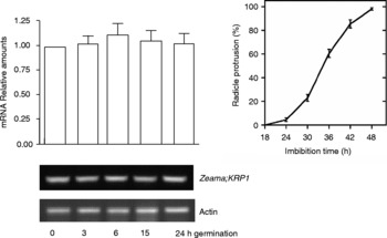
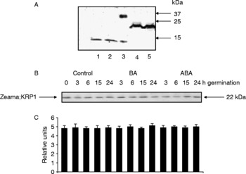
RT-PCR experiments were performed to follow the expression of the gene coding for Zeama;KRP1 during germination; as a control actin mRNA was also measured. Figure 1 shows that the amount of Zeama;KRP1 mRNA did not change during the 0–24 h period measured, which is up to the time when radicle protrusion is first evident (Fig. 1). Thus, the next step was to study the behaviour of the corresponding protein. For this, it was necessary to produce homologous antibodies. Therefore, the carboxy end (355 nt) of Zeama;KRP1 was cloned into the pGEX-4T2 expression vector which, when expressed, produces a protein fused to GST. The fused protein was overproduced, separated at the thrombin cleavage site, and then used to raise antibodies. These were prepared against both the fused protein and the 13 kDa KRP fragment. Only the antibody against the 13 kDa protein gave clear results in Western blot experiments, and this recognized a 22 kDa protein band in maize protein extracts (Fig. 2A). This protein band was not detected if the antibody was pre-treated with recombinant Zeama;KRP1, nor using the pre-immune serum (results not shown).
Expression of the Zeama;KRP1 gene during germination. Maize embryonic axes were imbibed for 0, 3, 6, 15 and 24 h and total RNA was extracted. RT-PCR was carried out, using the expression of the actin gene as a loading control. The bar graph shows a densitometry analysis relating Zeama;KRP1 mRNA band intensity to actin mRNA band intensity. The graph on the right illustrates a typical maize germination curve (completion is recorded as radicle protrusion).

Zeama;KRP1 protein accumulation during germination of maize embryonic axes. (A) Validation of the anti-Zeama;KRP1 antibody by Western blotting. Protein extracts were from 6 h germinating axes. Lanes 1 and 2, KRP1 13 kDa recombinant polypeptide; lane 3, GST–KRP1 recombinant protein; lanes 4–5, Zeama;KRP1 from 6 h-imbibed seed axes. (B) Maize embryonic axes were imbibed for 0, 3, 6, 15 and 24 h in the presence of benzyladenine (BA), abscisic acid (ABA) or water (control), and protein extracts were used to determine KRP1 protein amounts by Western blotting. (C) Densitometry analysis relating the Western blot signal to the amount of protein loaded.

The kinetics of accumulation of Zeama;KRP1 protein during germination was evaluated by Western blot experiments. This protein was present in extracts from dry embryonic axes, and its amount did not vary during the 24 h germination period (Fig. 2B, C). Neither the addition of cytokinin (BA) nor abscisic acid (ABA) modified this behaviour in the same time period (Fig. 2B, C); in contrast, Arabidopsis KRPs are induced by addition of ABA (Wang et al., Reference Wang, Qi, Schorr, Cutler, Crosby and Fowke1998; Stals and Inzé, Reference Stals and Inzé2001).
Zeama;KRP1 inhibits kinase activity in cyclin-CDK complexes
KRPs are inhibitors of the kinase activity in cyclin/Cdk complexes. Thus, to test whether Zeama;KRP1 has KRP properties, kinase activity in Zeama;CycD2;1 and proliferating cell nuclear antigen (PCNA) immunoprecipitates, or in p13Suc1-pulled-down complexes, was evaluated. First, a dose–response experiment was performed, adding 0.5, 1.5 and 2.5 μg of Zeama;KRP1 13 kDa peptide, using as a target the kinase activity present in the complexes described above. PCNA- and CycD2;1-associated kinase activities were obtained from 6 h germinating axes, and the p13Suc1-associated kinase activity was from 15 h germinating axes, because at these times kinase activity was maximal (Sánchez et al., Reference Sánchez, Torres, Boniotti, Gutierrez and Vázquez-Ramos2002; Gutiérrez et al., Reference Gutiérrez, Quiroz and Vázquez-Ramos2005).
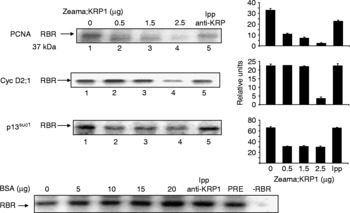
PCNA- and Zeama;CycD2;1-associated kinase activities were reduced in a dose-dependent manner, and KRP at 2.5 μg reduced activity almost completely. The p13Suc1-associated kinase activity was reduced around 50% with 0.5 μg peptide, and no further effect was observed at higher peptide amounts (Fig. 3). Exposure of kinase activity-containing immunoprecipitates to an anti-KRP immunoprecipitate did not produce an inhibitory effect (Fig. 3). Pre-immune serum, addition of 2.5 μg of GST–KRP or GST had no effect (not shown). Addition of increasing amounts of BSA (5–20 μg), instead of the KRP polypeptide, did not inhibit kinase activity (Fig. 3).
Zeama;KRP1 concentration-dependent inhibition of kinase activity from maize embryonic axes. Zeama;KRP1 polypeptide was added at 0, 0.5, 1.5 and 2.5 μg (lanes 1–4) to PCNA or Zeama;CycD2;1 immunoprecipitates, or to p13Suc1-pulled-down proteins, and protein kinase activity was measured. The carboxy terminal domain of maize RBR (retinoblastoma related) protein was used as substrate. A KRP1 immunoprecipitate was also used as a source of inhibitory activity (lane 5). Boxes at the right represent the densitometry analysis relating kinase activity to the amount of the RBR polypeptide added in kinase assays. Lowermost blot: as a control, bovine serum albumin (BSA) was added to PCNA immunoprecipitates to demonstrate the specificity of the KRP polypeptide inhibitory activity. PRE, pre-immune serum; − RBR, no RBR protein added.

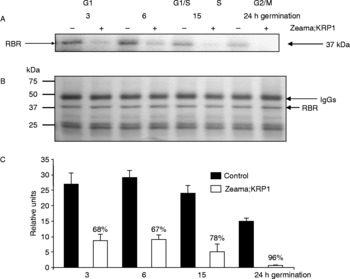
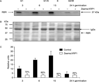
KRP1 inhibition of kinase activity associated with PCNA, Zeama;CycD2;1 or p13Suc1 was followed during maize germination, using the highest KRP amount (2.5 μg). In control samples, PCNA-associated kinase activity was highest during early germination and decreased by 24 h (Sánchez et al., Reference Sánchez, Torres, Boniotti, Gutierrez and Vázquez-Ramos2002) (Fig. 4). KRP1 strongly inhibited kinase activity during germination, from 68% at 3–6 h to 96% by 24 h (when PCNA-associated kinase activity was minimal).
Zeama;KRP1 inhibition of PCNA-associated kinase activity. Maize embryonic axes were imbibed for 0, 3, 6, 15 and 24 h, and the PCNA-associated kinase activity was obtained from the corresponding protein extracts by immunoprecipitation using the anti-PCNA antibodies. (A) Kinase activity measured in the presence or absence of Zeama;KRP1 polypeptide at different germination times; (B) stained gel showing the amount of RBR (retinoblastoma related) protein loaded; (C) densitometry analysis relating the kinase activity signal to the amount of the RBR protein loaded; numbers indicate the percentage of inhibition by the KRP polypeptide.

Zeama;CycD2;1-associated kinase activity increased between 0 and 15 h germination and then decreased (Gutiérrez et al., Reference Gutiérrez, Quiroz and Vázquez-Ramos2005) (Fig. 5). KRP1 strongly decreased kinase activity, by 70 to 92% between 0 and 15 h; by 24 h, inhibition was marginal, but kinase activity was also very low.
Zeama;KRP1 inhibition of CycD2;1-associated kinase activity from maize embryonic axes, using the anti-CycD2;1 antibodies. (A) Kinase activity measured in the presence or absence of the Zeama;KRP1 polypeptide at different germination times; (B) stained gel showing the amount of RBR (retinoblastoma related) protein loaded; (C) densitometry analysis relating the kinase activity signal to the amount of the RBR protein loaded; numbers indicate the percentage of inhibition by the KRP polypeptide.

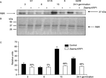
Inhibition of kinase activity in p13Suc1-pulled-down proteins was different. In control samples, kinase activity was low during early germination and increased by 15 h (Fig. 6). The inhibitory effect of KRP1 was low between 3 and 6 h germination and increased by 15 h, reaching 68% inhibition; by 24 h, inhibition was 48%.
Zeama;KRP1 inhibition of p13Suc1-associated kinase activity. Maize embryonic axes were imbibed for 0, 3, 6, 15 and 24 h, and the p13Suc1-associated kinase activity was obtained from the corresponding protein extracts using agarose beads containing bound p13Suc1. (A) Kinase activity was measured in the presence or absence of Zeama;KRP1 polypeptide at different germination times; (B) stained gel showing the amount of RBR (retinoblastoma related) protein loaded; (C) densitometry analysis relating the kinase activity signal to the amount of the RBR protein loaded; numbers indicate the percentage of inhibition by the KRP polypeptide.

Discussion
Expression of Zeama;KRP1 was followed both at the transcriptional and protein accumulation levels during maize germination. Because KRP1 is a cyclin/Cdk inhibitor, it was expected that its amount would decrease to allow cell cycle advance; thus, it was surprising to observe that there were no changes in either the mRNA or protein during the first 24 h of germination. It was also unexpected that ABA did not modify KRP protein accumulation. In Arabidopsis and Medicago cells, ABA stimulates KRP accumulation, and it would appear that the cell cycle and growth are inhibited in this way (Wang et al., Reference Wang, Qi, Schorr, Cutler, Crosby and Fowke1998; Pettkó-Szandtner et al., Reference Pettkó-Szandtner, Mészaros, Horvath, Bakó, Csordás-Toth, Blastyák, Zhiponova, Miskolczi and Dudits2006).
Demonstration of Zeama;KRP1 as a cyclin/Cdk inhibitor was obtained by measuring kinase activity in immunoprecipitates using anti-Zeama;PCNA or anti-Zeama;CycD2;1 antibodies, or activity in p13Suc1-pulled-down proteins. PCNA, the DNA polymerase δ sliding clamp, binds cyclin/Cdk complexes in animal and plant cells (Xiong et al., Reference Xiong, Zhang and Beach1992; Sánchez et al., Reference Sánchez, Torres, Boniotti, Gutierrez and Vázquez-Ramos2002), and p13Suc1 is a protein that binds strongly to Cdk1 (Cdk-A)-type kinases (Hayles et al., Reference Hayles, Aves and Nurse1986).
Maize PCNA associates with Zeama;CycD2;1 and Cdk-A type kinases, and in this association protein kinase activity can be measured. This and CycD2;1-associated kinase activity are present mainly during 0–15 h of germination, while activity is lower at later germination times (Sánchez et al., Reference Sánchez, Torres, Boniotti, Gutierrez and Vázquez-Ramos2002; Gutiérrez et al., Reference Gutiérrez, Quiroz and Vázquez-Ramos2005). The opposite occurs with the p13Suc1-associated kinase activity, which reaches a peak during late germination (by 24 h), but has low activity in the earlier hours. As the beginning of the S phase during maize germination occurs by 12–15 h of imbibition, and the first labelled mitotic figures appear by 24–28 h (previous to or concomitant with radicle protrusion) (Baíza et al., Reference Baíza, Vázquez-Ramos and Sánchez de Jiménez1989; Sánchez et al., Reference Sánchez, Gurusinghe, Bradford and Vázquez-Ramos2005), PCNA- and Zeama;CycD2;1-associated kinase activities must exert their actions in the G1 phase and at the G1–S transition, while activity in p13Suc1 complexes must correspond to a G2–M transition kinase.
Zeama;KRP1 very strongly inhibited the kinase activity associated with both PCNA and Zeama;CycD2;1 in in vitro assays, when 2.5 μg peptide were added. On the other hand, the p13Suc1-associated kinase activity was reduced to only about half at any concentration of peptide tested. This may indicate that p13Suc1 associates with more than one cyclin/Cdk complex, and only one (or some) of these complexes is sensitive to the KRP action. Association of p13Suc1 to Cdk-A and Cdk-B kinases has been demonstrated (Coelho et al., Reference Coelho, Dante, Sabelli, Sun, Dilkes, Gordon-Kamm and Larkins2005).
Zeama;KRP1 inhibited kinase activity in a differential fashion during maize germination. Kinase activity associated with either PCNA or Zeama;CycD2;1 was strongly inhibited virtually at all times, but a distinction observed was that the PCNA-associated kinase became more sensitive by 24 h, reflecting the predominance of only one type of cyclin/kinase bound to PCNA as germination advances. On the other hand, the CycD2;1-associated kinase activity importantly declined by 24 h of germination, and the remaining activity was only marginally inhibited, perhaps indicating the gradual loss of the sensitive moiety while the remaining kinase activity would be resistant to KRP1. An opposite trend was observed for kinase activity associated with p13Suc1, in which there was only a marginal effect during early germination, suggesting that at these times the associated cyclin/Cdk was not a good target for KRP1. Inhibition was observed only after 15 h germination, a time at which there must be another cyclin/Cdk-associated complex that is sensitive to KRP1. By 24 h, p13Suc1 may associate with more than one kinase complex. On the whole, it would appear that different Cdks and cyclins form different complexes with PCNA, with CycD2;1, or with p13Suc1, and they may participate in regulating different stages of the cell cycle, germination, or both.
Acknowledgements
We acknowledge receiving grants PAPIIT IN-206799 and IN-200706 and Conacyt grant 42 896-Z. N.J.J. and A.M. received grants from the Sistema Nacional de Investigadores, level III researchers.