Introduction
The longstanding dopamine (DA) hypothesis of psychosis suggests that hyperdopaminergic activity in the mesolimbic circuit is associated with cortical hypodopaminergia, which may represent a mechanism of cortical DA failing to inhibit DA release in the striatum (Deutch, Reference Deutch1992). The nucleus accumbens (NAcc) represents a key structure within the mesocorticolimbic (MCL) system in which DA neurotransmission is regulated by top-down cognitive control from frontal regions and bottom-up sensory experiences from limbic structures to maintain an updated representation of the social world (van Os et al. Reference van Os, Kenis and Rutten2010). It is suggested that especially the ventral striatum (NAcc) is involved in the pathway towards psychotic symptoms, whereas the dorsal striatum (caudate/putamen) may contribute to cognitive impairments (Fornito et al. Reference Fornito, Harrison, Goodby, Dean, Ooi, Nathan, Lennox, Jones, Suckling and Bullmore2013).
Resting-state functional magnetic resonance imaging (rs-fMRI) reflects the intrinsic organization of the brain (Fox et al. Reference Fox, Snyder, Vincent, Corbetta, Van Essen and Raichle2005) and can be used to study functional connectivity (fc) in brain networks (Smith et al. Reference Smith, Vidaurre, Beckmann, Glasser, Jenkinson, Miller, Nichols, Robinson, Salimi-Khorshidi, Woolrich, Barch, Ugurbil and Van Essen2013). A few studies have attempted to visualize the MCL circuit by means of rs-fMRI in healthy controls (Di Martino et al. Reference Di Martino, Scheres, Margulies, Kelly, Uddin, Shehzad, Biswal, Walters, Castellanos and Milham2008) and active cocaine users (Gu et al. Reference Gu, Salmeron, Ross, Geng, Zhan, Stein and Yang2010). These studies used the NAcc as a region of interest to image brain regions that are related to the MCL system [i.e. orbitofrontal cortex (OFC), prefrontal cortex (PFC), midbrain, amygdala, hippocampus, parietal and temporal cortices]. In addition, studies have shown that DA neuromodulation initiated by a pharmacological challenge can be measured by means of rs-fc. Kelly et al. (Reference Kelly, de Zubicaray, Di Martino, Copland, Reiss, Klein, Castellanos, Milham and McMahon2009) showed that administration of a DA agonist (l-DOPA) led to increased fc within frontostriatal circuits (i.e. inferior ventral striatum and ventrolateral PFC; Kelly et al. Reference Kelly, de Zubicaray, Di Martino, Copland, Reiss, Klein, Castellanos, Milham and McMahon2009). Cole et al. (Reference Cole, Beckmann, Oei, Both, van Gerven and Rombouts2013) provided evidence that both DA agonistic and antagonistic manipulations affect rs-fc in the opposite direction, i.e. fc between the basal ganglia and frontal regions (especially the motor cortex) was increased after administration of l-DOPA and decreased after haloperidol administration (Cole et al. Reference Cole, Beckmann, Oei, Both, van Gerven and Rombouts2013). The question remains whether increased endogenous synaptic DA levels in patients with schizophrenia, as shown by Abi-Dargham et al. (Reference Abi-Dargham, Rodenhiser, Printz, Zea-Ponce, Gil, Kegeles, Weiss, Cooper, Mann, Van Heertum, Gorman and Laruelle2000), are associated with altered fc during rest in comparison with controls.
It has been hypothesized that (repeated) environmental exposure, in interaction with genetic vulnerability, contributes to the development of psychosis through a final common pathway of changes in DA regulation, especially in the mesolimbic circuit (Howes & Kapur, Reference Howes and Kapur2009; van Os et al. Reference van Os, Kenis and Rutten2010; Howes & Murray, Reference Howes and Murray2014). The environmental exposures that have been associated with the risk for psychotic disorder include: cannabis use, developmental trauma and urban upbringing (van Os, Reference van Os2004; Henquet et al. Reference Henquet, Di Forti, Morrison, Kuepper and Murray2008; Varese et al. Reference Varese, Smeets, Drukker, Lieverse, Lataster, Viechtbauer, Read, van Os and Bentall2012). Developmental trauma and urban upbringing may induce (chronically) increased stress levels, although other mechanisms can be envisaged. Chronic stress may result in permanent changes in the hypothalamic–pituitary–adrenal axis, which may augment the dopaminergic abnormalities that are thought to underlie psychosis (Walker & Diforio, Reference Walker and Diforio1997; Read et al. Reference Read, Perry, Moskowitz and Connolly2001; van Winkel et al. Reference van Winkel, Stefanis and Myin-Germeys2008). Furthermore, studies have shown that experimentally induced stress (directly) influences DA release in the brain (van Winkel et al. Reference van Winkel, Stefanis and Myin-Germeys2008; Lataster et al. Reference Lataster, Collip, Ceccarini, Haas, Booij, van Os, Pruessner, Van Laere and Myin-Germeys2011; Mizrahi et al. Reference Mizrahi, Addington, Rusjan, Suridjan, Ng, Boileau, Pruessner, Remington, Houle and Wilson2012), especially mesocortical DA (Finlay & Zigmond, Reference Finlay and Zigmond1997). With regard to cannabis use, animal research and positron emission tomography studies in humans have shown that Δ9-tetrahydrocannabinol (THC) may increase mesolimbic DA through a set of mechanisms primarily mediated by CB1 cannabinoid receptor agonists (Quickfall & Crockford, Reference Quickfall and Crockford2006; Bossong et al. Reference Bossong, van Berckel, Boellaard, Zuurman, Schuit, Windhorst, van Gerven, Ramsey, Lammertsma and Kahn2009; Kuepper et al. Reference Kuepper, Ceccarini, Lataster, van Os, van Kroonenburgh, van Gerven, Marcelis, Van Laere and Henquet2013), although other studies have yielded inconclusive results (Thompson et al. Reference Thompson, Urban, Slifstein, Xu, Kegeles, Girgis, Beckerman, Harkavy-Friedman, Gil and Abi-Dargham2013; Bloomfield et al. Reference Bloomfield, Morgan, Egerton, Kapur, Curran and Howes2014).
Alterations in rs-fc in different networks are observed in patients with psychotic disorder and their relatives (Whitfield-Gabrieli & Ford, Reference Whitfield-Gabrieli and Ford2012), though MCL connectivity in relation to psychosis (vulnerability) has never been examined. In addition, differential sensitivity to the psychosis-inducing effects of environmental factors may be mediated by genetic factors (van Os et al. Reference van Os, Rutten and Poulton2008, Reference van Os, Kenis and Rutten2010). Therefore, we examined MCL connectivity during rest in relation to familial risk for psychotic disorder and environmental exposures (i.e. to cannabis, developmental trauma and urban upbringing). We hypothesized that (i) familial risk for psychotic disorder is associated with increased connectivity between striatal and midbrain regions and decreased connectivity between striatal and frontal regions, and that (ii) the effect of familial risk on MCL-fc is conditional on exposure to environmental risks.
Method
Participants
Data were collected from a longitudinal MRI study in Maastricht, the Netherlands. For recruitment and inclusion criteria of patients, siblings and healthy controls, see Habets et al. (Reference Habets, Marcelis, Gronenschild, Drukker and van Os2011). The sample comprised 73 patients with psychotic disorder, 83 siblings of patients with psychotic disorder and 72 controls. In all, 46 families participated. There were 25 families with one patient and one sibling, three families with one patient and two siblings, one family with two patients, six families with two siblings, and two families with one patient and three siblings. In the control group, there were nine families with two siblings. Furthermore, 41 independent patients, 34 independent siblings and 54 independent controls were included.
Diagnosis was based on Diagnostic and Statistical Manual of Mental Disorder-IV (DSM-IV) criteria (American Psychiatric Association, 2000), assessed with the Comprehensive Assessment of Symptoms and History (CASH) interview (Andreasen et al. Reference Andreasen, Flaum and Arndt1992). Patients were diagnosed with schizophrenia (n = 47), schizo-affective disorder (n = 9), schizophreniform disorder (n = 4), brief psychotic disorder (n = 2) and psychotic disorder not otherwise specified (n = 11). The CASH was also used to confirm the absence of a diagnosis of non-affective psychosis in the siblings and absence of a lifetime diagnosis of any psychotic disorder or current affective disorder in the healthy controls. The occurrence of any psychotic disorder in first-degree family members constituted an exclusion criterion for the controls. Schizotypy was assessed with the Structured Interview for Schizotypy-revised (SIS-r) (Vollema & Ormel, Reference Vollema and Ormel2000). In total, 10 controls and 16 siblings were diagnosed (lifetime) with major depressive disorder, but none of them presented in a current depressive state.
Before MRI acquisition, participants were screened for the following exclusion criteria: brain injury with unconsciousness of greater than 1 h, meningitis or other neurological diseases that might have affected brain structure or function, cardiac arrhythmia requiring medical treatment, and severe claustrophobia. Additionally, participants with metal corpora aliena were excluded from the study, as were women with intra-uterine device status and (suspected) pregnancy.
The standing ethics committee approved the study, and all the participants gave written informed consent in accordance with the committee's guidelines.
Behavioural measures
Psychopathology assessments were done at the time of scanning using the Positive and Negative Syndrome Scale (PANSS) (Kay et al. Reference Kay, Fiszbein and Opler1987; van der Gaag et al. Reference van der Gaag, Hoffman, Remijsen, Hijman, de Haan, van Meijel, van Harten, Valmaggia, de Hert, Cuijpers and Wiersma2006) in all three groups.
Educational level was defined as the highest accomplished level of education. Handedness was assessed using the Annett Handedness Scale (Annett, Reference Annett1970).
In the patient group, antipsychotic (AP) medication use was determined by patient reports and verified with the treating consultant psychiatrist. Best estimate lifetime (cumulative) AP use was determined by multiplying the number of days of AP use with the corresponding haloperidol equivalents and summing these scores for all periods of AP use [including the exposure period between baseline assessment for the Genetic Risk and Outcome of Psychosis (G.R.O.U.P.) study and the moment of baseline MRI scanning], using the recently published conversion formulas for AP dose equivalents described in Andreasen et al. (Reference Andreasen, Pressler, Nopoulos, Miller and Ho2010).
Substance use
Substance use was measured with the Composite International Diagnostic Interview (CIDI; World Health Organization, 1990). Cannabis use and other drug use were based on the lifetime number of instances of use. CIDI frequency data on lifetime cannabis use were available for 220 participants (4% missing data). Additionally, cannabis was tested in urine (18% missing data). The two measures were combined into one variable, which was coded as follows: never used cannabis = 0, ever used cannabis = 1 (0% missing data). Data on other drug use were available for 223 participants (2% missing data). Data on cigarette smoking and alcohol use were available for 212 (7% missing data) and 206 participants (9% missing data), respectively.
Childhood trauma
Childhood trauma was assessed with the Dutch version of the Childhood Trauma Questionnaire Short Form (CTQ). The short CTQ consists of 25 items rated on a five-point Likert scale (1 = never true to 5 = very often true) inquiring about traumatic experiences in childhood. Five types of childhood maltreatment were assessed: emotional, physical and sexual abuse and emotional and physical neglect (Bernstein et al. Reference Bernstein, Ahluvalia, Pogge and Handelsman1997). A general measure of childhood trauma was created by calculating the mean of the 25 items. The CTQ data were missing for two participants (1% missing data).
Level of developmental urbanicity
A historical population density record for each municipality was generated from 1930 onwards using the Dutch Central Bureau of Statistics (CBS) and equivalent Belgium database (Central Bureau of Statistics, 1993; Vanhaute & Vrielinck, Reference Vanhaute and Vrielinck2013). It was determined where the subject lived at birth, between the ages of 0 and 4 years; between 5 and 9 years; 10 and 14 years; 15 and 19 years; 20 and 39 years; 40 and 59 years; and 60+ years up to the actual age. For each of these records, the average population density was computed (by square kilometre, excluding water) of the municipality. Average population density was categorized in accordance with the Dutch CBS urbanicity rating (1 = ≤500/km2; 2 = 500–1000/km2; 3 = 1000–1500/km2; 4 = 1500–2500/km2; 5 = 2500+/km2). The periods 0–4 years, 5–9 years and 10–14 years were merged to average urbanicity exposure between 0 and 14 years. This variable, reflecting developmental urbanicity exposure, was collapsed a priori into five intervals (1–1.49 = 1; 1.5–2.49 = 2; 2.5–3.49 = 3; 3.5–4.49 = 4; 4.5–5 = 5) to reflect the same categories as used by the Dutch CBS (Central Bureau of Statistics, 1993). Data on developmental urbanicity were available for all participants (0% missing data).
Additionally, following the same CBS classification, five levels of current urbanization were defined (Central Bureau of Statistics, 1993). Data on current urbanicity were available for 159 participants (30% missing data).
MRI acquisition
Functional and anatomical MRI images were acquired using a 3T Siemens scanner. The functional rs data were acquired using an echo-planar imaging sequence: 200 volumes; echo time (TE) = 30 ms; repetition time (TR) = 1500 ms; voxel size = 3.5 × 3.5 × 4.0 mm3; flip angle = 90°; total acquisition time = 5 min. During the scan, participants were instructed to lie with their eyes closed, think of nothing in particular, and not fall asleep. Additionally, anatomical MRI scans had the following acquisition parameters: (1) Modified Driven Equilibrium Fourier Transform (MDEFT) sequence [176 slices; voxel size = 1 mm isotropic; TE = 2.4 ms; TR = 7.92 ms; inversion time (TI) = 910 ms; flip angle = 15°; total acquisition time = 12 min 51 s]; (2) Magnetization Prepared Rapid Acquisition Gradient-Echo (MPRAGE; Alzheimer's Disease Neuroimaging Initiative) sequence (192 slices; voxel size = 1 mm isotropic; TE = 2.6 ms; TR = 2250 ms; TI = 900 ms; flip angle = 9°; total acquisition time = 7 min 23 s). For both anatomical scans the matrix size was 256 × 256 and field of view was 256 × 256 mm2. Two sequences were used because of a scanner update during data collection. The MPRAGE and MDEFT are very similar, but to prevent any systematic bias, the total proportion of MPRAGE scans (44%) was balanced between the groups.
Data preprocessing and analysis
Image preprocessing was carried out on a Macintosh using both Analysis of Functional NeuroImages (AFNI, version 2011_12_21_1014) (Cox, Reference Cox1996) and the Oxford Centre for Functional MRI of the Brain Software Library (FSL, version 5.0.4) (Jenkinson & Smith, Reference Jenkinson and Smith2001; Jenkinson et al. Reference Jenkinson, Bannister, Brady and Smith2002). The first four volumes of each rs dataset were removed to eliminate the non-equilibrium effects of magnetization. Preprocessing steps included slice-time correction, motion correction, despiking of the functional data (removing artifactual outliers in voxelwise time series), temporal bandpass filtering (0.02–0.1 Hz), co-registration to structural scan, spatial normalization to standard space and spatial smoothing (6-mm full width at half maximum Gaussian kernel). Several sources of spurious variance (nuisance variables) were removed from the data through linear regression: six motion-correction parameters and their first temporal derivatives, and cerebrospinal fluid signal from ventricular regions of interest.
Analysis of fc
BrainVoyager QX (Goebel et al. Reference Goebel, Esposito and Formisano2006) and routines in Matlab (The Mathworks, USA) were used [NeuroElf toolbox (www.neuroelf.net) and custom routines] to estimate fc for each participant using seed-based correlation analysis. First, whole-brain signal intensity averaged across all brain voxels and white matter signal were removed from the rs data via linear regression. Next, two seed regions were defined by placing bilateral spherical regions of interest with a 3.5-mm radius in the NAcc (Montreal Neurological Institute coordinates: ±9, 9, −8) based on a previous study (Di Martino et al. Reference Di Martino, Scheres, Margulies, Kelly, Uddin, Shehzad, Biswal, Walters, Castellanos and Milham2008). For each hemisphere, Pearson's correlation coefficients were computed between the time courses of the NAcc seed and all other brain voxels and normalized by applying Fisher's r-to-z transformation. Visualization of group effects was restricted to those voxels that empirically were associated with the NAcc seed connectivity in all participants. For this purpose, a NAcc connectivity mask was created by thresholding a one-sample t test map of the NAcc connectivity across all participants using a false-discovery rate of q = 0.05 (Genovese et al. Reference Genovese, Lazar and Nichols2002). We then performed an analysis of covariance (ANCOVA) with group as the between-subject factor, controlling for the subject-level confounders sex, age, handedness, level of education, tobacco, alcohol, cannabis and other drugs, because previous rs studies in healthy controls have suggested that drug use could potentially influence fc measures (Volkow et al. Reference Volkow, Ma, Zhu, Fowler, Li, Rao, Mueller, Pradhan, Wong and Wang2008; Roberts & Garavan, Reference Roberts and Garavan2010; Tomasi et al. Reference Tomasi, Volkow, Wang, Carrillo, Maloney, Alia-Klein, Woicik, Telang and Goldstein2010; Ma et al. Reference Ma, Liu, Fu, Li, Wang, Zhang, Qian, Xu, Hu and Zhang2011; Ding & Lee, Reference Ding and Lee2013; Fischer et al. Reference Fischer, Whitfield-Gabrieli, Roth, Brunette and Green2014). Including these confounders resulted in a final sample of 195 participants with complete data (59 controls, 73 siblings, 63 patients). Significant group effects were visualized using a statistical threshold (p = 0.05, uncorrected) and a cluster-size threshold (left NAcc: 405 mm3; right NAcc: 486 mm3). The cluster-size threshold was estimated using a simulation procedure that incorporates the spatial smoothness of the statistical map (1000 Monte Carlo simulations) and corrects for multiple comparisons (Forman et al. Reference Forman, Cohen, Fitzgerald, Eddy, Mintun and Noll1995; Goebel et al. Reference Goebel, Esposito and Formisano2006). The simulated maps were thresholded at a false-positive rate (α) of 5% and surviving clusters were tabulated. Additionally, the minimum cluster size was selected by taking a false-positive rate of 5%.
Analyses of between-group differences in MCL connectivity
The mean individual fc coefficients of the voxel clusters that showed a significant group effect were transported to STATA version 12 (StataCorp., 2011) for post-hoc analyses (corrected for the above-mentioned confounders). Since analyses were performed using voxel-level ANCOVA, which assumes independency of groups, post-hoc analyses on mean individual fc coefficients of the voxel clusters were done using multiple linear regression analyses in STATA. Group was the independent variable (dummy variables: controls = 0, siblings = 1, patients = 2). Because of the non-independency of the groups (familial relationships), additional multilevel random regression analyses were carried out using the XTREG command in STATA.
Analyses of associations between environmental exposure and MCL connectivity
Main effects of the a priori hypothesized environmental exposures (cannabis, trauma and urbanicity) on fc were examined with multiple linear regression analyses using the ANCOVA selected regions with a significant between-subject (group) effect. The environmental exposures were entered as linear variables and as dummy variables [never used cannabis or ever used cannabis; childhood trauma score divided by its tertiles (low, medium, high trauma groups); five levels of population density].
Three two-way interaction analyses were modelled between group and the environmental exposures with MCL connectivity as the dependent variable. Interactions were evaluated by the Wald test (Clayton & Hills, Reference Clayton and Hills1993). Stratified effect sizes for all levels of environmental exposure for each group were calculated by combining the effects from the model containing the interactions using the STATA MARGINS routine. Analyses were adjusted for the a priori hypothesized confounders age, sex, handedness, level of education, tobacco, alcohol, cannabis (not included in the analyses with cannabis use) and other drugs. Previous research has shown that, although developmental urbanicity and current urbanicity are correlated, they have distinct effects on neural activity (Lederbogen et al. Reference Lederbogen, Kirsch, Haddad, Streit, Tost, Schuch, Wust, Pruessner, Rietschel, Deuschle and Meyer-Lindenberg2011). Therefore, in the current study, analyses with developmental urbanicity were performed with and without current urbanicity as an additional covariate.
Additionally, associations between AP medication and fc were analysed in patients, with AP medication as the independent variable and age, sex, illness duration, tobacco, alcohol, cannabis and other drugs as confounders.
All significant p values were subjected to correction for multiple testing using the Simes correction (Simes, Reference Simes1986).
Ethical Standards
All procedures contributing to this work comply with the ethical standards of the relevant national and institutional committees on human experimentation and with the Helsinki Declaration of 1975, as revised in 2008.
Results
Participant characteristics
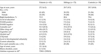
The study comprised a relatively stable patient group as reflected by the low PANSS scores (Table 1). Patients had a mean age of onset of 21.1 years and an illness duration of 6.4 years. Patients and siblings reported more lifetime cannabis and other drug use than controls, with siblings showing intermediate values between patients and controls. Childhood trauma was more frequently experienced in patients than in siblings and controls, with no differences between the latter two. The three groups did not differ in developmental level of urban upbringing. Out of 73 patients, 64 used AP medication at the time of scanning (second generation: n = 60; first generation: n = 4). The mean current dosage of AP medication in terms of standard haloperidol equivalents was 5.3 mg (s.d. = 4.8 mg). Furthermore, 12 patients used antidepressants, three used benzodiazepines, five used anticonvulsants, and one used lithium. Two siblings and two control participants used antidepressants and one control participant used benzodiazepines.
Subject demographics

Values are given as mean (standard deviation) unless otherwise indicated.
PANSS, Positive and Negative Syndrome Scale; CTQ, Childhood Trauma Questionnaire; AP, antipsychotics.
a Lifetime number of instances of cannabis use.
b Lifetime number of times of other drug use.
c Average number of daily consumptions over the last 12 months.
d Average number of weekly consumptions over the last 12 months.
e Cumulative exposure to AP medication, expressed in haloperidol equivalents.
NAcc connectivity maps
Bilateral NAcc connectivity maps revealed positive correlations with frontal regions [i.e. middle frontal gyrus (MFG), OFC, superior frontal gyrus, inferior frontal gyrus], the cingulate cortex, subcortical regions (i.e. putamen, caudate nucleus, globus pallidus, thalamus), parietal regions (i.e. precuneus), temporal regions (i.e. amygdala, hippocampus, parahippocampal gyrus) and the midbrain (see online Supplementary Fig. S1).
Between-group differences in MCL connectivity
NAcc left hemisphere
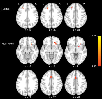
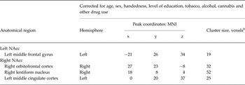
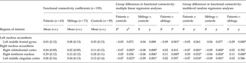
The voxel-level ANCOVA of the left NAcc yielded an effect of group in the MFG (cluster-level corrected based on 1000 Monte Carlo simulations) (Table 2, Fig. 1). Post-hoc analyses showed that NAcc connectivity with the left MFG was decreased in patients and increased in siblings compared with controls, yielding a significant difference between patients and siblings (the significant finding was upheld after Simes correction; p Simes < 0.033) (Table 3).
Areas showing significant between-group connectivity differences with the left nucleus accumbens (NAcc) (i.e. left middle frontal gyrus) and right NAcc (i.e. right orbitofrontal cortex, right lentiform nucleus and left middle cingulate cortex) seed regions during rest. Results are from voxel-level analysis of covariance corrected for age, sex, handedness, level of education, tobacco, alcohol, cannabis and other drugs. Statistical (p = 0.05, uncorrected) and cluster-size (left NAcc: 405 mm3; right NAcc: 486 mm3) thresholds were estimated using 1000 Monte Carlo simulations. Images are depicted along the z-axis. The red–yellow colour map refers to F values. L, Left; R, right.

Significant regions of the NAcc connectivity network showing a between-subject (group) effect in ANCOVA a

NAcc, Nucleus accumbens; ANCOVA, analysis of covariance; MNI, Montreal Neurological Institute.
a Results represent regions with significant group differences using a statistical threshold of p = 0.05 (uncorrected) and a cluster threshold (cluster-size threshold left NAcc: 405 mm3 and cluster-size threshold right NAcc: 486 mm3) based on 1000 Monte Carlo simulations.
b Voxel size equals 3 × 3 × 3 mm3.
Mean functional connectivity coefficients by group and post-hoc group differences in the ANCOVA selected regions with a significant between-subject (group) effect

ANCOVA, Analysis of covariance; s.d., standard deviation.
a The B's represent the regression coefficients from multiple linear regression and multilevel regression analyses in Stata corrected for age, sex, handedness, level of education, tobacco, alcohol, cannabis and other drug use.
b p Values refer to between-group differences.
* Areas that are significant after Simes correction (p Simes < 0.033).
NAcc right hemisphere
The voxel-level ANCOVA of the right NAcc connectivity maps revealed a between-subject (group) effect in three regions: the right OFC, the right lentiform nucleus (LN) (constituting the putamen and globus pallidus), and the left middle cingulate cortex (MCC) (cluster-level corrected based on 1000 Monte Carlo simulations) (Table 2, Fig. 1). Post-hoc multiple linear regression analyses showed significant patient–control differences in NAcc connectivity with the OFC, the left MCC [i.e. reduced connectivity in patients (patients<controls)] and right LN (patients>siblings). Comparing siblings with controls, there was reduced fc between the NAcc seed and the right OFC, the left MCC and the right LN. Patients differed significantly from siblings in NAcc connectivity with the right LN (patients>siblings). All significant findings were upheld after Simes correction (p Simes < 0.033), except for the NAcc–right LN connectivity in patients (Table 3).
Post-hoc multilevel random regression analyses with XTREG (taking into account familial clustering) did not change the pattern of results (Table 3).
Environmental exposure and NAcc-fc: main and interaction effects
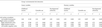
The fc between the bilateral NAcc seeds and aforementioned regions (i.e. MFG, OFC, LN, MCC) was used to evaluate main and interaction effects of environmental exposure on fc. No significant associations were derived between fc in the whole group and developmental trauma, urbanicity and cannabis use. There was a significant group × urbanicity (linear variable) interaction for the left MFG (F = 4.50, p = 0.012) and right OFC (F = 3.27, p = 0.041), and also when urbanicity was entered as a dummy variable for the left MFG (F = 2.02, p = 0.047), but not for the right OFC (F = 1.72, p = 0.097). The group × trauma interaction (dummy variable) was significant for the right LN (F = 2.82, p = 0.027) but not when entered as a linear variable (F = 2.31, p = 0.102), indicating that the effect of group NAcc–LN connectivity varied as a function of developmental trauma exposure.
These interactions did not hold after correction for multiple testing (p Simes < 0.004) (Table 4).
Associations between genetic risk level of psychotic disorder (group) and functional connectivity, corrected for additional confounders a

a The F and p values represent the results of the Wald test. No interactions were significant after Simes correction (p Simes < 0.004).
AP medication
There was a significant association between lifetime AP exposure and NAcc connectivity with the right LN (B < 0.00, p = 0.013), though not with the left MFG (B < 0.00, p = 0.277), right OFC (B < 0.00, p = 0.450), and the left MCC (B < 0.00, p = 0. 214). The significant association did not hold after Simes correction (p Simes < 0.013).
Discussion
This is the first study that examined MCL-fc and environmental correlates in individuals at different levels of psychosis risk. Both patients and siblings displayed reduced NAcc–OFC/MCC connectivity compared with controls. Siblings had reduced NAcc connectivity with the right LN compared with patients and controls, whereas patients had increased NAcc connectivity with the right LN compared with siblings. Additionally, left NAcc connectivity with the MFG was reduced in patients compared with siblings, with a trend-significant reduced fc compared with controls. The effect of familial risk on MCL connectivity was not conditional on environmental exposure.
MCL-fc and familial risk for psychotic disorder
Experimental studies examining the relationship between DA and fc, using a DA challenge, found that DA antagonistic activity between basal ganglia and frontal regions was associated with reduced fc, whereas DA agonistic activity in frontostriatal regions (i.e. ventral striatum and ventrolateral PFC) was associated with increased connectivity (Kelly et al. Reference Kelly, de Zubicaray, Di Martino, Copland, Reiss, Klein, Castellanos, Milham and McMahon2009; Cole et al. Reference Cole, Beckmann, Oei, Both, van Gerven and Rombouts2013). As hypothesized, the present study revealed an overall decreased rs-fc between the NAcc seed and OFC in patients and siblings. This could indicate that reduced fc reflects DA hypoactivity, assuming that endogenous DA activity is associated with fc. This is in line with evidence showing that reduced PFC activation (and hyperactivity of striatal DA) is associated with psychosis risk (Meyer-Lindenberg et al. Reference Meyer-Lindenberg, Miletich, Kohn, Esposito, Carson, Quarantelli, Weinberger and Berman2002). Moreover, this is in accordance with evidence from animal studies suggesting that lower DA levels in frontal areas may have a negative impact on the break function on the striatum, which in turn leads to increased levels of DA in the striatum (including NAcc) (Finlay & Zigmond, Reference Finlay and Zigmond1997). Furthermore, the shared decreased NAcc connectivity with the OFC between patients and unaffected siblings suggests that OFC dysfunction is associated with genetic risk for psychotic disorder and not associated with illness-related factors such as medication use, illness duration or illness severity. The OFC is associated with complex social and emotional decision-making processes (Wallis, Reference Wallis2007), which suggests that both siblings and patients might have problems with assigning affective values to the consequences of their decisions (Larquet et al. Reference Larquet, Coricelli, Opolczynski and Thibaut2010). In addition, evidence for an intermediate phenotype was not only apparent for NAcc–OFC connectivity, but also for NAcc–MCC connectivity. This suggests that NAcc–MCC is also associated with familial risk for psychotic disorder. The cingulate cortex is involved in attentional, executive and emotional processes (Cohen et al. Reference Cohen, Kaplan, Moser, Jenkins and Wilkinson1999). The shared reduced connectivity between the NAcc and MCC may indicate that altered goal-directed behaviour is expressed in both patients and siblings (Fornito et al. Reference Fornito, Yucel, Wood, Bechdolf, Carter, Adamson, Velakoulis, Saling, McGorry and Pantelis2009).
Group-specific findings
In addition to shared alterations with the patient group, siblings had reduced NAcc–LN connectivity compared with controls, which was not observed in the patient group. The LN is part of the basal ganglia and implicated in psychotic symptoms, such as visual and auditory hallucinations (Hibar et al. Reference Hibar, Stein, Ryles, Kohannim, Jahanshad, Medland, Hansell, McMahon, de Zubicaray, Montgomery, Martin, Wright, Saykin, Jack, Weiner, Toga and Thompson2013). Patients had trend-significant increased NAcc–LN connectivity compared with controls, which is in agreement with the assumption that increased DA in the striatum is associated with risk for psychotic disorder. It is speculated that decreased NAcc–LN connectivity in siblings reflects a compensatory mechanism by which illness expression in individuals at genetic risk for psychotic disorder may be prevented.
Lastly, patients had lower left NAcc–MFG connectivity compared with siblings, driven by an opposite direction of non-significant effects with respect to controls in both groups. The directionally opposite effects may be explained by non-shared genetic variants, whereas illness or treatment-related factors could also make an impact on brain connectivity. The trend-significant reduced NAcc–MFG connectivity in patients compared with controls may have implications for working memory performance (Barbey et al. Reference Barbey, Koenigs and Grafman2013).
Association with environmental exposure: evidence for a final common pathway?
A recent systematic review (Geoffroy et al. Reference Geoffroy, Etain and Houenou2013) has shown that so far only eight structural MRI studies have investigated gene × environmental exposure interactions (G × E) in the pathology of schizophrenia. Results of these studies reveal that the association between specific environmental risks (i.e. obstetric complications, cannabis use) and neural correlates may be conditional on a person's genetic liability to psychosis. Several rs-fMRI studies have shown that altered fc is associated with (increased) risk for psychotic disorder (Fornito et al. Reference Fornito, Harrison, Goodby, Dean, Ooi, Nathan, Lennox, Jones, Suckling and Bullmore2013). However, associations between altered fc and environmental factors, such as cannabis use, developmental urbanicity and trauma, have only been examined in healthy individuals without mental disorder (Bluhm et al. Reference Bluhm, Williamson, Osuch, Frewen, Stevens, Boksman, Neufeld, Theberge and Lanius2009; Lederbogen et al. Reference Lederbogen, Kirsch, Haddad, Streit, Tost, Schuch, Wust, Pruessner, Rietschel, Deuschle and Meyer-Lindenberg2011), with the exception of a recent rs-fMRI study of patients with schizophrenia and cannabis use disorder (Fischer et al. Reference Fischer, Whitfield-Gabrieli, Roth, Brunette and Green2014). In this pilot study reduced NAcc–PFC fc was found in patients compared with controls, with cannabis smoking and orally administered THC increasing the connectivity between these areas.
As the three environmental factors under examination in the current study have been associated with risk for schizophrenia, rs-fc would be a candidate outcome measure to assess G×E in psychotic disorder. However, the current analyses did not provide conclusive evidence for a differential impact of environmental exposures on MCL-fc in individuals with (risk for) psychotic disorder. This may suggest that functional rs brain abnormalities in psychotic disorder may not be an expression of these specific G × E interactions or, alternatively, that this particular cerebral phenotype, as an indirect measure of DA alterations, is not sensitive for the analysis of G × E. In addition, the interactions under investigation resulted in relatively small sample sizes per group (average of 20 individuals per group with unequal male/female proportions in the subgroups), which could have resulted in a loss of power to detect significant interactions.
Methodological considerations
The MCL DA system consists of several areas including the ventral tegmental area (VTA), ventral striatum (NAcc), cingulate cortex, PFC, hippocampus, amygdala, and the temporal and parietal cortices (Guillin et al. Reference Guillin, Abi-Dargham and Laruelle2007). Although the main projection area to the mesocortical and mesolimbic circuits is the VTA, this study provides evidence that the NAcc can be used to reveal this circuit as well. In fact, for the purpose of our study, the NAcc might serve as a better seed in seed correlation analyses because research has emphasized its role in the process leading to psychotic symptoms (van Os et al. Reference van Os, Kenis and Rutten2010).
Many G × E studies have relied on self-report or other measures with considerable inaccuracy (e.g. retrospective reports). Therefore, assessment of developmental trauma in patients with psychotic disorder may be biased. However, recent work suggests that patient reports of environmental exposures such as childhood trauma have good reliability and validity (Fisher et al. Reference Fisher, Craig, Fearon, Morgan, Dazzan, Lappin, Hutchinson, Doody, Jones, McGuffin, Murray, Leff and Morgan2009; Habets et al. Reference Habets, Marcelis, Gronenschild, Drukker and van Os2011).
Most of the patients in this study were receiving second-generation AP medication at the time of scanning. The effect of AP medication on intrinsic networks is still unclear, although some studies suggest that these medications tend to normalize aberrant connectivity (Stephan et al. Reference Stephan, Magnotta, White, Arndt, Flaum, O'Leary and Andreasen2001; Schlosser et al. Reference Schlosser, Gesierich, Kaufmann, Vucurevic, Hunsche, Gawehn and Stoeter2003). However, in the current study there was no main effect of AP on fc. Furthermore, unaffected siblings, who were unmedicated, showed similar patterns of connectivity as the patient group, which argues against this interpretation.
Neuroimaging studies indirectly measure molecular activity, which makes it difficult to draw direct conclusions with regard to DA activity. Translational research could help identify cellular and molecular mechanisms underlying brain activity. For example, animal studies can generate targets with regard to relevant brain circuits in human neuroimaging research (Hyde et al. Reference Hyde, Bogdan and Hariri2011; Geoffroy et al. Reference Geoffroy, Etain and Houenou2013).
Supplementary material
For supplementary material accompanying this paper visit http://dx.doi.org/10.1017/S0033291715000161
Acknowledgements
This work was sponsored by the Dutch organization for scientific research NWO (G.R.O.U.P.) and the European Community's Seventh Framework Programme under Grant Agreement no. HEALTH-F2-2009-241909 (European Network of National Schizophrenia Networks Studying Gene–Environment Interactions Consortium). The funders had no role in study design, data collection and analysis, decision to publish, or preparation of the manuscript. We wish to thank all participants and contributing staff of the participating mental health care centres. We thank Truda Driesen and Inge Crolla for their coordinating roles in the data collection, as well as the G.R.O.U.P. investigators: Rene Kahn, Don Linszen, Jim van Os, Durk Wiersma, Richard Bruggeman, Wiepke Cahn, Lieuwe de Haan, Lydia Krabbendam and Inez Myin-Germeys.
Declaration of Interest
J.v.O. is or has been an unrestricted research grant holder with, or has received financial compensation as an independent symposium speaker from, Lilly, BMS, Lundbeck, Organon, Janssen, GlaxoSmithKline, AstraZeneca, Pfizer and Servier. M.M. has received financial compensation as an independent symposium speaker from Lilly and Janssen. All other authors report no biomedical financial interests or potential conflicts of interest.