


Introduction
The strategic and tactical importance of underwater warfare and the need to protect critical undersea infrastructure has become increasingly apparent in recent years. Reference Lambert1 Naval divers play a critical role in this domain, conducting missions such as explosive ordnance disposal, infrastructure sabotage or repair, reconnaissance, and recovery operations. In parallel, the civilian sector requires a similar body of expertise, as commercial and industrial diving operations frequently take place in remote and austere underwater environments. Enabling on-site, real-time decompression stress monitoring could improve operational efficiency and enhance dive safety in both a civilian and a military context.
The validation of diving tables relies on assessing decompression stress, determined by measuring the presence of inert gas bubbles in the venous bloodstream. Reference Hugon2 Diving tables specify or recommend maximum dive times at certain depths based on prior determined decompression stress. Reference Pollock3,Reference Le, Dayton and Tillmans4 The gold standard is non-graphical Doppler ultrasound, but it requires experts and controlled conditions, making it unsuitable for waterside use. Reference Le, Dayton and Tillmans4,Reference Eftedal, Lydersen and Brubakk5 When operational military dive teams are able to perform on-site decompression stress monitoring, they will be able to assess decompression stress in real time and make real-time decisions on dive profiles, reducing reliance on external support. Reference Le, Dayton and Tillmans4,Reference Germonpré, Papadopoulou and Hemelryck6,Reference Fichtner, Brunner, Pohl, Grab, Fieback and Koch7
When using graphical analysis as opposed to just Doppler audio, two-dimensional echocardiography serves as the preferred method for the assessment of decompression stress. Reference Germonpré, Papadopoulou and Hemelryck6 The initial step in evaluating the feasibility of self-monitoring decompression stress by operational dive teams at the waterside entails assessing the ability of military combat medics to record high-quality echocardiographic recordings. A previous paper by this study group shows that combat medics of the Netherlands Armed Forces were able to acquire qualitative recordings of the inferior vena cava (IVC) and popliteal vein (PV) after a microteaching session. The primary aim of this study is to assess the feasibility of training combat medics to perform four-chamber cardiac ultrasound recordings using a concise microteaching program.
Methods
This study was approved by the institutional review board of the Alrijne Hospital (Leiderdorp, The Netherlands) under the approval number ALR/RVB/2024/UIT44438.
Participants
The group of participants consisted of a convenience sample of 22 combat medics of the Netherlands Armed Forces. A combat medic is a servicemember who is tasked with providing emergency medical care in combat. The participant group contained combat medics and North Atlantic Treaty Organization (NATO; Brussels, Belgium) Special Operations Combat Medics (NSOCMs).
Microteaching Program
Each participant was provided a microteaching session by a military anesthesiologist (RW). The course consisted of a ten-minute theoretical session including the background, basic principles, and techniques of (cardiac) ultrasonography and anatomy of the heart, followed by a five-minute demonstration on how to obtain the cardiac four-chamber view.
Materials
A Lumify (Philips Medical Systems International B.V.; Best, The Netherlands) S4-1 handheld ultrasound probe was used, connected to a Panasonic FZ-A2 tablet (Panasonic; Kadoma, Japan) or a Samsung Galaxy Tab A (Samsung; Suwon, South-Korea).
Procedure
The test station consisted of a stretcher and ultrasound probe, tablet, and ultrasound gel. The participants entered the test station in randomly assigned buddy pairs. Participants were allowed to adjust the grain and depth settings according to their own insight.
The buddy took place on the examination bench in the left lateral decubitus position. First, the performing participant was allowed two minutes of practice time with the cardiac ultrasound probe on their buddy, receiving direct feedback from the instructor (RW). The participant was then instructed to remove the probe from the patient and start the official procedure. The performer was allowed a maximum of three minutes to obtain a four-chamber cardiac view recording. Once the performer stated that they felt confident that a sufficiently clear recording was in view, a five-second video was recorded and saved.
Scoring
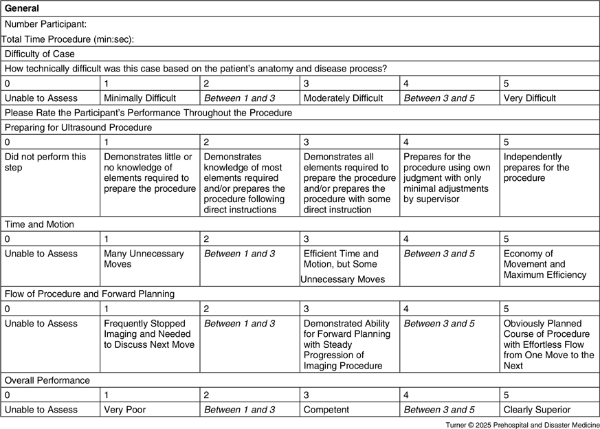
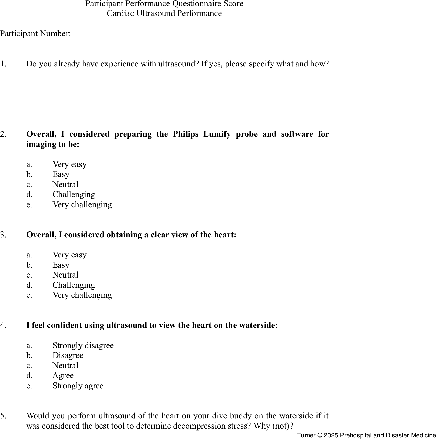
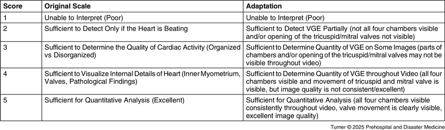
Three outcomes were measured in this study: (1) observer assessment of the performance; (2) self-perceived procedure experience; and (3) video recording quality. Performers were assessed by the instructor on preparation of the procedure, time and motion, the progression of the procedure and forward planning, ability to adapt to individual anatomical circumstances, and their overall performance (Table 1). This scoring list was adapted from the Objective Structured Assessment of Technical Skills (OSATS). Reference Niitsu, Hirabayashi and Yoshimitsu8 Participants filled out a scoring sheet regarding their self-perceived experience, visibility of the heart, and opinion on performing cardiac ultrasound on the waterside. Scoring options ranged from one (strongly disagree/very challenging) to five (strongly agree/very easy); Figure 1. Recording quality was scored blinded, in a random order, individually by an anesthesiologist (RW) and radiology resident (CO). Due to the absence of a standardized scale to assess quality of the four-chamber view, a Likert scale proposed by Gaspari, et al was adapted (Table 2), inspired by findings of Papadopoulou, et al. Reference Papadopoulou, Germonpré and Cosgrove9 Each assessor first completed a practice round by scoring each video once, followed by scoring each video twice in blinded, randomized order during the main evaluation phase.
Objective Structured Assessment of Technical Skills (OSATS) Adjusted for Ultrasound Procedure

Note: The OSATS scoring system adjusted for ultrasound procedures, as utilized in this study.
Participant Performance Questionnaire Score.

Utilized Image Quality Scale Adapted from Gaspari, et al

Abbreviation: VGE, vascular gas emboli.
Statistical Analysis
All analyses were performed using R (version 4.4.2; R Foundation for Statistical Computing; Vienna, Austria) and RStudio (version 2024.12.0+467 on macOSX). Normality testing consisted of visual inspection and a Shapiro-Wilk test, due to the small sample size. The median and interquartile ranges were reported for non-normally distributed data. A weighted kappa was calculated to determine intra- and inter-observer variability.
Results
All 21 combat medics took part in the microteaching program. Three recordings were lost due to technical difficulties with the Samsung Tablet. These recordings were excluded from the image quality assessment. All participants were included in the questionnaire and performance assessment.
Performance Assessment
All participants obtained at least a partial cardiac view. Recordings were completed within three minutes, with a median time of 61 seconds (Q1 = 35, Q3 = 100). In 18 of 21 cases, the test subject’s anatomy was rated as minimally difficult by the observing anesthesiologist. Median scores indicated adequate preparation (3.0; Q1 = 3, Q3 = 3), with time and motion rated at 2.5 (Q1 = 2, Q3 = 3), flow and procedure at 2.0 (Q1 = 2, Q3 = 3), and overall performance was close to competent at 2.5 (Q1 = 2, Q3 = 3).
Questionnaire
Nine participants had no prior ultrasound experience. Six had attended a microteaching session on vascular echography (vena puncture or diving medicine). Four had taken the NSOCM course which includes echo-FAST training. One reported ultrasound use in an international course (unspecified) and one only reported “some experience.”
Most participants (n = 13) found the Lumify device and software easy to prepare; one found it very easy and seven were neutral. When asked about obtaining a clear cardiac view, responses were mostly neutral (n = 9), followed by easy (n = 7) and challenging (n = 4). Thirteen felt confident in using echocardiography at the waterside, two lacked confidence, and six were neutral. Among the neutral group, two expressed concerns about time constraints.
Image Quality
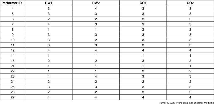
All videos were scored twice by authors RW and CO on a scale of one to five (Table 3). The weighted kappa for RW and CO was 0.904 and 0.960, indicating almost perfect agreement. The inter-observer variability was substantial (0.671). The median average scores were 2.75 (for RW) and 3.0 (for CO). In total, RW and CO respectively awarded nine and 13 of the videos with an average score of 3.0 or higher. Figure 2 depicts an overview of representative recording-stills obtained by the participants for each awarded score.
Image Quality Score per Performer

Note: RW1 = first score awarded by RW; RW2 = second score awarded by RW. CO1 = first score awarded by CO; CO2 = second score awarded by CO.
Depiction of Video-Stills Obtained in the Study per Score.
Note: All depicted videos were awarded the respective score by both assessors.
1 = Unable to interpret (poor);
2 = Sufficient to detect vascular gas emboli [VGE] partially (not all four chambers visible and/or opening of the tricuspid/mitral valves not visible);
3 = Sufficient to determine the quantity of VGE on some images (parts of chambers and/or opening of the tricuspid/mitral valves may not be visible throughout video);
4 = Sufficient to determine the quantity of VGE throughout video (all four chambers visible and movement of tricuspid and mitral valve is visible, but image quality is not consistent/excellent);
5 = Sufficient for quantitative analysis (all four chambers visible consistently throughout video, valve movement is clearly visible, excellent image quality).

Discussion
This study shows that after a microteaching session consisting of ten minutes of theory and two minutes of individual practice time, all participants were able to achieve a view of the heart, one-half of which would at least be partially suitable to determine vascular gas emboli (VGE) load. The authors’ microteaching is a feasible initial approach for introducing combat medics to cardiac echography. However, to ensure effective application of this technique in real-world scenarios, further training and hands-on practice is required.
This is the first study of microteaching for echocardiography in a military setting; prior studies have been conducted on civilian participants. Reference Booth, Reed and Brady10 This study extends the understanding of microteaching effectiveness for ultrasonographic procedures. In this study group’s previous research, the effectiveness of a microteaching program for teaching vascular ultrasonography of the IVC and PV was demonstrated. Reference Turner, van de Voort, van der Burg, Fransen, van Ooij and Hoencamp11 The current study’s hypothesis was that echocardiography would be significantly more challenging to perform for combat medics, which led to a focus on the predictive value of bubble loads in these veins to assess decompression stress in the field. This study’s findings demonstrate that combat medics can quickly learn to perform echocardiography. Given its extensive prior usage as a predictor of decompression stress, echocardiography may therefore offer a more reliable tool than IVC and PV imaging in austere settings. Future research should explore its trainability and applicability at the waterside in more real-time scenarios with external, distracting factors.
These findings are promising in the light of enabling operational dive teams to be more autonomous in determining their decompression stress during diving operations. No difference in performance was found between participants with and without prior ultrasound experience, suggesting that microteaching can serve as a valuable first step in exposing combat medics to echocardiography. Several participants expressed a desire for additional ultrasound training, and the authors agree that performance improvement is a necessity for reliable real-life application. Integrating ultrasound with telemedicine could allow remote physicians to give real-time feedback. Reference Madsen, Poropatich and Koehlmoos12 Another area of interest is adding artificial intelligence capabilities to on-site echocardiography, which could possibly provide instantaneous feedback to operators about bubble loads in the future. Reference Hamza, Pellikka, Abdulla and Ahmad13
The current risk of emerging armed conflicts and ever-present combat zones world-wide highlight the need for technical advancements in the field of austere medicine in a military context. Providing frontline servicemembers with enhanced equipment, skills, and training enables more effective and timely medical interventions, ultimately improving morbidity and mortality outcomes. This research represents an initial step in establishing the feasibility of echocardiography performed by combat medics at the waterside.
Limitations
Limitations include the use of an unvalidated adaptation of the image quality scale from Gaspari, et al. The authors felt it was essential to construct their own adaptation, since screening for the presence of VGE on a four-chamber view requires different terms and considerations than other applications of echocardiography. A score of one represented a completely inadequate recording. Recordings scored as two may still have provided usable information within ordinal scoring frameworks. Reference Møllerløkken, Blogg, Doolette, Nishi and Pollock14 Further real-world applications will allow the refinement of the image quality assessment. The internal consistency of the approach in this study is well-supported, indicated by the near-perfect intra- and inter-observer variability.
Participants practiced briefly on the same test subject on whom they performed their image acquisition, which may have over-estimated proficiency. Future studies should separate practice and test subjects to better assess the operator’s level of skill.
Furthermore, the setting utilized in this microteaching does not fully translate to the complex environment of the waterside.
Future research should investigate whether images recorded with handheld ultrasound devices are suitable for more detailed diagnostic purposes, in addition to basic screening, by determining how the amount of observed VGE correspond to an increased level of risk for decompression stress (at the waterside). This should also include examining potential differences in image quality between handheld ultrasound devices and standard ultrasound machines, as this could impact their broader applicability in clinical and field settings. Reference Slader, Young and Barker15,Reference Karimpour, Brenner and Dong16 Further studies should also aim to assess the feasibility of performing these cardiac ultrasounds in real-world waterside environments, taking into account operational constraints, environmental conditions, and the potential impact on image quality and diagnostic accuracy.
Conclusion
This study demonstrates that a brief microteaching course represents a feasible first step in training combat medics to acquire cardiac ultrasound images for the assessment of VGE. While participants were able to obtain partially diagnostic views, additional training and practice will be necessary to achieve proficiency for application in operational settings.
Author Contributions
BT, JvdV, BBB, CF, RW, and RH contributed to study design. CF, RH, and BBB contributed to data acquisition. BT, JvdV, CO, and RW contributed to further data acquisition and management. BT conducted data analysis. BT and JvdV conducted the original draft. BBB, CO, RW, and RH contributed further to data interpretation. All authors contributed to revisions of the draft. RH supervised the research. All authors approved the final version of the manuscript. All authors contributed substantially to this research.
Use of AI Technology: ChatGPT (OpenAI; San Francisco, California USA) was used to aid in writing a few sentences in a compact manner (eg, asking for synonyms and/or different ways of phrasing a specific sentence).
Conflicts of interest/funding
All authors declare that they have no conflicts of interest relevant to the content of this article. No financial support, commercial sponsorship, or competing relationships influenced the design, execution, or reporting of this study.