Introduction
The ban of antibiotic growth-promoters in the European Union has increased the interest in alternative ways to control microbes, in the gastrointestinal tract of monogastric animals (Close, Reference Close2000; Doyle, Reference Doyle2001; Lallès et al., Reference Lallès, Bosi, Smidt and Stokes2007). Organic acids (OA) have been used as the first alternative to in-feed antibiotics by many feed companies, especially in diets for piglets (Kim et al., Reference Kim, Kil, Oh and Han2005; Mroz, Reference Mroz2005). Meta-analysis confirms that OA improve performance of weaned piglets, but the significance and magnitude of the effect appears to depend on the type and level of the OA used, as well as differences in feed composition and buffering (Partanen and Mroz, Reference Partanen and Mroz1999; Partanen, Reference Partanen2001). Formic acid (FA, 90%) is one of the most commonly used OA, which have been shown to exert positive effects on animal performance and gastrointestinal health (Bolduan et al., Reference Bolduan, Jung, Schneider, Block and Klenke1988; Kirchgessner et al., Reference Kirchgessner, Gedek, Wiehler, Bott, Eidelsburger and Roth1992; Canibe et al., Reference Canibe, Højberg, Højsgaard and Jensen2005; Franco et al., Reference Franco, Fondevila, Lobera and Castrillo2005; Partanen et al., Reference Partanen, Jalava and Valaja2007). The mode of action of OA still remains unclear, even though it is commonly accepted that after weaning OA help the piglets to maintain a low pH in the stomach, thus increasing proteolytic activity and feed digestion. There are also reports on shifting of the microbiota composition in various parts of the intestine by OA, to the direction considered beneficial (Ravindran and Kornegay, Reference Ravindran and Kornegay1993; Roth and Kirchgessner, Reference Roth and Kirchgessner1998; Overland et al., Reference Overland, Granli, Kjos, Fjetland, Steien and Stokstad2000). It is worth noting that in the small intestine the mechanism of microbial modulation is hardly based on pH reduction, since pancreatic secretions neutralise the free acids. However, not only undissociated, but also neutralised acids inhibit microbial growth (Dijkhuizen and Harder, Reference Dijkhuizen and Harder1975; Pasricha et al., Reference Pasricha, Bhalla and Sharma1979). There are reports showing that OA applied in feed are not quantitatively absorbed in the proximal intestine, but are still found in ileum, and may thus account for the microbial modulation effects reported (Canibe et al., Reference Canibe, Højberg, Højsgaard and Jensen2005; Malik et al., Reference Malik, Drew, Hoehler, Rademacher and Van Kessel2006).
Diets based on cereal grains and oil-seed meals do not sufficiently supply all the essential amino acids for the requirements of growing pigs. To meet the requirements, free amino acids are routinely added in the diets. dl-methionine (DLM, 99% purity) is one of the amino acids added in feeds. However, the liquid form of methionine hydroxy analogue-free acid (MHA, containing 88% of the compound 2-hydroxy-4-methylthiobutanoic acid) can also be used as a methionine source for pigs, provided that a sufficient compensation is given for the lower methionine concentration and potential lower availability for the host (Knight et al., Reference Knight, Atwell, Wuelling, Ivey and Dibner1998; Kim et al., Reference Kim, Lindeman, Rademacher, Brennan and Cromwell2006). Being a strong acid with a pKa value of 3.53, MHA could theoretically act as a feed acidifier similar to FA, which is a slightly weaker acid with a pKa value of 3.75. Indeed, from commercial viewpoint, it appears appealing to partially replace FA, or some other OA, by MHA, which would thus serve a dual purpose in piglet feed. However, the evidence on the efficiency of MHA as a microbial modulator in the complex microbial system remains inconclusive. The capability of MHA to control Salmonella spp. in feed appears weaker than that of FA (Enthoven et al., Reference Enthoven, van den Hoven and Wiltenburg2002; Creus et al., Reference Creus, Pérez, Peralta, Baucells and Mateu2007), a finding which is likely to turn out increasingly relevant with a stricter future policy against food-borne pathogens.
In order to reduce invasive and high cost in vivo studies, and to rapidly screen for feed additives, many in vitro models have been developed to mimic digestion by host enzymes, as well as bacterial growth and metabolism in the gut (Knarreborg et al., Reference Knarreborg, Miquel, Granli and Jensen2002; Piva et al., Reference Piva, Casadei and Biagi2002; Højberg et al., Reference Højberg, Canibe, Knudsen and Jensen2003; Bauer et al., Reference Bauer, Williams, Bosch, Voigth, Mosenthin and Verstegen2004; Awati et al., Reference Awati, Williams, Bosch, Li and Verstegen2006). Most in vitro models are based on incubation of a test compound with faecal matter, which shows effects on large-intestinal microbial activities. In contrast, the model applied here is using the authentic medium and inoculum recovered from the small intestine, as this is the proposed target site of OA in the pig. Systematic in vitro dose–response studies, with single test products and product combinations, revealed the complexity of the phenomena and divergent effects of the tested acids on the growth and metabolism of the ileal bacteria. An in vivo study with weaned piglets and commercially relevant levels of FA, MHA and DLM was carried out to confirm the findings made in the laboratory simulation studies.
Material and methods
In vitro simulation studies
The conditions for in vitro intestinal simulation studies were adjusted to mimic small-intestinal conditions as accurately as possible. For the preparation of the growth medium, a section of the small intestine, ranging 16 to 20 m from proximal duodenum, of three adult pigs was cut off. The intestinal contents were collected, mixed and transferred into 500-ml centrifuge bottles placed on ice. This intestinal section was chosen as most of the simple nutrients have been absorbed proximal to that site, and the major bacterial fermentation takes place distal to the site. The digesta was centrifuged at 18 000 × g for 20 min to pellet the solids. The supernatant was used as the sole bacterial growth substrate in the subsequent ileal simulations. For the preparation of inoculum, the digesta from the most distal portion of the small intestine (4 m in length) of three adult pigs was collected, mixed and kept in completely filled glass jars on ice, to maintain the anoxic environment until used for the inoculation of the simulation vessels (in 2 h from killing the pigs). For the simulations, the acid solutions were neutralised and diluted in anaerobic Milli-Q water (Millipore Corp., Milford, MA, USA). The final concentrations of DLM, MHA and FA in the first simulation were 0.1%, 0.2%, 0.4%, 0.6% and 0.8%. In the second simulation, combinations of FA and MHA were tested, so that the total concentrations of the two compounds were 0.4%, 0.6% and 0.8%, respectively. The relative proportions of the two acids increased from 0% to 100%, in 25% increments.
The simulation was initiated by adding 12.5 ml of the intestinal substrate medium, 2.5 ml of the neutralised test-product solution (DLM, MHA or FA), 2.5 g of digesta inoculum and 6.5 ml of pH 6.5 buffer solution (0.02 M K2HPO4, 0.02 M NH4H2PO4, 0.0006 M MgSO4) in 50-ml serum bottles, under CO2 gas flow scavenged for O2, by passing through the column with hot copper shavings. Bottles were closed with thick butyl rubber stoppers, transferred to 37°C and continuously mixed in a gyratory shaker at 100 r.p.m. The simulations had four replicate vessels for each treatment, and the inoculation was done in a random order to avoid any potential systematic shifts. Incubation was continued for 12 h prior to sampling of the vessels for various analyses.
At samplings, the total gas production was measured by puncturing the rubber stopper with a needle connected to an accurate 20-ml glass syringe with a sensitive ground plunger. The volume of gas released from the vessels was recorded. After opening the simulation vessel, the pH of the fermentation medium was measured with a calibrated pH meter. This measurement was done immediately after the gas measurement to avoid pH shifts caused by the escape of CO2 from the medium. The medium was then sampled for the analysis of short chain fatty acids (SCFA) and bacterial density. The SCFA were analysed as free acids by gas chromatography, using pivalic acid as an internal standard, as described elsewhere in detail (Holben et al., Reference Holben, Williams, Gilbert, Saarinen, Särkilahti and Apajalahti2002). The acids measured were acetic, propionic, butyric, iso-butyric, 2-methyl-butyric, valeric, iso-valeric and lactic acid. For the determination of total bacterial counts, samples were fixed with formaldehyde (4% final concentration), stained with a fluorescent nucleic-acid binding dye, SYTO24 (Molecular Probes, Leiden, The Netherlands), and analysed by flow cytometry, as previously described (Apajalahti et al., Reference Apajalahti, Kettunen, Kettunen, Holben, Nurminen, Rautonen and Mutanen2002).
In vivo animal trial
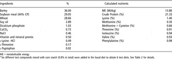
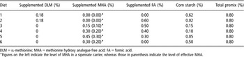
Thirty newly weaned 1-month-old piglets, from eight sows, were divided into six groups of five piglets (average initial BW of 9 kg) that were each fed one of six test diets, ad libitum, for 2 weeks. The experimental diets were based on barley, wheat and soybean meal, and no acidifier or antibiotic was supplemented. A methionine-deficient basal diet was first formulated (Table 1). The basal diet was supplemented with test compounds that were pre-mixed with corn starch (the total inclusion level was 0.8% per kg diet), to obtain six test diets, according to the experimental design described in Table 2. The analysed results confirmed that all test diets met the amino-acid requirements for the piglets used in this study. The DLM and MHA were provided by Evonik Degussa (Hanau-Wolfgang, Germany), and FA by Kemira Growhow (Helsinki, Finland), respectively.
Composition of the basal diet (%, as-fed)Footnote *

ME = metabolisable energy.
* Six different test compounds mixed with corn starch (0.8% in total) were added in the basal diet to obtain 6 test diets. See Table 2 for details.
Experimental design

DLM = dl-methionine; MHA = methionine hydroxy analogue-free acid; FA = formic acid.
*Figures on the left indicate the level of MHA in a sipernate carrier, whereas those in parenthesis indicate the level of effective MHA.
After the 2-week feeding period, all piglets were killed at random within 3 h (between 0800 and 1100 h). Ileal digesta from the most distal portion of the small intestine (4 m in length) was collected from individual piglets right after killing, and pH was immediately measured. For the total bacterial quantification, bacterial fraction was separated from the digesta solids by a protocol involving repeated washing and differential centrifugation, as described elsewhere for chicken ileal samples (Apajalahti et al., Reference Apajalahti, Särkilahti, Mäki, Heikkinen, Nurminen and Holben1998). The bacterial fraction was then counted for the total bacteria by flow cytometry, as was described above for samples from ileal simulation. The digesta subsamples were frozen for subsequent SCFA analysis, which was done as described in an earlier study (Holben et al., Reference Holben, Williams, Gilbert, Saarinen, Särkilahti and Apajalahti2002).
The killing and sampling of pigs was carried out according to the Finnish law and statute on Animal Experiments, and followed the ethical principles of the University of Helsinki.
Statistical analysis
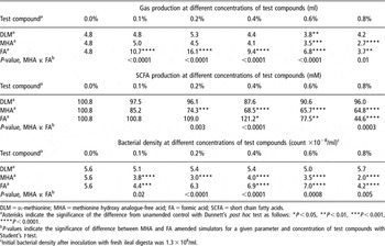
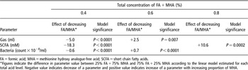
All data was assessed statistically using ANOVA, followed by post hoc tests with JMP 7.0 software (SAS Institute Inc., 2007), and multiple linear modelling combined with principal component analysis using Stata 9.2 (StataCorp), to reveal the effects of the test compounds in each data set. Dunnett’s post hoc tests were reported in cases where single compounds were tested in vitro (Table 3). For the in vitro trial, where FA was systematically replaced by MHA, linear modelling for the increasing concentration of MHA was carried out separately for each total MHA + FA level (0.4%, 0.6% and 0.8%) to illustrate that the total acid level affects the response to increasing proportion of MHA (Table 4). Dummy variables were constructed for the different levels of all three test compounds, and the explanatory power of all dummy variables was tested systematically. The block effects of different sows were also tested and taken into account, if they turned out to be significant.
Effect of DLM, MHA and FA supplementations on bacterial gas and acid production, and growth in ileal simulation

DLM = dl-methionine; MHA = methionine hydroxy analogue-free acid; FA = formic acid; SCFA = short chain fatty acids.
aAsterisks indicate the significance of the difference from unamended control with Dunnett’s post hoc test as follows: *P < 0.05, **P < 0.01, ***P < 0.001, ****P < 0.0001.
bP-values indicate the significance of difference between MHA and FA amended simulators for a given parameter and concentration of test compounds with Student’s t-test.
cInitial bacterial density after inoculation with fresh ileal digesta was 1.3 × 10⁸/ml.
Modelled effect of incremental replacement of FA by MHA on ileal fermentation parameters, within different total acid levels

FA = formic acid; MHA = methionine hydroxy analogue-free acid; SCFA = short chain fatty acids.
*Figures indicate the difference in parameter value between 25% FA + 75% MHA and 75% FA + 25% MHA according to the linear model estimated for each total acid level. Negative value indicates decrease of a parameter and positive value indicates increase of a parameter with increasing proportion of MHA.
Results
Effect of DLM, MHA and FA on ileal bacterial metabolism: in vitro studies
Table 3 shows the measured parameters and the statistical significances of the fermentation shifts. DLM supplementation had little or no effect on the bacterial fermentation parameters measured. The only statistically significant difference from the unsupplemented control was observed at 0.6% dose in bacterial gas production. MHA started to suppress bacterial gas production only at 0.6% concentration, but its suppressing effect on the total SCFA production was statistically significant already at 0.2%, and on bacterial growth already at 0.1% dose (Table 3). Effect of FA dose on bacterial fermentation was quadratic. At low doses, FA stimulated bacterial fermentation and growth, but suppressed it strongly at high doses. When the fermentation stimulation was measured as gas production, the effect was statistically significant at 0.1% to 0.6% inclusion levels of FA, whereas 0.8% level suppressed gas production significantly. When the bacterial activity was measured as the total SCFA production, the inhibitory effect of FA was significant already at 0.6% inclusion level, but the highest 0.8% dose caused almost 60% inhibition on the total SCFA production. Effect of FA on bacterial growth was highly inhibitory only at 0.8% inclusion level (Table 3). More than 97% of the SCFA was composed of lactic and acetic acid, the relative proportion of lactic acid being 79% with no acid supplementation. MHA tended to shift the proportion of lactic acid dose dependently towards 70%, but this effect was not statistically significant (data not shown).
Effect of MHA and FA combinations on ileal bacterial metabolism: in vitro studies
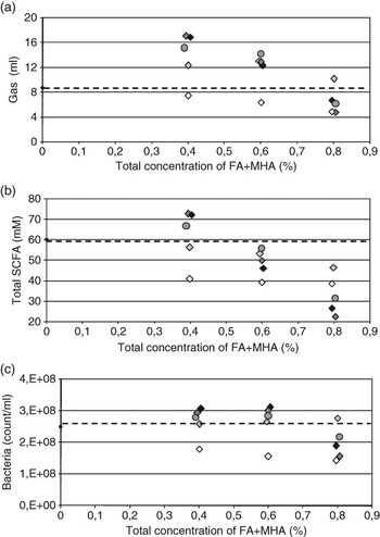
The results of the in vitro ileal simulation showed that, not only the total test compound doses (0.4%, 0.6% and 0.8%), but also the FA : MHA ratios, at the tested doses, significantly affected the microbial response. The FA and MHA inclusions at various doses showed similar responses to the independent simulation study described above. When FA was replaced by MHA in 25% increments at the total acid level of 0.4%, the stimulatory effect of FA dominated until the proportion of MHA was 100% (gas production; Figure 1a) or 75% (total SCFA and bacterial growth; Figure 1b and c). At the 0.6% total supplementation level, the inhibition profiles for gas production and bacterial growth were similar to that of 0.4% inclusion (Figure 1a and c). However, when measured as SCFA production, all combinations of FA and MHA were inhibitory, the 100% FA and 100% MHA giving relatively similar responses (Figure 1b). At the highest 0.8% total supplementation level, both FA and MHA at 100% proportion were highly inhibitory, but when partially replaced by each other, some neutralisation of the inhibitory effect occurred (Figure 1). Table 4 shows the outcomes of the linear regression models for the incremental replacement of FA by MHA at each total supplementation level. According to the model, it was apparent that at 0.4% total supplementation level, decreasing FA : MHA ratio led from FA mediated stimulation to MHA caused suppression. Accordingly, the well-fitting model was able to predict the reduction of the levels of bacterial metabolites with increased proportion of MHA (Table 4). At 0.8% total supplementation level, the effect of incremental replacement of FA by MHA was opposite for the total SCFA, according to the fitted regression model, suggesting that MHA cancelled the inhibition by the high dose of FA. The other parameters (gas production and bacterial count) did not follow any linear model at the 0.8% total supplementation level (Table 4). The SCFA were again entirely composed of lactic and acetic acid, but this time the relative proportion of lactic acid was 56% in the control vessels. The varying acid-ratios reflect the bacterial composition in the inoculum used. Similar to the simulation described above, the MHA tended to reduce the proportion of lactic acid (data not shown).
Modulation of swine ileal bacterial fermentation in vitro with acid additives containing various proportions of formic acid (FA) and methionine hydroxy analogue-free acid (MHA). Figure shows the effect of FA, MHA and various mixtures of the two, on bacterial fermentation in swine ileum. The concentrations of the test compounds were adjusted so that the summed levels of FA and MHA were 0.4%, 0.6% and 0.8%. Fermentation parameters shown are: (a) bacterial gas production, (b) total short chain fatty acids (SCFA) production and (c) the total bacterial density. The proportions of FA and MHA at each total concentration were changed stepwise from 100% FA to 100% MHA. Increasing darkness of the symbols indicates increasing proportion of FA, and the broken line indicates the level of a parameter obtained in control simulators with no amendments. Black diamond = 100% FA; dark grey diamond = 75% FA + 25% MHA; grey circle = 50% FA + 50% MHA; light grey diamond = 25% FA + 75% MHA; white diamond = 100% MHA.

Effect of MHA and FA combinations on ileal bacterial metabolism: in vivo studies
It is obvious that even the most advanced in vitro simulation system will never completely mimic the conditions prevailing in the gastrointestinal tract of live animals. Therefore, we conducted an animal trial with six experimental diets that were each fed to five piglets, for 2 weeks. Two of the diets had no FA, but either 0.18% DLM or 0.2% MHA, the reasoning being that the ingredients provided about the same amount of methionine for the animals. The other diets had FA that was incrementally replaced by MHA, from 0.6% FA + 0.18% DLM, to 0.3% FA + 0.3% MHA (Table 2).
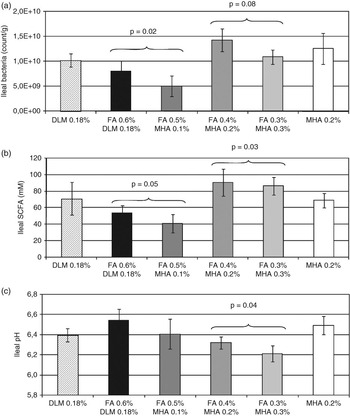
As shown in Figure 2, the diets with 0.5% and 0.6% FA inhibited ileal microbiota when measured as the total bacterial density or total SCFA produced by ileal bacteria. Furthermore, the pH in the ileal digesta of piglets, fed the diet with 0.6% FA, tended to be higher than with the other diets, most likely reflecting suppressed production of SCFA by intestinal bacteria. The diets with 0.3% and 0.4% FA stimulated the growth and acid production of ileal bacteria (Figure 2). In the ileal digesta of piglets fed diets with low FA inclusion, pH was reduced probably as a reflection of the increased level of SCFA. Due to the animal-to-animal variation between the five replicate piglets, the statistical significance of the described effects was improved when the data from animals fed 0.3% and 0.4% FA diets, and those fed 0.5% and 0.6% FA diets (Figure 2), were combined. Combining the data from animals fed MHA at different combinations, systematically, did not result in statistically valid models for any MHA combinations tested. Consequently, at the diet inclusion levels used in this trial, MHA had no effect on the microbial parameters measured. It is worth noting that MHA at the same 0.2% level had an inhibitory effect on bacterial growth and metabolism in vitro. In the ileal digesta, 98% of SCFA was composed of lactic and acetic acid. The relative proportion of lactic acid was 91%, suggesting that homofermentative lactic acid bacteria dominated the fermentation. None of the test compounds in the diet tended to shift the relative proportion of lactic acid (data not shown).
In vivo modulation of swine ileal fermentation with acid additives containing various proportions of formic acid (FA) and methionine hydroxy analogue-free acid (MHA). Figure shows the effect of FA, MHA and various mixtures of the two, on bacterial fermentation in swine ileum. dl-methionine (DLM) and MHA, with no FA amendment, were dosed as recommended when used as a source of methionine. The other diets contained FA and MHA at varying proportions so that their sum was always 0.6% of the diet, as specified in the column labels. Increasing darkness of the columns indicates increasing proportion of FA. Fermentation parameters shown are: (a) total bacterial density,, (b) total short chain fatty acids (SCFA) and (c) pH in the ileal digesta. Error bars indicate standard error of the mean between the replicate animals. Indicated P-values represent the significance of the linear model using dummy variables FA 0.5% and 0.6%, and FA 0.3% and 0.4% as independent variables.

Multivariate analysis of in vitro and in vivo results
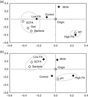
As described above, the effect of low FA level was opposite to that of the high FA level. Therefore, we created dummy variables, referred to as Low FA and High FA, which comprised the data from the treatments with ⩽0.4% FA and >0.4% FA, respectively. Figure 3 shows the results of the principal component analysis (PCA) of the data from the in vitro simulation (Figure 3a) and the piglet trial (Figure 3b).
Summary of effects of acid amendments on ileal fermentation in in vitro and in vivo studies. (a) Data from in vitro and (b) in vivo parts of the work was processed by principal component analysis. Black diamonds indicate various treatments of the studies, and white ones indicate different fermentation parameters. Parameters and treatments that cluster together correlate positively with each other, whereas those on the opposite sides of the origin correlate negatively. X- and Y-axes are principal components mathematically derived from the experimental data. Low formic acid (FA) refers to ⩽0.4% and High FA to >0.4% inclusion level of FA. The parameters used for the analysis of both in vitro and in vivo studies were pH, bacterial count, total short chain fatty acids (SCFA) and lactic and acetic acids. In addition, gas production was included in the analysis of the in vitro study.

Figure 3 shows how each measured parameter correlated with low FA, high FA or MHA inclusion. Low FA inclusion correlated positively with the concentration of SCFA and the bacterial density, and negatively with pH, in both in vitro and in vivo studies. In contrast, high FA correlated negatively with SCFA and bacterial density, but positively with pH (Figure 3). The results suggest that bacterial fermentation and especially the SCFA produced were the major determinants of digesta pH. In in vitro studies, the pH in unsupplemented vessels dropped during the simulation, from the initial value of 6.5 to 5.9. With the highest doses of neutralised FA, a drop in pH was not observed during the simulation, most likely, due to marked inhibition of bacterial fermentation. In contrast, the low doses of FA led to the most pronounced pH drop, 0.9 units, during the 12-h simulation. The effects of Low and High FA were highly consistent between the in vitro simulation study and the in vivo animal trial. In the in vitro study, MHA appeared to correlate negatively with the indicators of bacterial activity (Figure 3a); this did not appear to be the case in the in vivo study (Figure 3b).
Discussion
In this paper, we have shown that both MHA and FA were efficient modulators of bacterial growth and metabolism in the simulation system that has been developed to mimic the conditions prevailing in the swine ileum. In the in vitro simulation system, the added FA shifted bacterial growth and metabolism in the same way as was measured in live animals. However, MHA effects that were detected in vitro could not be confirmed in vivo at the commercially relevant doses.
The effect of FA on microbial growth and metabolism was consistently quadratic in all in vitro and in vivo studies, whichever bacterial parameter was used as an indicator. Indeed, its effect on microbiota was highly stimulatory at low concentrations, but inhibitory at high concentrations. In the different studies presented in this paper, we observed that while the shape of the dose–response curve was roughly the same, the FA concentration at the turning point varied, being different for different bacterial metabolites. For instance, the turning point for bacterial gas production was different from that of SCFA production, suggesting that FA had effects on the regulation and activity of different metabolic pathways. Most likely, the point in the dose–response curve where the bacterial stimulation turns to inhibition, is also affected by the density and composition of the resident microbiota. This quadratic dose–response curve makes the use of FA as a feed additive challenging. This is because the practice of dosing FA at lower than recommended levels leads to stimulatory, rather than inhibitory, effects on intestinal bacteria. It is beyond the scope of this paper to evaluate in detail which microbiota-modulating mode of FA is beneficial for the performance and health, the stimulation of lactic acid fermentation, or general inhibition of bacterial activities in the small intestine of swine. Whichever mode is better for performance, the existence of two opposite outcomes of FA inclusion in feed explains why the results of animal trials testing the effects of FA on animal performance, have been inconclusive (Partanen and Mroz, Reference Partanen and Mroz1999; Close, Reference Close2000; Doyle, Reference Doyle2001; Mroz, Reference Mroz2005). Differences in the diet composition, buffering capacity and farming practices make it difficult to compare results from different studies. However, in general, significant effects on performance and intestinal microbiota were observed in trials with higher than 0.5% inclusion level of FA (Bolduan et al., Reference Bolduan, Jung, Schneider, Block and Klenke1988; Partanen and Mroz, Reference Partanen and Mroz1999; Canibe et al., Reference Canibe, Højberg, Højsgaard and Jensen2005; Franco et al., Reference Franco, Fondevila, Lobera and Castrillo2005), whereas studies using less than 0.5% FA did not report significant effects (Gabert et al., Reference Gabert, Sauer, Schmitz, Ahrens and Mosenthin1995; Knarreborg et al., Reference Knarreborg, Miquel, Granli and Jensen2002). This, together with the observations made in the present investigation, strongly suggest that it is the inhibitory, rather than stimulatory, effect of FA that improves the animal performance.
The MHA proved to be antibacterial according to our in vitro studies. It did not show any stimulatory effect at low concentrations as FA did, but appeared to have a linearly dose-dependent inhibitory effect. Already, at 0.1% concentration, it suppressed bacterial growth and production of SCFA. The MHA is a stronger acid than FA, having a pKa value of 3.53, whereas that of FA is 3.75. This means that at pH 6.5, both acids would be in highly dissociated form, MHA even more than FA. Thus, the passive diffusion across the bacterial cell membrane should be more efficient with FA than with MHA. In their energy metabolism, bacteria maintain a proton gradient across the membrane, the pH always being higher inside the cell than in the surrounding medium. Acids that are lipophilic in their protonated form transport protons across the membrane, thus, uncoupling the proton gradient, which prevents ATP synthesis and inhibits the growth of bacteria (McLaughlin and Dilger, Reference McLaughlin and Dilger1980). It is possible that MHA transport into bacterial cells is facilitated owing to its molecular characteristics other than acid strength, or its ability to use an ATP driven or facilitated transport system of lactic acid, methionine etc., with relaxed specificity.
It has been suggested that MHA could be used as an in-feed microbial modulator due to its antibacterial properties, thus, partially eliminating the need of using FA or other feed acidifiers. Based on the results of the current study, the partial replacement of FA by MHA may be challenging due to the non-linear, quadratic dose–response curve of FA. If, at commercially relevant inclusion level, part of FA is replaced by MHA, there is a fair chance that the initially inhibitory effect of FA turns to stimulation of intestinal microbiota, as was obvious in the piglet study reported in this paper. The findings from our in vitro studies, in turn, suggested that the inhibitory effect of MHA was either reduced or cancelled totally when even a small part of MHA was replaced by FA. The results of this study, therefore, suggest that if the target is to ensure the inhibitory effect of FA, with even modest safety margin, FA level in feed should be kept above 0.6%, and not reduced, if MHA is used as a methionine source instead of DLM.
The MHA inhibited bacterial growth and metabolism effectively under in vitro conditions, but this effect was not detected in vivo. Furthermore, in in vitro studies, MHA tended to shift bacterial metabolism towards heterolactic fermentation (increased proportion of acetic acid), but in the animal trial no such trend was seen. These observations suggest that MHA did not reach the ileum of live pigs in the original form or at the concentration used in the simulation studies. Richards et al. (Reference Richards, Atwell, Vázquez-Añon and Dibner2005) showed that MHA, in its native form, disappeared from the intestine of broiler chickens even more rapidly than DLM. In studies where DLM and MHA in the intestine of broiler chickens were traced both as intact substances and as tritium label, showed that a significant proportion of the original MHA had been converted to unknown tritiated compounds; only 10% of the retained label was found as intact MHA (Maenz and Engele-Schaan, Reference Maenz and Engele-Schaan1996). Malik et al. (Reference Malik, Drew, Hoehler, Rademacher and Van Kessel2006) showed that liquid MHA was, in fact, more slowly absorbed throughout the digestive tract of pigs than l-methionine, and thus more exposed to the degradation by intestinal microbes. It thus appears, that even though MHA is highly antibacterial, it fails to function as a microbial modulator in vivo due to its rapid disappearance and bacterial modification in the upper intestine. In order to gain from its potential to serve as a microbial modulator, a slow release formulation of MHA should be developed. This, however, would most likely reduce its value as a methionine source for the host. In studies with FA, a significant level of FA added in feed was found intact in the distal small intestine (Canibe et al., Reference Canibe, Højberg, Højsgaard and Jensen2005). This explains why FA acted similarly both in in vitro and in vivo studies.
Banning of in-feed antibiotics has led to impaired animal health and increased usage of therapeutic antibiotics, particularly in farms with challenging conditions, suboptimal management and poor hygiene. Under such conditions, microbial management products such as OA might aid in maintaining health and productivity. However, as we showed in this paper, different OA with varied doses may have major differences in their efficiency and mode of action, and should, therefore, not be randomly chosen and dosed. Rational screening and testing of the product candidates by relevant screening methods should be used for product selection and dose–response evaluation. As exemplified in the present study, even the best product may express unpredictable effects when wrongly dosed.
Acknowledgments
The authors thank Seppo Peuranen for his valuable help in organising the animal trial, Osmo Siikanen for skilfully conducting the in vitro simulations, Susanna Alaja for her contribution to statistical analysis and Marko Lauraeus for chemical analysis and fruitful discussions on data interpretation.