Case report
A 31-year-old woman was referred at 21 weeks of gestation for foetal cardiology assessment with an abnormal four-chamber view on a routine anomaly scan. Past history included one normal pregnancy resulting in a live birth and no family history of congenital cardiac disease. She had taken Citalopram for the treatment of depression throughout pregnancy. Early pregnancy serum screening was rated as low risk and nuchal translucency screening was not performed. The detailed anomaly scan at 20 weeks of gestation had confirmed no extracardiac abnormalities.
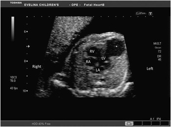
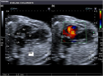
Foetal echocardiogram showed a non-hydropic foetus with normal arrangement of abdominal vessels. The stomach and apex were on the left. There was a large, poorly contractile, and thin-walled aneurysmal sac arising from the left ventricular apex (Fig 1). In addition, there was a large muscular inlet ventricular septal defect shunting left to right (Fig 2). Assessment of the outflow tracts showed great artery disproportion with a large bifurcating pulmonary artery (4.9 millimetre, z score plus 2.35Reference Schneider, McCrindle, Carvalho, Hornberger, McCarthy and Daubeney1) and a smaller aortic valve (2.6 millimetre, z score minus 1.35Reference Schneider, McCrindle, Carvalho, Hornberger, McCarthy and Daubeney1) and ascending aorta. There was no continuity of the aortic arch but rather a straight superior course from the proximal aorta to the right-sided head and neck vessels consistent with interruption of the aortic arch. There was no evidence of arrhythmia.
Four-chamber view of the foetal heart showing aneurysm of the left ventricle. LA=left atrium; RA=right atrium; RV=right ventricle; LV=left ventricle, *aneurysm of the left ventricle.

(a) Two dimensional four-chamber view of the foetal heart showing the VSD. (b) Colour flow Doppler across VSD. LA=left atrium; RA=right atrium; RV=right ventricle; LV=left ventricle; VSD=ventricular septal defect.

Owing to the aortic arch interruption and inlet ventricular septal defect, amniocentesis was performed and returned a normal female karyotype and a negative fluorescent in situ hybridisation screen for the 22q11 deletion. Our counselling focused on an uncertain and guarded prognosis for a newborn needing neonatal cardiac surgery for the aortic arch interruption on a background of a poorly contractile left ventricle and an extensive aneurysm.
This couple elected to continue the pregnancy, but unfortunately spontaneous intra-uterine death occurred in the foetus at 26 weeks of gestation.
Post-mortem examination of the foetus confirmed the antenatal findings of left ventricular aneurysm and interruption of the aortic arch with ventricular septal defect. Histologically, there were areas of patchy fibrosis in the left ventricular myocardium. In addition, there was evidence of micrognathia and a high arched palate.
Discussion
Left ventricular aneurysm or diverticulum is a rare finding in the foetus. Despite the terms being often used interchangeably, it has been suggested that they should be distinguished on the basis of the morphology of the communication with the ventricle. An aneurysm has a wide communication with the ventricle, whereas the diverticulum communicates with the ventricle through a narrow sleeve.Reference Papagiannis, Van Praagh and Schwint2 Their presence has been described in association with pentalogy of Cantrell, in which there are abdominal and thoracic wall defects, through which the ventricular myocardium can herniate to form an aneurysm.Reference Grethel, Hornberger and Farmer3 However, left ventricular aneurysms can occur in isolation. Interruption of the aorta has a reported incidence of less than 1.3% of all congenital cardiac disease,Reference Fyler4 although it is likely to be even lower due to the increased spectrum of congenital cardiac disease recognised today. It is usually seen in association with a posteriorly malaligned ventricular septal defect and may occur in combination with other congenital cardiac defects, in particular the common arterial trunk and aorto-pulmonary window.
Left ventricular aneurysms are thought to be a developmental anomaly, with idiopathic dysplasia of the left ventricular endocardium and myocardium.Reference Cavalle-Garrido, Cloutier, Harder, Boutin and Smallhorn5 The patient reported here had received the selective serotonin reuptake inhibitor antidepressant citalopram throughout her pregnancy. Despite the possibility of finding anecdotal reports of a link between the use of such anti-depressants in pregnancy and congenital cardiac defects in the literature, this has not been confirmed in larger studies.Reference Wichman, Moore, Lang, St Sauver, Heise and Watson6
The natural history of ventricular aneurysm or diverticulum diagnosed in foetal life is not well established due to the rarity of cases, although smaller lesions appear to have a good outlook.Reference Cavalle-Garrido, Cloutier, Harder, Boutin and Smallhorn5 Some foetuses may develop impairment of left ventricular function, pericardial effusions, and foetal arrhythmias. The presence of impaired left ventricular function seen on foetal echocardiography may have contributed to intra-uterine death in this case.
Left ventricular aneurysm in the foetus is a rare abnormality. To our knowledge, this is the first report of a left ventricular aneurysm in association with interruption of the aortic arch.