Introduction
The extinct superfamily Elcanoidea is an unusual group of orthopteran insects that range from the early Permian to Late Cretaceous (Tillyard, Reference Tillyard1937; Sharov, Reference Sharov1968; Gorochov, Reference Gorochov1995; Grimaldi and Engel, Reference Grimaldi and Engel2005). Due to the lack of fossils with well-preserved body structures, its systematic position remains controversial (Fang et al., Reference Fang, Zhang, Wang, Wang, Zheng, Zhang, Li and Liu2015). The long antennae and nymphal characters of elcanoids are similar to those of the Ensifera (Grimaldi and Engel, Reference Grimaldi and Engel2005; Peñalver and Grimaldi, Reference Peñalver and Grimaldi2010), but their wing venation and other adult characters are more consistent with the Caelifera (Béthoux and Nel, Reference Béthoux and Nel2002). The superfamily consists of two families (Gorochov, Reference Gorochov1995): Permelcanidae and Elcanidae. Permelcanids differ from elcanids in having CuA+CuPa1 secondary branches (e.g., two branches in Promarlynovia venicosta Tillyard, Reference Tillyard1937 and three branches in Meselcana madygenica Sharov, Reference Sharov1968), and RP ending in MA rather than in MA1. Elcanids have so far been reported only from Triassic–Cretaceous deposits in Eurasia and Brazil (Sharov, Reference Sharov1968; Gorochov et al., Reference Gorochov, Jarzembowski and Coram2006; Peñalver and Grimaldi, Reference Peñalver and Grimaldi2010), whereas permelcanids occur in Permian–Triassic strata of Europe, Central Asia, and North America (Gorochov, Reference Gorochov1995).
Fossil Elcanoidea in North America are generally rare. The earliest record is represented by the permelcanid Promartynovia venicosta Tillyard, Reference Tillyard1937, which was reported from the Permian Wellington Formation of Kansas, but no species of the Elcanidae have been formally described from North America. Nonetheless, two unnamed elcanoid specimens from the Cow Branch Formation of the Solite Quarry in Virginia, USA have been illustrated (Liutkus et al., Reference Liutkus, Beard, Fraser and Ragland2010, fig. 3D; Muscente and Xiao, Reference Muscente and Xiao2015, fig. 6). Here, we describe a new genus and species based on the well-preserved forewing illustrated in Muscente and Xiao (Reference Muscente and Xiao2015). This fossil is the earliest definitive record of the family Elcanidae, and the first formally described orthopteran from the Triassic of North America.
Geological setting
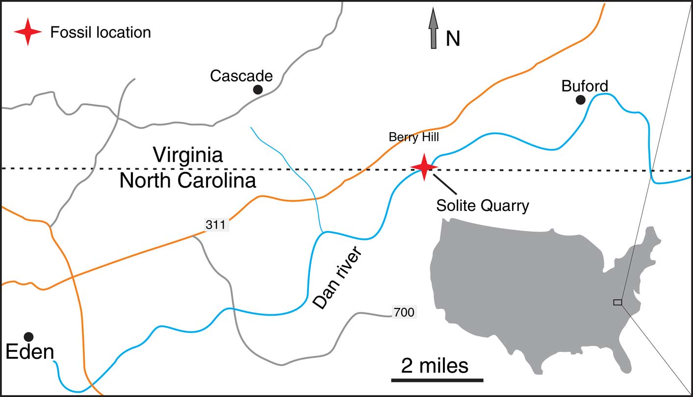
The specimen described in this study was collected by Dr. Nicholas Fraser from the Cow Branch Formation in the Solite Quarry, Dan River-Danville Basin, near the North Carolina-Virginia boundary, United States (Fig. 1). The Dan River-Danville Basin is part the Mesozoic rift system in eastern North America that formed during the breakup of the Pangaea (Olsen et al., Reference Olsen, Froelich, Daniels, Smoot and Gore1991). The basin fill consists of thick sediments of the Dan River Group that unconformably overlie Proterozoic–Paleozoic basement (Liutkus et al., Reference Liutkus, Beard, Fraser and Ragland2010; Liutkus-Pierce et al., Reference Liutkus-Pierce, Fraser and Heckert2014).
Map showing the fossil locality. See Liutkus et al. (Reference Liutkus, Beard, Fraser and Ragland2010) for stratigraphic column of the Cow Branch Formation and the stratigraphic horizon of the insect layer.

The Cow Branch Formation was previously considered Carnian in age (Olsen et al., Reference Olsen, Froelich, Daniels, Smoot and Gore1991), but is now regarded as Norian (~227–208 Ma; Liutkus et al., Reference Liutkus, Beard, Fraser and Ragland2010), although uncertainty remains (Fraser et al., Reference Fraser, Grimaldi, Axsmith, Heckert, Liutkus-Pierce, Smith and Dooley2017). At the Solite Quarry, which is located in the middle of the Dan River-Danville Basin, the upper part of the Cow Branch Formation is exposed. The exposed section of the Cow Branch Formation mainly consists of lacustrine shales, mudstones, and sandstones. The depositional environment was interpreted as a deep and chemically stratified lake with an oxygenated surface layer but anoxic deep waters (Olsen et al., Reference Olsen, Froelich, Daniels, Smoot and Gore1991; Fraser et al., Reference Fraser, Grimaldi, Olsen and Axsmith1996). However, recent studies by Liutkus et al. (Reference Liutkus, Beard, Fraser and Ragland2010) and Criscione and Grimaldi (Reference Criscione and Grimaldi2017) suggested that the Cow Branch Formation was deposited in a relatively shallow, saline, and alkaline lake. Exceptionally preserved fossils, including insects, plants, and vertebrates, have been found in multiple horizons of organic-rich and dolomitic black shale in the Cow Branch Formation at the Solite Quarry (Fraser et al., 1996, Reference Fraser, Grimaldi, Axsmith, Heckert, Liutkus-Pierce, Smith and Dooley2017). One fossiliferous horizon, dubbed ‘the insect layer,’ is particularly rich in insect fossils (Liutkus et al., Reference Liutkus, Beard, Fraser and Ragland2010). Insects found in this layer are diverse, including members of the Blattodea, Coleoptera, Hemiptera, Diptera, Heteroptera, Thysanoptera, and Trichoptera (Fraser et al., Reference Fraser, Grimaldi, Olsen and Axsmith1996; Grimaldi et al., Reference Grimaldi, Shmakov and Fraser2004; Blagoderov et al., Reference Blagoderov, Grimaldi and Fraser2007; Liutkus et al., Reference Liutkus, Beard, Fraser and Ragland2010; Criscione and Grimaldi, Reference Criscione and Grimaldi2017). The fossil described in this paper was collected from this insect layer.
Plant and insect fossils in the Solite Quarry Lagerstätte are preserved as carbonaceous compressions encrusted by silicate minerals, possibly biotite (Kearns and Orr, Reference Kearns and Orr2009; Muscente and Xiao, Reference Muscente and Xiao2015). They appear silvery in color under reflected light microscopy, perhaps because of the presence of silicate minerals. However, carbonaceous material is preserved in these fossils, and carbonaceous films constituting distinct tissues within Solite Quarry plant and insect fossils generally differ in thickness; such thickness variations may correspond to taphonomic variations among anatomical features (Muscente and Xiao, Reference Muscente and Xiao2015).
Materials and methods
The specimen was examined under reflected light microscopy and scanning electron microscopy. Interpretive drawing of wing venation pattern was based upon light and electron microscopic images, using image-editing software packages CorelDraw 12.0 and Photoshop CS6. Backscattered electron (BSE) images, secondary electron (SE) images, and EDS elemental maps of the part and counterpart were acquired at different accelerating voltages (VA) using a FEI Quanta 600F low-vacuum environmental scanning electron microscope (SEM) housed in the VT Institute of Critical Technology and Applied Science Nanoscale Characterization and Fabrication Laboratory (VT-ICTAS-NCFL). The SEM was equipped with a field-emission gun electron source, pole piece BSE solid state detector (SSD), BSE and SE Everhart-Thornley detector (ETD), and Bruker AXS QUANTAX 400 with a high-speed EDS silicon drift detector. The shale pieces containing the part and counterpart were trimmed to dimensions no more than 6×6×2 cm so that they could fit in the sample chamber of the SEM system. Because this study made use of a low vacuum system, in which the sample chamber is held at a pressure (~10–2 Torr) allowing for the ionization and electrical conduction of surface charge by gases, conductive coating was not deposited on the pieces prior to imaging. To minimize charging during electron imaging, the shale pieces were wrapped in copper foil tape with only the fossils exposed, and mounted so that the foil was in contact with the sample stage, thereby grounding surface electrical charge (Orr et al., Reference Orr, Kearns and Briggs2002). High-resolution composite SEM images of specimens larger than the imaging area at the lowest magnification level (horizontal field width ~4 mm) were assembled using Adobe Photoshop from multiple high-magnification images acquired under identical operating conditions (focus setting, brightness, contrast, dwell time, probe spot diameter, working distance, and VA). The EDS elemental maps in this study were collected at an accelerating voltage of 3 keV and a working distance of 12 mm for 400 s live time with ~200 counts/s and ~27% dead time. Elemental peaks were identified with the Bruker Esprit 1.9.2 software.
To analyze variations in the relative mass and thicknesses of the materials comprising the specimen, we imaged the part and counterpart using the BSE SSD in the compositional (atomic number, or Z) contrast imaging mode at successively higher beam energies (e.g., 5, 6, 7 keV, etc.) in order to acquire a series of images (see Muscente and Xiao, Reference Muscente and Xiao2015, for a technical background on the acquisition of BSE SSD Z-contrast images and interpretation of beam-energy image series). Variations in the mass and thickness of materials in the uppermost few microns of a sample can be inferred from a beam-energy image series via consideration of ‘mass-thickness contrast,’ which manifests because, at each point in an electron beam raster scan, the number of beam electrons backscattered from the sample and used in image formation depends on the masses and thicknesses of the materials within the volume of the sample interacting with the electron beam (see Muscente and Xiao, Reference Muscente and Xiao2015, fig. 1F). Because BSE emission depth increases with VA, electron beam energy also affects mass-thickness contrast, and changes in mass-thickness contrast with VA follow predictable patterns, which are evident in a beam-energy image series. Thus, observations of changes in image contrast through beam-energy image series can be used to reconstruct the subsurface microstructure of samples (Muscente and Xiao, Reference Muscente and Xiao2015; Tang et al., Reference Tang, Pang, Yuan and Xiao2017).
Repository and institutional abbreviation
The material consists of the part and counterpart of the same specimen deposited in the Virginia Polytechnic Institute Geosciences Museum (VPIGM).
Systematic paleontology
Nomenclature for wing venation and abbreviations
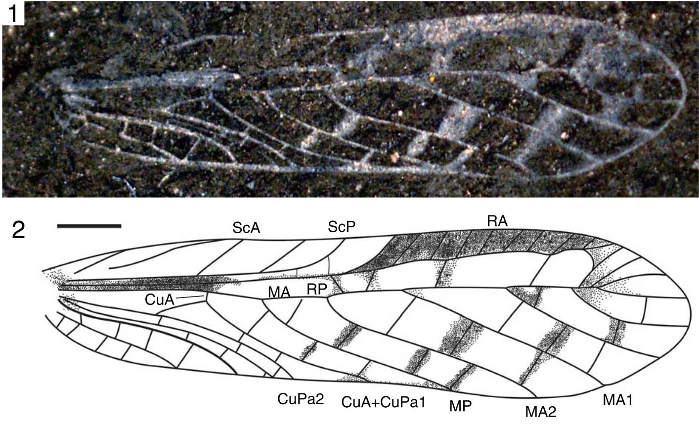
The description of wing venation follows terminology proposed by Béthoux and Nel (2001, Reference Béthoux and Nel2002). Abbreviations of forewing venation: ScA, ScP, anterior, posterior sub-costa vein; RA, RP, anterior, posterior radial vein; MA, MP, anterior, posterior media veins; MA1, MA2, first, second branch of anterior media vein; CuA, CuP, anterior, posterior cubitus veins; CuPa, anterior branch of cubitus vein; CuPa1, first branch of CuPa; CuA+CuPa1, fusion part of anterior cubitus vein and first branch of CuPa; CuPa2, posterior branch of CuPa; CuPb, second branch of CuP; ScA, ScP, anterior, posterior sub-costa vein; A, anal vein; 1A, first branch of anal vein.
Class Insecta Linnaeus, Reference Linnaeus1758
Order Orthoptera Olivier, Reference Olivier1789
Family Elcanidae Handlirsch, Reference Handlirsch1906
Subfamily Archelcaninae Gorochov, Jarzembowski and Coram, Reference Gorochov, Jarzembowski and Coram2006
Genus Cascadelcana new genus
Type species
Cascadelcana virginiana n. gen. n. sp.; by present designation.
Diagnosis
As for type species.
Occurrence
Upper Triassic (Norian), Cow Branch Formation; Solite Quarry, near the North Carolina-Virginia boundary, United States.
Etymology
The generic epithet is derived from Cascade, in reference to the town of Cascade near the Solite Quarry, and the common genus name Elcana.
Composition
Only the type species, Cascadelcana virginiana n. gen. n. sp.
Remarks
Casadelcana n. gen. differs from all other known genera of the Elcanidae in its forewing venation, in which the short CuA is almost vertical against the posterior margin and RP+MA1 have fewer branches.
Cascadelcana virginiana new genus new species
Light photomicrograph (1) and interpretive drawing (2) of the holotype, 06/L4BH/2-1/107, VPIGM-4698. Interpretive drawing combines observation of the part and counterpart. Scale bar=1 mm. Abbreviations of forewing venation: ScA and ScP, anterior and posterior sub-costa vein, respectively; RA, anterior radial vein; MA, anterior media veins; MA1 and MA2, first and second branch of anterior media vein, respectively; CuA, anterior cubitus veins; CuPa, anterior branch of cubitus vein; CuPa1, first branch of CuPa; CuPa2, posterior branch of CuPa; CuA+CuPa1, fused part of anterior cubitus vein and first branch of CuPa; RP, posterior radial vein; MP, posterior media vein.

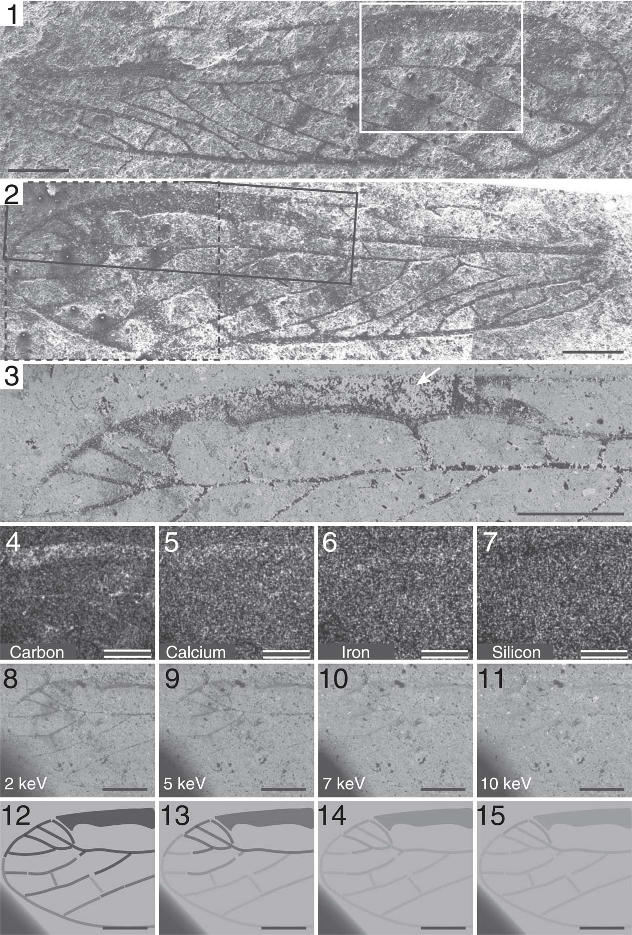
SEM images and EDS elemental maps of the forewing of C. virginiana n. gen. n. sp., holotype (06/L4BH/2-1/107, VPIGM-4698). (1) Composite SE ETD image (VA=10 keV) of counterpart; (2) composite SE ETD image (VA=10 keV) of part; (3) composite BSE SSD Z-contrast image (VA=3 keV) of area denoted by solid black line in (2); arrow in (3) indicates relatively high-Z material encrusting carbonaceous material (most likely silicate materials such as biotite); (4–7) EDS elemental maps of pterostigma-like structure in area denoted by solid white line in (1); (8–11) beam-energy BSE SSD Z-contrast image series of area denoted by dashed black line in (2); (12–15) illustrative diagrams of (8–11). Scale bars=1 mm.

Holotype
06/L4BH/2-1/107, VPIGM 4698, sex unknown, complete forewing, part and counterpart (Figs. 2, 3).
Diagnosis
RP+MA1 with six branches; M with 2 branches before stem MA1 fused with RP; CuA short, almost vertical against the posterior margin.
Occurrence
Upper Triassic (Norian), Cow Branch Formation; Solite Quarry, near the North Carolina-Virginia boundary, United States.
Description
Forewing: length 9.7 mm, width 2.3 mm at mid-length. ScA slightly S shaped, ending in anterior margin before 1/3 of total wing length. ScP ending in anterior margin close to half-length of forewing, giving off two or three distinct oblique branches ending in anterior margin. Stem R very strong, branched near wing mid-length, RA trending up after split from R, and slightly waved in distal part. RA with 10 branches ending in anterior margin. Stem RP+MA1 nearly straight, ending in wing apex, RP+MA1 with six simple branches, MA1 and MA2 diverging at 4.2 mm distal of forewing base. Area between RA and RP broad. MP simple, originates after ScA ending in anterior margin, MP ending in posterior margin before 2/3 distal of forewing base. CuA short, originates before ScA ending in anterior margin, and almost vertical against the posterior margin. CuPa1 fused with CuA opposite M+CuA branch, CuPa2 simple, ending in posterior margin opposite to where M branches. Area between CuA+CuPa1 and CuPa2 narrow. CuPb simple, ending in posterior margin before ScA ending in anterior margin. Anals simple, ending in posterior margin. Color occurrence between RA and anterior margin (pterostigma-like area), also along crossveins between branches of RP (and RP+MA1). The pterostigma-like area ~4.0 mm long, and covered with seven branches of RA.
Etymology
The species name is after the state of Virginia.
Remarks
The holotype was previously figured by Muscente and Xiao (Reference Muscente and Xiao2015, fig. 6A, B). An additional specimen with unclear forewings was reported in Liutkus et al. (Reference Liutkus, Beard, Fraser and Ragland2010, fig. 3D). The latter specimen probably belongs to the same species, but the poor preservation of the basal part of its forewings prevented a definitive assignment to this species.
Discussion
Preservation of the wing
Results of combined SEM and EDS are consistent with previous studies showing that, like other fossils from the Solite Quarry Lagerstätte, the specimen is preserved as carbonaceous compressions with minimal topographic relief (Kearns and Orr, Reference Kearns and Orr2009; Muscente and Xiao, Reference Muscente and Xiao2015). In general, the specimen was divided evenly between its part and counterpart during rock splitting (Fig. 3.1, 3.2). The Z-contrast in BSE SSD images of the part and counterpart indicates that the fossil is predominantly comprised of a lower Z material than the silicate minerals constituting the underlying shale; EDS elemental mapping confirms that this low Z material contains relatively higher concentrations of carbon than the shale, and thus is consistent with the presence of organic matter (Fig. 3.4–3.7). In some BSE SSD images, this carbonaceous material is encrusted by a material with a high Z relative to the carbonaceous material and shale (Fig. 3.3; see also Muscente and Xiao, Reference Muscente and Xiao2015, fig. 6L–P). This relatively high Z material most likely consists of silicate minerals (e.g., biotite), which reportedly encrust organic matter of fossils in the Solite Quarry (Kearns and Orr, Reference Kearns and Orr2009; Muscente and Xiao, Reference Muscente and Xiao2015). The high Z material is not readily apparent in EDS elemental maps, suggesting that the silicate minerals are too thin for detection with EDS (Fig. 3.7–3.11).
Beam-energy BSE SSD Z-contrast image series of the specimen (Fig. 3) indicates that the various veins and structures in the wing are preserved as carbonaceous films of different thicknesses. The beam-energy image series displays notable changes in contrast with VA. In images acquired at low VA (2 keV), whereas the contrast between fossil and the substrate is high, contrast between the medial sector and radial sector (including pterostigma-like structure) veins is relatively low (Fig. 3.8). The contrast between the veins in the medial and radial sectors, however, increases with VA (Fig. 3.9–3.11), and in images acquired at VA of 10 keV, only the radial sector veins and pterostigma-like structure are evident (Fig. 3.11). Because mass-thickness contrast in BSE SSD Z-contrast images of the Solite Quarry fossils primarily corresponds to differences in the thicknesses of the carbonaceous material, and thicker carbonaceous layers appear darker in BSE SSD images than thinner carbonaceous layers on relatively higher Z substrates (Muscente and Xiao, Reference Muscente and Xiao2015), the beam-energy image series suggests that the pterostigma-like structure is preserved as a relatively thicker carbonaceous layer than those of veins in other parts of the wing. Such differences in the thicknesses of these features most likely correspond to taphonomic differences among the tissues because the ultimate thickness of a carbonaceous compression depends upon its original thickness and composition (Muscente and Xiao, Reference Muscente and Xiao2015).
Systematic position
Cascadelcana virginiana n. gen. n. sp. can be attributed to the Elcanidae based on the simple CuA+CuPa1 and RP ending in MA1. The family Elcanidae is divided into two subfamilies: Archelcaninae Gorochov, Jarzembowski and Coram, Reference Gorochov, Jarzembowski and Coram2006 and Elcaninae Handlirsch, Reference Handlirsch1906. Cascadelcana virginiana n. gen. n. sp. belongs to Archelcaninae in having a broad area between RA and RP, as well as free (rather than fused) distal parts of CuPa2, CuPb, and 1A. It clearly differs from Archelcana Sharov, Reference Sharov1968 and Sibelcana Gorochov, Reference Gorochov1990 in its short CuA that is almost vertical against the posterior margin, and its RP+MA1 with fewer branches. It can also be distinguished from Parelcana Handlirsch, Reference Handlirsch1906 and Synelcana Zessin, Reference Zessin1988 by its MA with fewer branches before RP ending in MA1 (Parelcana with 4 branches). The relationship between CuA and CuPa1 was considered an important character for the classification of fossil orthopterans (Béthoux and Nel, Reference Béthoux and Nel2002). The character of short but measurable CuA is also found in all ancient species of Permelcanidae, in contrast with some Elcanidae species that have CuPa1 fused with the point of origin of CuA. Thus, C. virginiana n. gen. n. sp. may have a mosaic combination of features characteristic of the Permelcanidae and Elcanidae. These characters may help to clarify the phylogenetic relationship of the Elcanoidea (Fig. 4).
Hypothesized phylogenetic relationships of the Elcanoidea. Modified from Gorochov (Reference Gorochov1995). The number ‘1’ marks the geological age (Upper Triassic, Norian, ~227–208 Ma; Liutkus et al., Reference Liutkus, Beard, Fraser and Ragland2010) of Cascadelcana virginiana n. gen. n. sp. from the Cow Branch Formation in the Solite Quarry.

Pterostigma structure
The pterostigma is a thickened, sclerotized area on the anterior margin of the insect wing near the tip (Grimaldi and Engel, Reference Grimaldi and Engel2005). This structure is particularly noticeable on both wings of the Odonata and on the forewings of many species of the Mecoptera, Hymenoptera, Psocoptera, and Megaloptera (Chapman, Reference Chapman1998). Similar to the Odonata, C. virginiana n. gen. n. sp. and some other elcanids (such as Panorpidium yixianensis Fang et al., Reference Fang, Zhang, Wang, Wang, Zheng, Zhang, Li and Liu2015) also have thickened or colored parts close to the leading edge far out on the wing (Fig. 2). According to the Z-contrast image series (Fig. 3.8–3.11), this pterostigma-like area is characterized by relatively thick carbonaceous material, indicating that the leading edge on the wing of C. virginiana n. gen. n. sp. was probably thickened and sclerotized.
The pterostigma structure in the wings of elcanids differs from the pterostigmae of Recent dragonflies and hymenopterans in that it includes several crossveins (branches of RA) in the anterior margin of the wing. The normal pterostigma structure of Recent dragonflies and hymenopterans is often a thickened or highly pigmented cell in the outer wing with special micro-structures, such as a spinous protuberance and/or a network-like structure of the micro arrays (Bechly, Reference Bechly1995; Grimaldi and Engel, Reference Grimaldi and Engel2005). Nevertheless, some fossil species of these clades also have crossveins in the pterostigma area, including some species of the Aeschnidiidae (Neoanisoptera, Odonata) (e.g., Wigbtonia araripina Carle and Wighton, Reference Carle and Wighton1990; Bechly, Reference Bechly1998), and some species of the Nanosialidae (Siarapha, Panmegaloptera) (e.g., Hymega rasnitsyni Shcherbakov, 2013). The pterostigmae in those fossils differ from that of elcanids in that the pterostigma area of the latter is traversed by more crossveins (e.g., seven crossveins are visible in C. virginiana n. gen. n. sp. and more than fifteen in Panorpidium yixianensis Fang et al., Reference Fang, Zhang, Wang, Wang, Zheng, Zhang, Li and Liu2015). In addition, the pterostigma area of elcanids is larger in size relative to the whole wing. Due to its relatively greater mass than other nearby sections of wings, the pterostigma may function to facilitate gliding. The mass and location of pterostigma has an important influence on the speed limits of the pitching moments during the acceleration phases of wings flapping (Norberg, Reference Norberg1972). The pterostigma structures have not been previously described from other orthopterans. Elcanids may have evolved a particular flight mechanism distinct from those of other orthopterans.
Conclusions
The earliest known Elcanidae, Cascadelcana virginiana n. gen. n. sp., is described based on a forewing specimen from the Upper Triassic (Norian) Cow Branch Formation at the Solite Quarry Lagerstätte near the North Carolina-Virginia boundary, USA. It represents the first orthopteran described from the Triassic of North America. The pterostigma-like structure on the wing of C. virginiana n. gen. n. sp. was analyzed using scanning electron microscopy (SEM) and energy dispersive X-ray spectroscopy (EDS). Similar to pterostigma in modern insects, the pterostigma-like structure in elcanids was probably thickened and sclerotized, but it differs from true pterostigmae on the outer wings of other insects in that it is traversed by more crossveins. Elcanids may have evolved a particular flight mechanism distinct from those of other orthopterans.
Acknowledgments
This research was supported by the National Natural Science Foundation of China (41572010, 41622201, 41688103, 41730317), Chinese Academy of Sciences (XDPB05), U.S. National Science Foundation (EAR-1528553), Clay Mineral Society, Geological Society of America, Sigma Xi Scientific Research Society, and Virginia Tech Department of Geosciences. Analyses were conducted at the VT-ICTAS-NCFL with technical assistance from A. Giordani, S. McCartney, and C. Winkler. We thank N.C. Fraser for giving us permission to study this specimen, D.R. Zheng for discussion, and S. Chatzimanolis, B. Hunda, and three anonymous reviewers for their constructive comments on an earlier draft of the paper.