Implications
Diverse range of heavy metal removing methods have been suggested, that is usage of algae, agricultural by-products and microorganisms. Here, we showed that lead (Pb) can be effectively biosorbed by two lactic acid bacteria species (LABs) both under in vitro and in vivo conditions. Animal feed producer may incorporate these LABs in feed to benefit from both probiotic function and heavy metal removal.
Introduction
Pb is a widespread environmental pollutant causing broad range of physiological and biochemical dysfunctions in animal. It is normally released to environment from number of anthropogenic sources such as agricultural fertilizer, sewage sludge, metal mine and smelting (Scheuhammer, Reference Scheuhammer1987). Animal accumulate Pb in various tissues and subsequently transfer it via food chain to human (Fox, Reference Fox1987). Mahesar et al. (Reference Mahesar, Sherazi, Niaz, Bhanger and Abdul Rauf2010) reported that amount of Pb in most of commercial poultry feed samples is higher than the minimum harmful levels. Chickens particularly during the younger age are susceptible to as little as 1.0 mg Pb/kg diet (Bakalli et al., Reference Bakalli, Pesti and Ragland1995). It has been suggested that the biochemical and molecular mechanisms of Pb toxicity involve the induction of oxidative stress in target cells and generation of reactive oxygen species, followed by DNA damage and apoptosis, partly via (i) direct effect of Pb on cell membranes, (ii) Pb–hemoglobin interactions, (iii) consistent reduction in blood Δ-aminolevulinic acid dehydratase, an erythrocyte enzyme sensitive to Pb and (iv) effect of Pb on the antioxidant defence systems of cells (Ercal et al., Reference Ercal, Guerer-orhan and Aykin-burns2001). Thus, Pb exposure is manifested with high lipid peroxidation and poor animal growth performance (Erdogan et al., Reference Erdogan, Erdogan, Aksu and Baytok2004).
Various approaches has been suggested to remove heavy metal pollution such as usage of algae (Davis et al., Reference Davis, Volesky and Mucci2003), agricultural by-products (Demirbas, Reference Demirbas2008), garlic (Senapati et al., Reference Senapati, Dey, Dwivedi and Swarup2001), yeast (see review by Soares and Soares, Reference Soares and Soares2012), lactic acid bacteria (Teemu et al., Reference Teemu, Seppo, Jussi, Raija and Kalle2008) and fungi (Kapoor et al., Reference Kapoor, Viraraghavan and Cullimore1999). Although heavy metal removal ability of microorganisms has been reported previously in wastewater filtration, no report is available on their functionality within in vivo biological systems. Animal’s digestive system is a dynamic microenvironment that can affect stability of reversible surface binding between microorganism and heavy metal. Successful excretion of heavy metal from animal’s body require a stable bound throughout the digestive system. Thus, here we aimed to investigate in vitro and in vivo potential of two Lactobacillus isolates (LABs) from mulberry silage in biosorption of Pb and reduction of its preceding toxicity in broiler chickens. Previously, we have reported strong probiotic properties for these LABs (Altaher et al., Reference Altaher, Jahromi, Ebrahim, Zulkifli and Liang2015; Jahromi et al., Reference Jahromi, Altaher, Shokryazdan, Ebrahimi, Ebrahimi, Zulkifli, Goh, Tufarelli and Liang2016).
Material and methods
Bacterial isolates
The isolation, identification, characterization and screening procedure of probiotic bacteria used in this study has been elucidated in previous publication from our laboratory (Shokryazdan et al., Reference Shokryazdan, Liang, Jahromi and Abdullah2015). Briefly, a total of 50 initial isolates from mulberry (Morusalba) silage were tested and two isolates of Lactobacillus pentosus ITA23 (GenBank accession number: KF297814) and Lactobacillus acidipiscis ITA44 (GenBank accession number: KF297816) were selected as highly resistant to bile and acid, and reduced the pH level of culture medium to 3.2 and 3.7, respectively. The pH reduction was mainly due to production of lactic acid (534 and 433 mM for L. pentosus ITA23 and L. acidipiscis ITA44, respectively) and acetic acid (91 and 93 mM for L. pentosus ITA23 and L. acidipiscis ITA44, respectively).
In vitro study
Approximately 3 ml of deMan–Rogosa–Sharpe broth (with or without Pb) was transferred to 5-ml tubes. The concentration of Pb was 155 mg/l by addition of lead acetate (200 mg/l; Nacalai Tesque, Kyoto, Japan). The medium samples were inoculated by 5% of inoculums (overnight culture of each bacterium) and incubated at 37°C for 24 h. At the end of incubation, the culture broth was quantitatively assessed using spectrophotometer at 650 nm. In order to determine the absorption rate of Pb by the cells, 1 ml of each culture was centrifuged at 10 000 r.p.m. for 5 min and the cell-free supernatant was assayed for Pb concentration using microwave digestion and inductively coupled plasma mass spectrometry (ICP-MS) (Ahmed et al., Reference Ahmed, Abdullah, Wood, Hamza and Othman2013).
In vivo study
Experimental design, birds and diets
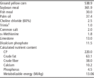
The experiments were conducted in accordance to procedures approved by the Universiti Putra Malaysia Institutional Animal Care and Use Committee. Male broiler chicks aged 108 days (Cobb500) were purchased from a commercial hatchery, weighed and assigned at random to 18 battery cages of six chicks each in an open-sided house under natural tropical environment. Equal number of birds (six cages) were randomly allocated to one of three dietary treatments as follows: (i) basal diet (no lead acetate supplementation; control) (Table 1), (ii) basal diet +200 mg lead acetate/kg (equal to 155 mg Pb/kg) and (iii) basal diet +200 mg lead acetate/kg +1000 mg LABs/kg. Diets were prepared as mash form using raw ingredients according to Table 1. Lead acetate was product of Nacalai Tesque and the probiotic was an equal combination of two species of Lactobacillus (L. pentosus ITA23 and L. acidipiscis ITA44) with final cell viability of 109/g. The inclusion level of Pb was selected based on the National Research Council (1994) recommendations for minimum toxic level of Pb in chicken’s diet. No feed additives or antimicrobials were used throughout the study. Chicks were maintained on a continuous light schedule and allowed ad libitum access to feed and water.
Ingredient composition and nutrient content of the basal diet (g/kg as fed unless otherwise stated)

1 Trimix (per kg Trimix): iron 100 g; manganese 110 g; copper 20 g; zinc 100 g; iodine 2 g; selenite 0.2 g; cobalt 0.6 g; santoquin 0.6 g; folic acid 0.33 g; thiamin 0.83 g; pyridoxine 1.33 g; biotin 2% 0.03 g; riboflavin 2 g; cyanocobalamin 0.03 g; d-calcium pantothenate 3.75 g; niacin 23.3 g; retinol 2000 mg; cholecalciferol 25 mg; α-tocopherol 23 000 mg IU.
Data collection
Chicks were weighed individually on the 1st day and following 21 days of feeding experimental diets. Feed intake (FI) was recorded and feed conversion ratio (FCR) was calculated. On day 21, two birds from each cage were decapitated and blood, cecal content, jejunum and liver samples were collected.
Determination of Pb
Microwave digestion was used for determination of Pb concentrations (Ahmed et al., Reference Ahmed, Abdullah, Wood, Hamza and Othman2013). Briefly, 0.2 g of feed, liver, cecal content or 0.2 ml of serum were added to a solution of 6 ml nitric acid (17 M), 2 ml hydrogen peroxide (8.8 M) and 2 ml hydrogen fluoride (0.02 M) and digested in a microwave oven (Model MARS-5; CEM, Matthews, NC, USA). All standard and blank solutions were prepared as above and were analyzed using ICP-MS system (PE SCIEX ELAN 6000; PerkinElmer, Beaconsfield, Buckinghamshire, UK).
Histopathological study
Liver and jejunum samples were fixed in 10% formalin, dehydrated for 16 h in an automatic tissue processor (Leica ASP 3000, Leica Microsystems Nussloch GmbH, Heidelberg, Germany) and embedded in paraffin wax using a paraffin embedding system (Leica EG 1160, Leica Microsystems Nussloch GmbH). Each sample was cut to 4-μm thick sections using a rotary microtome (Leica RM 2155, Leica Microsystems Nussloch GmbH). The sections were fixed on a glass slide, heated at 57°C until dried, stained with hematoxylin and eosin and examined under a light microscope (Dialux, LeitzWetzlar, Germany) as described previously (Grenier et al., Reference Grenier, Bracarense, Schwartz, Trumel, Cossalter, Schatzmayr, Kolf-Clauw, Moll and Oswald2012). A lesion score per animal was recorded in a blind way by a pathologist, taking into account the severity type and its intensity extent (scored from 0 to 3).
Antioxidant activity assay
Preparation of the liver tissue for antioxidant assay was according to the method of Bourogaa et al. (Reference Bourogaa, Nciri, Mezghani-Jarraya, Racaud-Sultan, Damak and El-Feki2013). Briefly, 1 g of liver tissue was homogenized in 2-ml tris buffered saline (TBS) (50 mM Tris, 150 mM NaCl, pH 7.4) in a homogenizer and centrifuged at 10 000 r.p.m. for 10 min. The supernatants were transferred into new 2-ml tube and stored at −80°C for subsequent analysis. The antioxidant activity of liver was determined using two methods of ferric reducing antioxidant power (FRAP) and Trolox equivalent antioxidant capacity (TEAC). The TEAC method is also known as 2, 2′-azinobis-3-ethylbenzothiazoline-6-sulfonic acid radical cation (ABTS) method. Different concentrations (0 to 100 µmol) of Trolox (water-soluble analogue of vitamin E) were used as standard and results expressed as µmol Trolox equivalent (TE)/g liver tissue.
Lipid peroxidation (malondialdehyde (MDA))
The lipid oxidation in liver samples was measured using thiobarbituric acid-reactive substances (TBARS). One gram of liver samples was homogenized in 4 ml 0.15 M KCl and 0.1 mM butylated hydroxytoluene (BHT). Homogenate samples (200 µl) were incubated with 1% (w/v) 2-thiobarbituric acid in 50 mM NaOH (0.25 ml) and 2.8% (w/v) trichloroacetic acid (0.25 ml) in water bath at 95°C for 60 min until pink color development. After cooling, 1 ml of distilled water and 3 ml of n-butyl alcohol were added to extracts and vortexed. The mixtures were centrifuged at 5000 r.p.m. for 10 min. Absorbance of supernatant was read against an appropriate blank at 532 nm using spectrophotometer (Secomam, Domont, France). The TBARS were calculated from a standard curve of 1, 1, 3, 3-tetraethoxypropane and expressed as microgram MDA per gram of liver.
Microbial quantification using quantitative real-time PCR
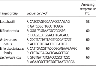
The populations of selected microbiota in the cecal content were determined using method described by Navidshad et al. (Reference Navidshad, Liang and Jahromi2012). Total DNA was extracted using QIAamp DNA Stool Mini Kit (Qiagen Inc., Valencia, CA, USA) according to manufacturer’s instructions. The extracted DNA were then stored at −20°C until use. The standard curves were constructed using number of copies of the 16S ribosomal RNA (rRNA) gene plotted against quantification cycle obtained from 10-fold serial dilutions of PCR products from pure culture of each bacterial group. For preparation of standard curves, DNA was extracted from the pure culture of each target bacteria (Lactobacillus, Bifidobacterium, Enterococcus, Enterobacteriaceae and Escherichia coli) and conventional PCR with specific primers were used to amplify bacterial DNA (Table 2). PCR products of the target bacteria were run in 1% agarose gel and specific bands were purified using the MEGAquick-spinTM purification kit (iNtRON Biotechnology, Seoul, Gyeonggi-do, Korea). Purity and concentration of 16S rRNA gene in each sample was measured using Nanodrop ND-1000 spectrophotometer (Implen NanoPhotometerTM, Munich, Germany). Copy number of 16S rRNA gene/ml of elution buffer was calculated using the following formula:
 $${\rm Number}\,{\rm of }\,{\rm copies}\,{\rm {\equals}}\,{{{\rm Amount}\,{\rm of }\,{\rm DNA}\,{\rm (\mu g\,/\,ml){\times}6}{\rm .022{\times}10}^{{23}} } \over {{\rm Length}\,{\rm (bp){\times}10}^{{\rm 9}} {\rm {\times}650}}}$$
Primers sequences and annealing temperatures for quantified microorganismsFootnote 1

1 Primers and annealing temperatures were according to Navidshad et al. (Reference Navidshad, Liang and Jahromi2012).
Real-time PCR was performed with BioRad CFX96 Real-time PCR system (Bio-Rad Laboratories, Hercules, CA, USA). Each reaction consisted of 12.5 μl of 2× SYBR Green Master Mix (Thermo Scientific, PA, USA), 1 μl of 10 µM forward and reverse primers, 2 μl DNA and 8.5 μl nuclease-free water. Each sample was assayed in duplicate. No-template control was included to rule out any cross-contamination. Real-time PCR cycling condition comprised initial denaturation at 94°C for 5 min, followed by 40 cycles of denaturation at 94°C for 20 s, primer annealing (according to Table 2) for 30 s and extension at 72°C for 20 s. Upon completion of amplification, specificity of the amplified product was confirmed using melting curve analysis. The real-time PCR products were incubated by raising the temperature from 65°C to 95°C in 0.5°C increments with a hold of 5 s at each increment.
Statistical analysis
Experimental data were analyzed using one-way ANOVA procedure of SAS program version 9.2 (version 9.3; SAS Institute Inc., Cary, NC, USA) followed by multiple comparison among the means using Duncan’s new multiple range test. Differences were considered significant if P<0.05.
Results and discussion
In vitro study
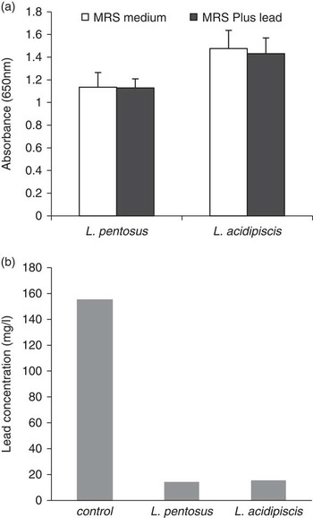
Addition of lead acetate had no negative effect on LABs growth (Figure 1a). The concentration of Pb in the control culture medium and after incubation in the presence LABs is presented in Figure 1b. Although the Pb concentration was 155 mg/l in control medium, it was significantly reduced by more than 90% to 14.29 mg/l and 15.49 mg/l in cultures of L. pentosus ITA23 and L. acidipiscis ITA44, respectively. The mechanism behind such biological Pb absorption could be attributed to negative charge of carboxyl and phosphoryl groups in cell membrane of LABs. These negative-charge molecules are able to bind with any positive-charge molecules such as Pb. The ion exchange mechanism of biosorption of heavy metal by microorganisms previously reported in Aspergillus niger (Kapoor and Viraraghavan, Reference Kapoor and Viraraghavan1997), Rhizopus arrhizus (Tobin et al., Reference Tobin, Cooper and Neufeld1990), Lactobacillus fermentum ME3 and Bifidobacterium longum 46 (Teemu et al., Reference Teemu, Seppo, Jussi, Raija and Kalle2008). In all of these studies, heavy metal biosorption was significantly reduced by chemical intervention that neutralize the negative charge of carboxyl and phosphoryl groups. Teemu et al. (Reference Teemu, Seppo, Jussi, Raija and Kalle2008) confirmed the existence of such biosorption using transmission electron micrographs showing the cell membrane binding to Pb.
Lead (Pb) reduction potential of two selected Lactobacillus species in vitro. Growth of either of Lactobacillus species in deMan–Rogosa–Sharpe (MRS) medium was not inhibited by Pb (a) and both species largely reduced Pb level in the culture medium (b). The reduction rate was 90.8% and 90.0% for Lactobacillus pentosus and Lactobacillus acidipiscis, respectively.

In vivo study
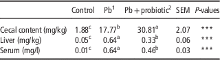
Concentration of Pb in cecal content, liver and serum is presented in Table 3. Although lead acetate was not added to control diet, our analysis showed some scarce level of Pb in the feed (0.155 mg /kg) and negligible amount in the cecal content, serum and liver samples. Low concentrations of Pb in feedstuff such as fish meal, soybean meal and corn has been frequently reported before (Nicholson et al., Reference Nicholson, Chambers, Williams and Unwin1999; Zhang et al., Reference Zhang, Li, Yang and Li2012). The concentration of Pb in cecal content of chickens supplemented with LABs was 1.7 times higher than the Pb-exposed chicks (P<0.05). This observation together with the in vitro results indicated that LABs biosorption of Pb maintained throughout the gastrointestinal tract (GIT) and resulted in by-pass transition of Pb from GIT absorption sites. This is also evident by the significant Pb reduction observed in liver and serum of chicks fed Pb+probiotic diet (P<0.05). Although probiotic was not able to reduce Pb accumulation in liver and serum to the same levels as in the control chicks, the reduction was sufficient to protect the chicks against the Pb adverse effects, as documented by other parameters such as liver lesion and performance. It is noteworthy to mention that the existence of interaction between microenvironment of GIT (i.e. pH) and stability of binding between Pb and LABs is still an open question to be answered.
Recovery of lead (Pb) in the cecal content and its accumulation in liver and serum of broiler chickens in different treatment groups

n=12.
a,b,cValues within a row with different superscripts differ significantly at P<0.05.
1 Pb was added in the form of lead acetate in the concentration of 200 mg/kg feed.
2 Probiotic was added in the concentration of 100 mg/kg feed (~109 cells/kg).
***P<0.001.
Oxidative parameters and tissue lesion
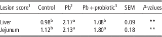
The effects of Pb and LABs supplementation on MDA and antioxidant activity (FRAP and ABTS) are presented in Table 4. The MDA level was increased (P<0.05) in liver of Pb received birds by nearly twofolds compared with those of control group (248.06 v. 130.66 µg MDA/g). MDA elevation represents a significant increase in lipid peroxidation. As measured by markers of oxidative (MDA) and antioxidative status (FRAP and ABTS), our results attribute the negative effects of Pb exposure to free radical generation. Ercal et al. (Reference Ercal, Guerer-orhan and Aykin-burns2001), Erdogan et al. (Reference Erdogan, Erdogan, Aksu and Baytok2004) and Prvulovic et al. (Reference Prvulovic, Kojic, Popovic and Grubor-Lajsic2015) reported the same finding. Therefore, the elevation of MDA or FRAP and ABTS could be explained by the massive production of free radicals and preceding exhaustion of antioxidant enzymatic activity (Sharma and Barber, Reference Sharma and Barber2012; Prvulovic et al., Reference Prvulovic, Kojic, Popovic and Grubor-Lajsic2015). Interestingly in our study, LABs supplementation completely restored oxidative and antioxidative status of the birds to its normal state (Table 4). This observation was despite the presence of Pb in liver and serum of Pb+probiotic birds. It seems that the accumulated Pb in tissues of these birds was below the toxicity level. This notion is supported by histopathological results obtained in liver samples (Table 5). Lesion score analysis revealed that Pb presence had detrimental effect on liver tissue (P<0.05), but LABs supplementation eliminated such tissue damage. The beneficial effect of LABs in reducing liver lesion score could be attributed to its role in lowering Pb levels to non-toxic amounts. However, we did not observe the same beneficial effect in jejunum tissue. It is possible that some proportions of Pb were still present in active form in the jejunum and the biosorption process was not fully completed.
Effect of lead (Pb) and probiotic supplementation on oxidative parameters in liver of broiler chickens

TE=µmol Trolox equivalent; ABTS=2, 2′-azinobis-3-ethylbenzothiazoline-6-sulfonic acid radical cation.
n=6.
a,bValues within a row with different superscripts differ significantly at P<0.05.
1 Pb was added in the form of lead acetate in the concentration of 200 mg/kg feed.
2 Probiotic was added in the concentration of 100 mg/kg feed (~109 cells/kg).
*P<0.05; **P<0.01.
Effect of lead (Pb) and probiotic supplementation on lesion score of liver and jejunum in broiler chickens

n=6
a,bValues within a row with different superscripts differ significantly at P<0.05.
1 The score for each lesion was obtained by multiplying the severity factor by the extent of the lesion. The organ score was then obtained by calculating the sum of each lesion score. Severity factor (or degree of severity), 1=mild lesions, 2=moderate lesions; the extent of each lesion (intensity or observed frequency) was evaluated and scored as 0=no lesion, 1=low extent, 2=intermediate extent, 3=large extent.
2 Pb was added in the form of lead acetate in the concentration of 200 mg/kg feed.
3 Probiotic was added in the concentration of 100 mg/kg feed (~109 cells/kg).
**P<0.01.
Performance parameters
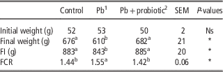
Effect of Pb exposure and LABs supplementation on BW gain, FI and FCR are shown in Table 6. Presence of Pb in the feed significantly reduced FI and weight gain and increased FCR. This is in agreement with previous reports by Damron et al. (Reference Damron, Simpson and Harms1969), Seven et al. (Reference Seven, Aksu and Tatli-Seven2012) and Yuan et al. (Reference Yuan, Song, Jiang, Azzam, Zhu and Zou2013). Previous study in our laboratory suggested that downregulation of major nutrient transporter genes in small intestine is one of the possible underlying mechanism for such observation (Ebrahimi et al., Reference Ebrahimi, Jahromi, Liang, Soleimani, Shokryazdan and Zulkifli2015). The FI reduction and preceding weight gain suppression of Pb-exposed birds could also be attributed to anorexia and anxiety caused by reduction of brain 5-hydroxytryptamine (serotonin) (Haider et al., Reference Haider, Saleem, Tabassum, Khaliq, Shamim, Batool, Parveen, Inam and Haleem2013). This is also in part explain the elevation observed in FCR. Furthermore, the negative effect of Pb exposure on FCR and weight gain maybe also associated with Pb effects on metabolic and endocrine responses (Scheuhammer, Reference Scheuhammer1987; Yuan et al., Reference Yuan, Song, Jiang, Azzam, Zhu and Zou2013). Exposure to Pb elevate plasma corticosterone (Cory-Slechta et al., Reference Cory-Slechta, Virgolini, Thiruchelvam, Weston and Bauter2004; Haider et al., Reference Haider, Saleem, Tabassum, Khaliq, Shamim, Batool, Parveen, Inam and Haleem2013), and consequently a diverse range of glucocorticoids effects take place from immune suppression to reduction of intestinal absorptive surface area (Hu et al., Reference Hu, Guo, Huang, Bun, Zhang, Li, Liu, Long, Yang and Jiao2010). Interestingly, LABs supplementation significantly eliminated the adverse effect of Pb on all performance parameters. The reason for such effect might come from the Pb biosorption by LABs, removing the Pb from intestinal absorption sites, and therefore reducing Pb accumulation in body (Table 6). Lowering Pb accumulation reduce the toxic effects of Pb exposure in birds, as documented here with lower liver lesion and lipid peroxidation, and higher antioxidant capacity. It is noteworthy to mention that the observed performance in our study was lower than the expectations from this strain of broiler chicks. This is probably due to the rearing environment of the chicks. Previous studies also reported such inferior performance in broiler chicks exposed to hot humid tropical condition (Yalcin et al., Reference Yalcin, Settar, Ozkan and Cahaner1997; Yodseranee and Bunchasak, Reference Yodseranee and Bunchasak2012).
Effect of lead (Pb) and probiotic supplementation on feed intake (FI), BW and feed conversion ratio (FCR) of broiler chickens

n=6.
a,bValues within a row with different superscripts differ significantly at P<0.05.
1 Pb was added in the form of lead acetate in the concentration of 200 mg/kg feed.
2 Probiotic was added in the concentration of 100 mg/kg feed (~109 cells/kg).
*P<0.05.
Microbial populations
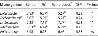
Table 7 showed the population of various microbiota in the cecum. There was no significant difference in microbial population between the control and Pb-exposed birds. However, supplementation of LABs significantly increased beneficial microbe population at the cost of pathogens. It has been reported that a modes of action of probiotics is their pathogen interference and antagonistic activity, whereby probiotic strains inhibit the growth and colonization of other microorganisms, such as E. coli (Mountzouris et al., Reference Mountzouris, Tsirtsikos, Kalamara, Nitsch, Schatzmayr and Fegeros2007; Meimandipour et al., Reference Meimandipour, Shuhaimi, Soleimani, Azhar, Hair-Bejo, Kabeir, Javanmard, Muhammad Anas and Yazid2010). This is a competitive exclusion where microbes are competing for nutrient and attachment sites on intestinal epithelial wall (Fuller, Reference Fuller2001). It is therefore reasonable to attribute some portion of the performance recovery in Pb+probiotics birds to effect of LABs supplementation in favoring microbes such as Lactobacillus and Bifidobacteria.
Effect of lead (Pb) and probiotic supplementation on population of microorganisms in the cecum of broiler chickens (log10 copy number/g cecal content sample)

n=12.
a,bValues within a row with different superscripts differ significantly at P<0.05.
1 Pb was added in the form of lead acetate in the concentration of 200 mg/kg feed.
2 Probiotic was added in the concentration of 100 mg/kg feed (~109 cells/kg).
*P<0.05.
Conclusions
Results of in vitro and in vivo studies suggested that supplementation of L. pentosus ITA23 and L. acidipiscis ITA44 besides the classical effect of favoring beneficial microbes at the cost of pathogens; it may also biosorb Pb and reduce its absorption by broiler chickens GIT. Therefore, LABs supplementation may protect the birds against the adverse effects of Pb exposure on growth performance, lipid peroxidation, antioxidant activity and tissue damage.
Acknowledgments
This study was sponsored by the Long-Term Research Grant Scheme (LRGS 2/2012) provided by the Ministry of Education Malaysia.