Introduction
The transduction of light by photopigments is an essential step in the initiation of vision, and these pigments thus serve as limiting intermediaries in the process of translating light into sight. In recent years, there have been significant advances in understanding the opsin genes that specify photopigment proteins, and that change, in conjunction with a growing accumulation of measurements of photopigments present in many different species, has provided an enhanced appreciation of the many ways in which photopigments can condition vision. One of the surprising results emerging from this work was the discovery that the opsin genes coding for short-wavelength-sensitive cone photopigments in some mammalian species have accumulated mutational changes that render them incapable of expressing fully functional photopigments. The loss of functional opsin genes can have important consequences for visual behavior; for instance, by restricting the overall photon capture, and thus modifying visual sensitivity, or by deleting a capacity for resolving a dimension of color vision. Here, I review the extent and causes of the losses of functional opsin genes that specify short-wavelength cone photopigments in mammals and also explore their possible implications.
Evolution of mammalian cone pigments: An overview
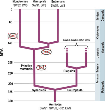
A general scheme that has emerged in recent years to account for the evolution of mammalian cone photopigments is outlined in Fig. 1 (for detailed reviews of this issue, see Yokoyama, Reference Yokoyama2008; Hunt et al., Reference Hunt, Carvallo, Cowing and Davies2009; Jacobs, Reference Jacobs2009). Vertebrate cone photopigments are specified by opsin genes derived from four gene families, designated as SWS1 (short-wavelength-sensitive opsin, type 1), SWS2 (short-wavelength-sensitive opsin, type 2), Rh2 (rod-like cone opsin), and LWS (long-wavelength-sensitive opsin), respectively. All four were present early in vertebrate history, apparently at least by the time that jawed and jawless representatives of the vertebrate lineage diverged, some 540 million years ago (Collin et al., Reference Collin, Davies, Hart and Hunt2009). No contemporary mammals have cone opsin genes drawn from more than two of the four gene families so the overall pattern of evolution of cone pigments among the mammals has been one of loss. Rh2 opsin genes are not found in any contemporary monotreme, marsupial, or eutherian mammal, and thus, representatives of that family must have disappeared in the period prior to the initial divergence of these three groups. SWS2 genes persist in monotremes (Davies et al., Reference Davies, Caravalho, Cowing, Beazley, Hunt and Arrese2007) but are absent from both marsupials and eutherians (Strachan et al., Reference Strachan, Chang, Wakefield, Marshall, Graves and Deeb2004; Cowing et al., Reference Cowing, Arrese, Davies, Beazley and Hunt2008); consequently, the SWS2 gene family must have been lost at some point prior to the divergence of the latter two lineages. Although it is not absent, the SWS1 gene has been found to be inactivated by mutational changes in the monotremes platypus (Ornithorhynchus anatinus) and echidna (Tachyglossus aculeatus) (Davies et al., Reference Davies, Caravalho, Cowing, Beazley, Hunt and Arrese2007; Wakefield et al., Reference Wakefield, Anderson, Chang, Wei, Kaul, Graves, Grutzner and Deeb2008). Similar arrangements have also been detected in a variety of eutherian species, and those constitute the focus of this paper.
A simplified cladogram showing the representation of the four vertebrate cone opsin gene families (SWS1, SWS2, Rh2, and LWS) in saurposids and synapsids and their modern descendents. All four of the cone opsin gene families are believed to date to at least ∼540 million years ago. Gene sequence comparisons and measurements of cone photopigments indicate that only two of these gene families are present in modern mammals. The pattern of loss of two gene families in the lines leading to the major mammalian groups is indicated (for further details, see the text).

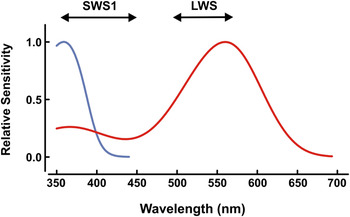
From comparisons of gene sequences, it is inferred that the cone pigments of ancestral mammals derived from the SWS1 and LWS gene families had respective peak absorptions (λ max) of ∼360 and 560 nm (Hunt et al., Reference Hunt, Wilkie, Bowmaker and Poopalasundaram2001; Yokoyama, Reference Yokoyama2008). Subsequent substitutions occurring at a limited number of sites in the opsin molecule served to expand the spectral ranges of pigments derived from these two gene families such that contemporary mammalian SWS1 pigments have λ max ranging from ∼360 to 440 nm, while those derived from the LWS opsin genes vary across the range from ∼500 to 565 nm (Fig. 2). No mammal derives more than a single type of cone pigment from each of these families, with the exception of various types of primate that have added uniquely a third type of cone photopigment, a change that arose either as a result of the duplication of the LWS opsin gene (all catarrhine species) or from the presence of LWS opsin gene polymorphisms (most platyrrhine and a few species of strepsirrhine primates) (Jacobs, Reference Jacobs2008, Reference Jacobs2010).
Spectral sensitivity curves for the cone pigments that have been inferred to be present in ancestral mammals that were derived respectively from the SWS1 opsin gene family (blue) and the LWS opsin gene family (red) and with peak values of ∼360 and 560 nm. Subsequent substitutions occurring at a limited number of sites in the opsin molecules allow contemporary mammalian SWS1 cone pigments to have λ max ranging from ∼360 to 450 nm, while those derived from the LWS gene family to have λ maxranging from ∼500 to 560 nm. The spectral ranges for each are indicated by the length of the lines and their arrow heads.

Based on a variety of indicators, early mammals are been assumed to have been nocturnal (Crompton et al., Reference Crompton, Taylor and Jagger1978; Kemp, Reference Kemp2005; Bickelmann, Reference Bickelmann2011) and that condition is usually credited as setting the stage for dramatic alterations in mammalian sensory systems. These alterations included numerous changes in visual systems as well as those that seem designed to support the enhancement of olfactory and auditory capabilities (Striedter, Reference Striedter2005). The modifications that occurred in the visual system included a fundamental reorganization of the retinofugal pathways as well as significant changes in eye shape and orbit orientation. In combination, these changes in the visual system are presumed to have served to enhance the likelihood of capturing the scarce light that is the characteristic of nocturnal habitats (Heesy & Hall, Reference Heesy and Hall2010). During this same period, there was also a striking remodeling of the retina that included the disappearance of photoreceptor-colored oil droplets, a simplification of retinal circuitry, and significant increases in rod/cone ratios (Jacobs & Rowe, Reference Jacobs and Rowe2004). A decreased dependence on daylight vision also may have set the stage for a reduction in the number of cone photopigment types, a change triggered by the disappearance of functional products for two cone opsin gene families in all three of the mammalian subclasses.
Although the belief that the early mammals were nocturnal for most of their early history has been widespread, that idea has not gone unchallenged. In one recent instance, an examination of the structure of the fossilized orbits derived from a collection of Mesozoic archosaurs led to the suggestion that different species in this group actually manifested variant photic rhythms—some apparently being diurnal, some nocturnal, and some arrhythmic. An implication that follows is that the general idea that dinosaurs were routinely diurnal and mammals routinely nocturnal may be incorrect (Schmitz & Motani, Reference Schmitz and Motani2011), and, if that is so, it remains possible that some lines of contemporary mammals may not have had nocturnal ancestors. We return below to a consideration of what it means to be nocturnal.
Genetic basis for human tritan color vision defects
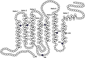
Nathans et al. (Reference Nathans, Thomas and Hogness1986) isolated and sequenced the opsin genes that are linked to the human cone pigments. The SWS1 gene coding for the short-wavelength opsin (a gene identified as OPN1SW) was mapped to chromosome 7, at 7q32. That gene codes for an opsin that is 348 amino acids in length and it shares a ∼40% nucleotide identity with the two other human cone opsin genes (OPN1MW and OPN1LW); the latter respectively specify the middle (M)- and long (L)- wavelength cone photopigments. The X-chromosome genes coding for the M and L cone pigments are believed to have arisen from an LWS opsin gene duplication that occurred some 35 million years ago. Results from psychophysical studies of human color vision defects had long implied that alterations in a gene such as OPN1SW caused tritanopia, a rare dichromatic condition with an incidence no greater (and perhaps much less) than about 1 in 500 individuals (Wright, Reference Wright1952; van Norren & Went, Reference van Norren and Went1981). Early pedigree studies had also established that inherited tritanopia follows an autosomal-dominant transmission pattern with incomplete penetrance (Kalmus, Reference Kalmus1955). Shortly after the human opsin genes were first characterized, Weitz et al. (Reference Weitz, Miyake, Shinzato, Montag, Zrenner, Went and Nathans1992Reference Weitz, Miyake, Shinzato, Montag, Zrenner, Went and Nathansa,b) examined OPN1SW genes that had been isolated from several tritanopic individuals. Their analysis detected the presence of three separate missense mutations that were associated with the loss of functional S-cone photopigment. Subsequently, three additional mutations have also been linked to tritanopic defects (Gunther et al., Reference Gunther, Neitz and Neitz2006; Baraas et al., Reference Baraas, Carroll, Gunther, Chung, Williams, Foster and Neitz2007, Reference Baraas, Hagen, Dees and Neitz2012). The causative pathway between these various gene defects (Fig. 3) and the absence of a viable S-cone pigment is not known certainly, but it is presumed that these mutations might perturb the folding, processing, or stability of the opsin. A study that featured a gene analysis of two tritan subjects, combined with direct imaging of their photoreceptor mosaics, suggests further that the loss of S-cones in individuals that are heterozygous for the mutant opsin genes may actually manifest as a progressive age-related condition, perhaps similar in character to that documented for autosomal-dominant retinitis pigmentosa (Baraas et al., Reference Baraas, Carroll, Gunther, Chung, Williams, Foster and Neitz2007). If that is so, then tritanopia, unlike the more common red/green dichromacies, is more properly considered as an inherited but not necessarily congenital color vision defect (Neitz & Neitz, Reference Neitz and Neitz2011). Whether or not that turns out to be the case, the examination of human tritanopes provided the first direct genetic explanation for loss of mammalian S-cone function.
Model of the topography of the human S-opsin with the circles representing the amino acids. The six colored circles (residues 56, 79, 190, 214, 264, and 283) show the locations of the amino acid substitutions identified in several studies as causing tritan color vision losses (see text). The lysine residue at position 293 is the chromophore binding site. (Figure modified from Gunther et al., Reference Gunther, Neitz and Neitz2006.)

Initial evidence for loss of S-cone function in nonhuman mammals
The first hints that there might be some nonhuman species similar to human tritanopes—in the sense of their lacking functional S cones—came from an opsin antibody labeling study of two species of nocturnal primate, the platyrrhine owl monkey (Aotus trivirgatus) and the strepsirrhine bushbaby (Galago garnettii) (Wikler & Rakic, Reference Wikler and Rakic1990). Although the retinas of both species had cones that could be labeled with an opsin antibody specific for M- or L-cone pigments, a second antibody that labels S cones in other primate retinas did not react with any of the cones in the retinas of these two species. A subsequent study of the Aotus monkey, involving both behavioral and electroretinogram (ERG) measurements, similarly failed to detect any functional evidence for the presence of S cones, and that observation provoked the proposal that the gene/pigment arrangement in this monkey might be analogous to that of human tritanopes in the sense that the SWS1 cone opsin genes in this species contained defects that obviated the production of functional S-cone pigments (Jacobs et al., Reference Jacobs, Deegan, Neitz, Crognale and Neitz1993). That suggestion was borne out when an examination of the sequences of the SWS1 cone opsin genes in both owl monkeys and bushbabies revealed that these genes were (a) highly homologous to the human S-cone opsin gene but (b) harbored mutational changes that should make the production of fully functional photopigments impossible (Jacobs et al., Reference Jacobs, Neitz and Neitz1996). The nature of the mutational changes was different in the two species but both featured the presence of nucleotide insertions and deletions that introduced premature stop codons.
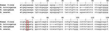
Aotus is the only nocturnal anthropoid species, and, as such, it offers an intriguing case for understanding the history surrounding the loss of S cones due to gene defect. Although this monkey is classified routinely as being nocturnal, its eye contains a number of features—including both the absence of a reflective tapetum and the sporadic presence of a fovea in some individual animals—which imply that its nocturnality was acquired secondarily, and if that is so, some ancestor of modern Aotus quite certainly had a functional population of S cones that was lost during its subsequent evolution, perhaps contingent on its transition to nocturnal life (Martin & Ross, Reference Martin, Ross and Kremers2005). Although the systematics of Aotus are still being refined, one recent analysis suggests that at least 11 different species can be reliably distinguished in this genus (Menezes et al., Reference Menezes, Bonvicino and Seuanez2010). A sequence analysis conducted on the S-cone opsin genes of two additional species (A. azarai and A. nancymaae) revealed that they harbored the same missense mutation earlier detected in A. trivirgatus (Fig. 4), a change leading to the introduction of a premature stop codon (Levenson et al., Reference Levenson, Fernandez-Duque, Evans and Jacobs2007). The lines leading to these three species are believed to have followed separate evolutionary pathways since the onset of divergence of this genus (Menezes et al., Reference Menezes, Bonvicino and Seuanez2010), implying that the loss of a functional S cone must date to at least that time. Importantly also, the three studies conducted on Aotus S-cone opsin genes involved examination of genes derived from a total of 11 different animals, and the genes from all the animals were found to share the same disabling mutation. This genetic uniformity across individuals implies that, unlike tritanopia, a condition detected in only a tiny minority of all humans, the loss of S cones here are species-wide traits.
Partial alignment of the S cone opsin sequences obtained from humans and three species of Aotus monkeys. Shown are first 114–118 bases of the first exon of this gene. In all three monkey species, there is a missense mutation (a substitution of guanine for adenine) at nucleotide 66 (boxed) that creates a translational alteration leading to a premature introduction of a stop codon. The S-cone opsin genes of all Aotus monkeys are thus pseudogenes, incapable of coding for a fully functional S-cone photopigment. (Sequence results derived from Levenson et al., Reference Levenson, Fernandez-Duque, Evans and Jacobs2007.)

Survey of the loss of S-cone function in nonhuman mammals
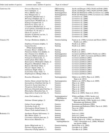
The list of mammalian species known to have lost S-cone function has expanded substantially over the past 15 years. Across various studies, the loss of S-cone function has been inferred from direct examination of SWS1 gene sequences, as well as from several functional measures, the most common of which is opsin antibody labeling of cone photoreceptors. In a few cases, loss of function has also been inferred from a failure to detect any S-cone signals in ERG recordings or as a result of evidence obtained from direct behavioral tests of vision. Strictly speaking, these functional measures do not require the absence of S cones to have resulted from opsin gene mutations, and of course, they may also fail to be definitive if they lack the sensitivity to detect contributions from the presence of only very small numbers of S cones; however, in the several instances where both genetic and functional measures have been obtained from the same species, the two agree, and thus, it seems reasonable to assume that all these types of result support the same conclusion. At present, there is evidence to indicate that several dozen mammalian species from five different orders have lost S-cone function as a result of opsin gene mutations. A list of these species appears in Table 1. Some specific lessons can be drawn from the distribution and nature of the losses that have been detected among animals falling into these various groupings.
Mammalian species known to have lost short-wavelength cone function as a result of opsin gene mutations

* ERG—electroretinogram; genetics—sequence analysis; staining—opsin immunocytochemistry; behavior—discrimination tests.
Carnivores
Representatives from all three of the pinniped families have been examined: in total, 13 of the 34 species that make up the order. None of these were found to have functional S cones. That commonality suggests that the widespread loss of S cones among the pinnipeds probably resulted from mutational changes that occurred near the basal point in this lineage—divergence in this group is believed to have commenced some 33 million years ago (Arnason et al., Reference Arnason, Gullberg, Janke, Kullberg, Lehman, Petrov and Vainola2006). The other carnivores known at present to lack S cones are three species from the family Procyonidae. These New World mammals, a group that totals only about 15 species, are mainly nocturnal. It is interesting that whereas the nocturnal species so far studied (the raccoons Procyon lotor and P. cancrivorus, along with the kinkajou, Potos flavus) were found to lack S cones, a species from this same family that is most often described as being diurnal (the coati, Nasua nasua) maintains a healthy population of S cones (Jacobs & Deegan, Reference Jacobs and Deegan1992; Peichl & Pohl, Reference Peichl and Pohl2000). Unlike the pinnipeds, then, the loss of S cones in the procyonids must have occurred in the more distal branches of the phylogeny of this family, perhaps contingent on their having embarked on separable patterns of photic rhymicity. Whether that is the case or not, these two examples underline the fact that SWS opsin gene loss in mammals has happened at various stages during evolutionary divergence.
Cetaceans
The first cetacean species examined, the bottlenose dolphin (Tursiops truncatus), was found to have an SWS1 opsin gene, which is homologous to that SWS1 opsin gene of other mammalian species. However, examination of the sequence of this gene revealed various disabling features, including a frameshift mutation that would obviate it being expressed in vivo (Fasick et al., Reference Fasick, Cronin, Hunt and Robinson1998). Subsequently, representative species from both suborders of Cetacea—Odontoceti (toothed whales, dolphins, porpoises) and Mysticeti (baleen whales)—have also been shown to lack functional S cones. In the most extensive study to date, Levenson and Dizon (Reference Levenson and Dizon2003) discovered that SWS1 opsin genes are not expressed in the retinas of any of 16 different species of cetacean. Identical disabling mutations were found in SWS1 opsin genes isolated from members of both the mysticete and odontocete suborders indicating that loss of a functional gene must have preceded their diversification, thus making it virtually certain that all modern cetaceans lack functional S-cones.
Chiropterans
Bats comprise some 20% of all mammals. Because of their long history of nocturnality, and their heavily rod-dominated retinas, relatively little has thus far been learned about the nature and utility of possible cone-based vision in bats. Of those bats for which information about cone pigments is now available, some 23 species have been shown to possess two types of functional cone pigments, SWS1 and LWS derived, respectively, while 16 species (Table 1) have been found to have an LWS pigment but lack functional SWS1 cone opsin genes (Wang et al., Reference Wang, Oakley, Mower, Shimmin, Yim, Honeycutt, Tsao and Li2004; Muller et al., Reference Muller, Goodman and Peichl2007; Feller et al., Reference Feller, Lagerholm, Clubwala, Silver, Haughey, Ryan, Loew, Deutschlander and Kenyon2009; Zhao et al., Reference Zhao, Rossiter, Teeling, Li, Cotton and Zhang2009). The classification of Chiroptera remains a topic of discussion. Traditionally, bats were classified into two suborders, Megachiroptera and Microchiroptera, but recent molecular data have been taken to identify two differently defined suborders—Yangochiroptera and Yinpterochiroptera—with megabats included in the former along with five families of microbat, while the latter group encompasses all the remaining families (Teeling et al., Reference Teeling, Springer, Madsen, Bates, O’Brien and Murphy2005). In the most extensive study of photopigment opsin genes in bats thus far, Zhao et al. (Reference Zhao, Rossiter, Teeling, Li, Cotton and Zhang2009) examined opsin genes derived from 33 species that included representatives from both major lineages. Both SWS1 and LWS opsin genes were intact in all the 12 species of Yinpterochiroptera studied; among the Yangochiroptera, some species featured a mutational loss of SWS1 gene loss, while others did not. These investigators offered some suggestions as to other features of the natural history of these various bat species that may correlate with retention or loss of SWS1 opsin genes. Such issues are considered below.
Primates
The singular case of Aotus, the only anthropoid in which the SWS1 opsin gene has acquired disabling mutations, was noted above. The other primates known to lack functional S-cone pigments are all from the suborder Strepsirrhini, a group comprised of some 125 species native to Africa and Southeast Asia. One of the two infraorders in this group, Lorisiformes, includes various galagos and lorises. Kawamura and Kubotera (Reference Kawamura and Kubotera2004) found that the S-cone opsin genes of galagos and lorises contain identical mutational changes, implying that all members of this infraorder lack functional S cones. The second strepsirrhine infraorder, Lemuriformes, is composed of a wide variety of different lemur species. It offers a quite different picture, as many of the species in this group have been shown to have intact SWS1 opsin genes and functional S cones (Tan & Li, Reference Tan and Li1999; Jacobs & Deegan, Reference Jacobs, Deegan, Mollon, Pokorny and Knoblauch2002, Reference Jacobs and Deegan2003; Veilleux & Bolnick, Reference Veilleux and Bolnick2009). The exceptions to the rule for this group, at least so far as has been established, are two species of dwarf lemur and three species of fork-marked lemur whose SWS1 opsin genes have acquired mutational defects that render the opsin nonfunctional (Tan et al., Reference Tan, Yoder, Yamashita and Li2005; Veilleux, Reference Veilleux2012).
Rodents
Rodents compose the largest group (at ∼40% of all species) of extant mammals, and partly because of the sheer number of species in this order, there is still much to learn about both the nature and distribution of rodent cone-based vision. The presence of functional SWS1-derived cone pigments has been established in a number of species, including some mice, some rats, various squirrels, guinea pigs, and gophers (Jacobs, Reference Jacobs1993). Those for which SWS1 cones are now known to be absent are listed in Table 1. Hamsters from the genus Mesocricetus have been particularly closely examined. The SWS1 opsin genes isolated from two of the four species from this group share in common mutations that obviate the expression of a fully functional SWS1 cone pigment, an absence confirmed by both electrophysiological and behavioral studies. A good example of the variable pattern of S-cone loss among the rodents is that the hamsters of another genus, Phodopus, have fully functional short-wavelength cones (Calderone & Jacobs, Reference Calderone and Jacobs1999). Particular attention has also been paid to the eyes and the visual pigments of the fossorial mole rat, Spalax ehrenbergi. These animals have a functional LWS-derived cone pigment, but their SWS1 opsin gene has accumulated a number of mutations that obviate its expression (David-Gray et al., Reference David-Gray, Bellingham, Munoz, Avivi, Nevo and Foster2002). Because these animals have severely regressed eyes and are believed to be blind (Cooper et al., Reference Cooper, Herbin and Nevo1993), the presence of functional photopigments in their eyes has focused attention on possible nonvisual roles of such pigments. Finally, the family Sciuridae has many species that have long been known to have functional SWS1 cones, for example, various tree squirrels, ground squirrels, chipmunks, and prairie dogs (Jacobs, Reference Jacobs1993), but it also includes the flying squirrels whose SWS1 cone opsin genes have acquired mutational changes that render them nonfunctional (Carvalho et al., Reference Carvalho, Cowing, Wilkie, Bowmaker and Hunt2006).
If the absence of functional S cones was as rare in individuals of other mammalian species as it is among people, the condition would most likely never have been detected, and even if it had, it would probably constitute little more than a minor curiosity. However, unlike the case of human tritanopia, the S-cone losses in these nonhuman mammals are species-wide traits. Table 1 identifies a total of 75 mammalian species now established as having lost S-cone function, and although that number is not trivial, it quite certainly significantly underestimates the total prevalence of such loss among the 5000 to 5500 species that compose this class. We know that, for example, there are some 85 additional species of pinnipeds and cetaceans yet to be examined, and given that the phylogeny of loss already established in these groups, it seems highly probable that all these additional marine mammals also lack functional S cones. Also, as noted above, there are almost certainly quite a number of other species of strepsirrhine primate that belong to this group. Similarly, the two orders containing the greatest number of mammalian species, Chiroptera and Rodentia, are already known to include species that have lost functional S cones, and both are likely to contain many more. In sum, although the total number of mammalian species that have lost S-cone function as a result of mutations in the SWS1 genes has not yet been established, the loss of that cone class clearly constitutes a significant trend in the evolution of mammalian vision.
Consequences for photoreceptors of SWS1 opsin gene mutations
When the loss of S-cone function resulting from opsin gene mutations was first described, it was unclear how those mutational changes might manifest themselves in the retina (Jacobs et al., Reference Jacobs, Neitz and Neitz1996). At that time, it was suggested that although such mutations might be expected to obviate gene expression, one alternative possibility was that a foreshortened protein would be expressed and that, by analogy to what happens in retinitis pigmentosa where mutations in the rhodopsin gene lead eventually to rod cell death (e.g., Marc & Jones, Reference Marc and Jones2003), the cones attempting to express the truncated S-cone opsin would die over time. Even though the years since have seen the identification of a greatly increased number of mammalian species in which SWS1 opsin gene defects have been documented (Table 1), there is still no clear picture of the photoreceptor consequences of these gene defects.
In recent years, there have been a number of investigations that have sought to understand how various human cone opsin gene mutations impact the organization of the photoreceptor mosaic. These experiments make clear that the outcomes can vary enormously depending on the nature of the gene alteration. Protan and deutan color vision defects constitute the most studied such cases, and from this work, it has become clear that these defects can arise in a number of different ways. In the simplest case, the X chromosome contains only a single opsin gene that codes for either an M- or an L-cone pigment. In such dichromatic individuals, the cone mosaic seems indiscriminable (except for the absence of one of pigment types) from that of the normal trichromat, both in terms of cone density and mosaic regularity (Wagner-Schuman et al., Reference Wagner-Schuman, Neitz, Rha, Williams, Neitz and Carroll2010). Alternatively, these same color vision defects can also be linked to individual missense amino acid substitutions or from unusual combinations of amino acid polymorphisms (Carroll et al., Reference Carroll, Neitz, Hofer, Neitz and Williams2004, Reference Carroll, Baraas, Wagner-Schuman, Rha, Siebe, Sloan, Tait, Thompson, Morgan, Neitz, Williams, Foster and Neitz2009). As assessed by adaptive optics imaging, such defects may lead to either little change in the organization of the cone mosaic or, alternatively, to a striking local losses of cones. The general interpretation offered is that cone opsin gene mutations produce varying effects on photoreceptor organization dependent on the developmental timing of their impact; for example, if a mutant gene is transcribed and translated, its deleterious effect accumulates over time producing a progressive loss of visual function and significant disruptions in the cone mosaic (Neitz & Neitz, Reference Neitz and Neitz2011). The latter is exactly what appears to happen in the tritan color vision defects described above linked to single amino acid substitutions (Fig. 3), where the loss of color vision becomes more pronounced as the individual ages accompanied by significant disruptions in the cone mosaic (Baraas et al., Reference Baraas, Carroll, Gunther, Chung, Williams, Foster and Neitz2007).
The developing understanding of the consequences of opsin gene mutations on photoreceptor organization in the human retina just described has been made possible largely through the development of adaptive optics imaging, a noninvasive technique, which allows high spatial resolution views of the photoreceptor mosaic (Williams, Reference Williams2011). These techniques have apparently not as yet been adapted for use with any of species listed in Table 1, although recent success in applying them to study the eye of the mouse (Geng et al., Reference Geng, Dubra, Yin, Merigan, Sharma, Libby and Williams2012) implies that the ability to do is within grasp. In the absence of such research, there appears to be no positive evidence to indicate that the mutational changes that eliminate S-cone function have had dramatic effects on organization of the photoreceptor mosaics in these species.
Potential roles for short-wavelength cones
To better appreciate the functional changes that might result from mutational inactivation of SWS1 opsin genes, it may prove useful to review the roles that short-wavelength cones are believed to play normally in supporting vision. Because of numerous differences between the S and the M/L cone classes and their visual consequences, human S cones have been the target of particularly intense study and debate (reviewed by Calkins, Reference Calkins2001). A basic fact is that S cones in humans (and in other primates) are sparsely represented across the photoreceptor mosaic. Although there are some well-documented regional variations in the density of S cones, they constitute, overall, no more than about 10% of the total cone population in most primate retinas. At least for the primates, the relative sparsity of S cones has often been causally linked to the chromatic aberration properties of the optics of the eye that serve to truncate the spatial frequency content of short-wavelength lights reaching the retina thus rendering high S-cone packing densities ineffective. One functional consequence of their sparse representation is that S cones cannot support as high spatial acuity as that of the longer-wavelength cones (Williams et al., Reference Williams, Collier, Thompson, Mollon and Sharpe1983). This same fact limits contributions of S cones to overall visual sensitivity but to what extent seems to depend both on the conditions of viewing and the nature of the sensitivity aspect being assessed. For example, although psychophysical tests conducted under some test conditions show that human S cones provide little or no contribution to luminance (Eisner & MacLeod, Reference Eisner and MacLeod1980), other circumstances, for example, when long-wavelength cones are simultaneously activated by light stimulation, reveal a clear contribution of S cones to luminance (Ripamonti et al., Reference Ripamonti, Woo, Crowther and Stockman2009). Of perhaps particular relevance to an understanding of the role of S cones to vision in many nonhuman species is the observation that contributions of S cones to spectral sensitivity appear to be more robust in dichromatic than in trichromatic human observers and that S-cone signals in these dichromatic subjects are more prominent under low light-level conditions than they are in comparably tested trichromats (Kurtenbach et al., Reference Kurtenbach, Meierkord and Kremers1999).
The traditional view is that the major functional role served by primate S cones is to provide inputs to spectrally opponent pathways in which signals derived from S-cone activation are contrasted to signals generated by longer-wavelength cones with the resultant forward projection then providing the neural basis for deriving a dimension of color vision (in humans, usually called “blue/yellow”). In recent years, the peripheral wiring that allows for this combination of signals has been particularly well studied, although it should be cautioned that attention has been restricted to the common laboratory species (reviewed by Wassle, Reference Wassle2004; Solomon & Lennie, Reference Solomon and Lennie2007). Such studies identify a class of cells (“S-cone bipolar cells”) that selectively contact the pedicles of S cones and whose signals are then fed onward to a class of small bistratified ganglion cells, in some instances indirectly mediated through a neural pathway involving amacrine cells (Sher & DeVries, Reference Sher and DeVries2012), where they are antagonistically combined with signals derived from diffuse bipolar cells, those having in turn gathered inputs from cones containing M- and L-cone pigment.
If the functional properties of S cones and the associated pathways of other mammals are similar to those found in primates, one might expect that a loss of this cone class would lead to some (probably modest) reduction in daylight visual sensitivity and to the loss of a dimension of color vision. To what extent is this true? Insofar as we now know, it does appear that S cones are relatively sparse in all mammalian retinas—for a variety of species in which antibody labeling has been undertaken, S cones constitute no more than 10–20% of the total cone population (Ahnelt & Kolb, Reference Ahnelt and Kolb2000; Szel et al., Reference Szel, Lukats, Fekete, Szepessy and Rohlich2000; Peichl, Reference Peichl2005). Importantly, that low proportion seems to hold irrespective of the overall cone representation; for example, the California ground squirrel (Spermophilus beecheyi) has a very cone-rich retina (∼86% of all receptors) with S cones making up only about 7% of all cones (Kryger et al., Reference Kryger, Galli-Resta, Jacobs and Reese1998), whereas in the rod-dominated retina of the flying fox (Pteropus rufus), cones comprise a scant ∼0.4–0.6% of all photoreceptors, yet, as it is in the ground squirrel, something less than 10% of this population are S cones (Muller et al., Reference Muller, Goodman and Peichl2007). Although many species remain unexamined, it seems likely that the relatively low representation of S cones is a primitive feature of mammalian retinas. It should be noted that there are some mammalian retinas, the house mouse being a good example, in which many cones coexpress pigments specified by SWS1 and LWS (Lukats et al., Reference Lukats, Szabo, Rohlich, Vigh and Szel2005). In those cases, it is believed that the population of “genuine” S cones is actually quite small, and that most of the cones coexpressing the two pigments are those that would normally be expected to express only the long-wavelength pigment (Haverkamp et al., Reference Haverkamp, Wassle, Dubel, Kumer, Augustine, Feng and Euler2005). Finally, although it has so far been much less broadly examined, the retinal pathways that allow S-cone signals to be selectively compared to signals originating in the LWS cones may also be common in contemporary eutherian mammals (Masland, Reference Masland2001; Wassle, Reference Wassle2004).
Even given these apparent similarities across all mammals in relative S-cone representation and retinal wiring, one should be cautious in automatically assuming that the loss of S cones in other mammalian species will impact vision in the same way that it would be expected to do for diurnal primates. A principal reason for this is that the species listed in Table 1 are mostly strongly nocturnal, thus having retinas that contain only a relatively small number of cones and, of that complement, even fewer would be short-wavelength-sensitive cones. It seems not unusual to find extremely low representation of S cones in nocturnal mammals; for example, the retina of the bat referred to above, Pteropus, contains no more than 200–300 S cones/mm2—over that same area, by way of context, there are some 400,000–800,000 rods (Muller et al., Reference Muller, Goodman and Peichl2007). Overall photon capture will necessarily be low in such a photoreceptor array, and that problem is exacerbated further because many species that have retinas like this are nocturnal, thus mostly active under conditions where light is a scarcer commodity. Despite what is assumed frequently—that the presence of two classes of cone automatically implies the presence of useful color vision—it is not at all clear that animals such as these have much of a color capacity. We know that in nocturnal animals such as the rat, a species in which about 1% of all the receptors are cones, it is difficult, though not impossible technically, to produce a clear laboratory demonstration of color vision (Jacobs et al., Reference Jacobs, Fenwick and Williams2001). The point is that a traditional color vision capacity in these nocturnal mammals, if it exists at all, may have limited utility—perhaps being very different in nature from what it is more color-dependent animals—and thus its loss through opsin gene mutation could have a much less of an impact on visual behavior than it would in, say, a primate whose retina contains a robust population of cones. In any event, all the animals listed in Table 1 stand as stark testimony to the fact that an absence of a traditional color vision capacity does not preclude success as a species.
There is one other potential role for S-cone signals. A little over a decade ago, it was discovered that a small fraction (in the human, ∼0.2%) of retinal ganglion cells are directly photosensitive by virtue of their expressing the photopigment melanopsin (Provencio et al., Reference Provencio, Rodriquez, Jiang, Hayes, Moreira and Rollag2000). Subsequently, Dacey et al. (Reference Dacey, Liao, Peterson, Robinson, Smith, Pokorny, Yau and Gamlin2005) showed that the melanopsin-containing ganglion cells found in the macaque monkey retina have large dendritic trees that gather afferent inputs from the rods and from all three classes of cone. In addition to providing signals that could be used to signal average irradiance levels over a large dynamic range, the melanopsin-containing ganglion cells also display a spectrally opponent property in which S-cone signals are opposed to those derived from stimulation of the M and L cones. That discovery prompted the suggestion that the opponency embodied in the responses of such cells may have evolved originally to provide a circadian entrainment mechanism, a device that might be specifically responsive to those changes in the spectral properties of ambient light that characterize the transitions in illumination occurring at dawn and dusk (Dacey et al., Reference Dacey, Liao, Peterson, Robinson, Smith, Pokorny, Yau and Gamlin2005; Neitz & Neitz, Reference Neitz and Neitz2011; Pauers et al., Reference Pauers, Kuchenbecker, Neitz and Neitz2012). If that is so, then one might expect that the evolutionary loss of S cones could hold some implications for circadian timing. Unfortunately, that possibility is hard to evaluate because there remains some uncertainty about the relative roles that rod and cone signals normally play in conditioning circadian light responses. For example, although some investigators find clear evidence for cone signal contributions to circadian timing behaviors (Dkhissi-Benyahya et al., Reference Dkhissi-Benyahya, Gronfier, De Vanssay, Flamant and Cooper2007; Dollet et al., Reference Dollet, Albrecht, Cooper and Dkhissi-Benyahya2010), others believe that only those signals derived from rods are important (Altimus et al., Reference Altimus, Guler, Alam, Arman, Prusky, Sampathl and Hattar2010). And even if cone signals do turn out to be important for circadian timing, there is so far no evidence that short-wavelength cones might play a specific role. Given all this, it is uncertain if or how the loss of S cones through opsin gene mutations might impact these crucial nonvisual behaviors. In this regard, it is perhaps worth noting that one species that has mutated SWS1 opsin genes and lacks functional S cones, the golden Syrian hamster (Mesocricetus auratus) (Table 1), has been very widely employed as a subject in studies of circadian rhythms.
Prospects for residual color vision
Although the loss of a short-wavelength pigment as a result of opsin gene mutation eliminates a dimension of daylight color vision in mammals, in the process shifting a species from dichromatic to monochromatic, it does not remove completely the possibility for the retention of some color vision capacities. That is because all mammalian retinas are duplex and so organized that rods and cones operate in common over a span of illuminances. This range (“mesopic”)—covering some four log units in the case of human vision—allows signals from rods and cones to interact in several different ways dependent on the conditions of stimulation (Buck, Reference Buck, Chalupa and Werner2004). It has long been known that at these mesopic light levels, rod and cone signals can be contrasted in the nervous system to yield an additional dimension of color vision, such that, for example, under these viewing conditions, dichromatic humans are capable of making trichromatic color matches (Smith & Pokorny, Reference Smith and Pokorny1977).
Since all the mammals that have lost cones containing SWS1 pigments retain the longer-wavelength cone pigment, along with abundant populations of rods, they could potentially derive some color vision from interactions of rod and cone signals, just as people have been shown to do. And there is circumstantial evidence to suggest that does happen. For example, in an early behavioral experiment, California sea lions (Zalophus californianus) were found to be able to make what appear to be true color discriminations (Griebel & Schmid, Reference Griebel and Schmid1992), despite the fact that a later genetic examination shows convincingly that the retina of this species contains only a single type of cone photopigment (Levenson et al., Reference Levenson, Ponganis, Crognale, Deegan, Dizon and Jacobs2006). The implication is that these animals must have been able to exploit jointly signals from rods and their single cone type to derive some color discrimination abilities. Mammals with visual adaptations similar to that of the sea lions, and that includes virtually all the animals identified in Table 1, should thus lack a capacity for color vision at both high and low light levels, yet potentially still able to derive some dichromatic color capabilities at intermediate light levels. Since many of these very animals are active during the twilight transitions, just when ambient light levels support mesopic vision, this residual color vision capacity could prove to be useful in some circumstances. Evaluating such possibilities provides an attractive topic for future research on species that have lost SWS1 opsin gene function.
Opsin pseudogenes
The term “pseudogene” was coined more than three decades ago to describe DNA sequences that were similar to known genes while lacking their protein-coding capability (Jacq et al., Reference Jacq, Miller and Brownlee1977). The inventory of pseudogenes has expanded enormously over the years and includes those cases now listed in Table 1. Pseudogenes are represented robustly in many genomes; in some mammalian species (e.g., humans), the number of documented pseudogenes approaches that of the number of protein-coding genes (Podlaha & Zhang, Reference Podlaha and Zhang2010). Although in some cases, pseudogenization is believed to have been adaptive (Kachane & Harrison, Reference Kachane and Harrison2009), it is usually thought to reflect the operation of neutral processes. Whatever the origin of the change, it has become clear that, in some instances, pseudogenes can themselves come to assume a number of functional roles, including involvement in gene expression and gene regulation (Balakriev & Ayala, Reference Balakriev and Ayala2003). The transition of a gene to pseudogene status is often said to have occurred when the function(s) supported by that gene becomes dispensable and that is certainly part of the reason why it has been particularly easy to identify pseudogenes that had been involved previously in the operation of sensory systems. In addition to the photoreceptor-related pseudogenes that are considered here, pseudogenes have also been found that are linked to olfactory, pheromone, and gustatory receptors (Gilad et al., Reference Gilad, Wiebe, Przeworski, Lancet and Paabo2004; Zhang & Webb, Reference Zhang and Webb2003; Go et al., Reference Go, Satta, Takenaka and Takahata2005).
Why have some mammalian SWS1 genes become pseudogenes?
In vertebrates, SWS1 opsin genes specify cone photopigments that have peak sensitivities ranging from ∼360 to 440 nm. In addition to enhancing the capture of short-wavelength lights, thus potentially impacting overall visual sensitivity, the signals derived from these cones play a pivotal role in supporting a dimension of color vision. As documented above, significant numbers of mammals have lost functional SWS1 cone pigments, and as a result, the visual capacities these pigments may have supported. What factors could have fostered the pseudogenization of mammalian SWS1 opsin genes? A number of possibilities can be suggested.
Nocturnality
The first mammals found to have SWS1 opsin pseudogenes, the owl monkey and the bushbaby, had been long identified as being quintessential nocturnal animals (Charles-Dominique, Reference Charles-Dominique1971; Wright, Reference Wright1989). Since both these species had been shown previously to be capable of making acute visual discriminations under viewing circumstances where luminance differences provided the relevant cues (Silver, Reference Silver1966; Jacobs, Reference Jacobs1977), it seemed reasonable to suppose that the principal impact of the loss of functional S cones in these animals would have been to make color vision impossible, and, given their nocturnality, it was suggested that the consequent loss of color vision might be expected to have minimal influence on overall fitness (Jacobs et al., Reference Jacobs, Neitz and Neitz1996). All the terrestrial mammals later added to the list (Table 1) are also usually classified as being nocturnal, and thus for them, there is at least a correlation between a nocturnal lifestyle and loss of S cones. For some other species listed in Table 1, however, that connection is less clear. For instance, various marine mammals have at times been listed as being either nocturnal or diurnal (Saayman et al., Reference Saayman, Tayler and Bower1973; Soldevillia et al., Reference Soldevillia, Wiggins and Hildebrand2010), but, in general, seals and whales are probably more accurately considered to be arrhythmic, displaying daily activity phases that vary by species and season, variously influenced by a host of factors, including food availability, reproductive behaviors, presence of predators, and tidal rhythmicity (e.g., Watts, Reference Watts1993; Bornemann et al., Reference Bornemann, Mohr, Plotz and Krause1998; Harkonen et al., Reference Harkonen, Jussi, Jussi, Vereykin, Dmitrieva, Helle, Sagnitov and Harding2008). Whatever their formal characterization, it is the case that in their underwater environments, these animals are often subject to lowered light levels, and, perhaps conditioned by that environmental constraint, the retinas of these marine mammals are most similar to those of more firmly nocturnal mammals in containing only sparse populations of cones (Peichl, Reference Peichl2005).
On the other side of the coin, there are many mammals classically considered nocturnal that have maintained functional SWS1 cone opsin genes and their associated photopigments. Common laboratory mice and rats serve as obvious examples of that fact (Jacobs, Reference Jacobs2010). In this regard, the aye-aye (Daubentonia madagascariensis), a small elusive primate native to Madagascar, is a particularly interesting nocturnal animal. A recent examination reveals that the aye-aye SWS1 gene seems fully functional, and, in fact, sequence comparisons indicate that the nucleotide diversity at coding sites in the aye-aye SWS1 opsin gene is lower than in comparison intergenic (neutral) regions, implying that this gene has been subject to purifying selection and thus presumably linked to the presence of some fitness advantage (Perry et al., Reference Perry, Martin and Verrelli2007). One suggestion is that the maintenance of a functional short-wavelength cone pigment in this primate may be an adaptation for crepuscular vision that is particularly linked to the harvest of a tree fruit that has high reflectivity in the shorter wavelengths (Melin et al., Reference Melin, Moritz, Fosbury, Kawamura and Dominy2012). And although blind mole rats have lost a functional SWS1 opsin gene (Table 1), there are other cases where the near absence of light has not led to the pseudogenization of SWS1 opsin genes. For example, pocket gophers (Thomomys bottae) are fossorial rodents observed to emerge from their near-lightless burrows no more than a few minutes per day on average, yet they too have a robust population of SWS1 cones (Williams et al., Reference Williams, Calderone and Jacobs2005). There are a number of other subterranean rodents known also to have intact SWS1 cones (Peichl, Reference Peichl2005). In sum, it seems apparent that a nocturnal lifestyle, at least as that state has normally been defined, has not necessarily led to pseudogenization of the mammalian SWS1 opsin gene; indeed, if there was such a link, one might have expected that functional SWS1 genes should be absent entirely from all contemporary mammals having undergone loss during that time when early representatives of this order are presumed to have experienced a long period of nocturnality.
Part of the difficulty in establishing a compulsive link between nocturnality and opsin gene loss may reflect a general problem inherent in the classification of photically linked rhythms. Over the years, there have been disputes about the assignment of particular species as being nocturnal (or, diurnal, etc.), and these uncertainties seem at least partially traceable to the fact that these categories were derived for the convenience of biologists and, as such, often underestimate the complexities of animal behavior. A related problem is that the judgments used to support such classifications have often been drawn from observations of laboratory species examined under highly unnatural light regimes (e.g., abrupt shifts from light to dark, the use of much lower daylight illuminance levels than are typical of natural environments, an absence of temporal variation in the spectral properties of the illuminant as occurs under natural lighting) or, alternatively, from casual observations made in the field. In this case, reality seems more complicated; for instance, Smale et al. (Reference Smale, Lee and Nunez2003) cite examples of cases where photic rhythms vary as a function of environmental context: some microtine rodents are nocturnal during the warm summer months but then become diurnal as winter approaches, while grass rats (Arvicanthis niloticus) show peak activity during daytime in the field but then become active at night or display crepuscular activity patterns under laboratory housing conditions. These condition-variant patterns are considered to be examples of “masking,” a term used in this context to include a range of stimulus conditions that can alter innate photic rhythms (Moore-Ede et al., Reference Moore-Ede, Salzman and Fuller1983). Known masking influences include things such as climate changes, food availability, presence of predators, etc.
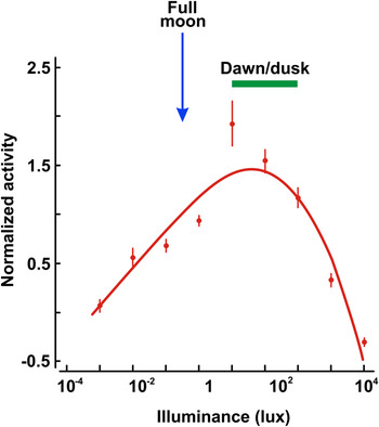
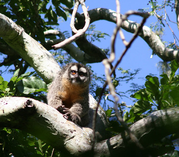
There has long been debate as to whether the early primates were nocturnal or diurnal and that concern has focused attention on the photic activity patterns of contemporary primates. In recent years, numerous examples have been reported of cases where certain primate species, mostly strepsirrhines, display highly variable distributions of daily activity (e.g., Bearder et al., Reference Bearder, Nekaris and Curtis2006; Curtis & Rasmussen, Reference Curtis and Rasmussen2006; Parga, Reference Parga2011). To encompass such behavior, the word “cathemeral” was coined to describe species whose activity rhythms occur during both the light and dark phases of the daily cycle (Tattersal, Reference Tattersal1987). Some particularly instructive observations have been made on the owl monkey, Aotus. Although this animal is typically considered as nocturnal, as was noted above, one species (A. azarae) native to portions of northern Argentina shows a considerable amount of daylight activity (Fernandez-Duque, Reference Fernandez-Duque2003). Detailed examinations of this species reveal that multiple factors influence its daily activity cycles, including ambient temperature, food availability, and illuminance levels (Fernandez-Duque & Erkert, Reference Fernandez-Duque and Erkert2006; Fernandez-Duque et al., Reference Fernandez-Duque, Iglesia and Erkert2010; Erkert et al., Reference Erkert, Fernandez-Duque, Rotundo and Scheideler2012). Averaged across an entire year, about 37% of the activity of this monkey occurs during the dark hours and about 20% during daylight hours, but the distribution is markedly uneven in that nocturnal activity is more prominent during the warmer months than during the months when the temperature drops to lower levels. Light level is also important in influencing behavior. The relationship between light level and activity for free-ranging A. azarae recorded in their native habitat is illustrated in Fig. 5, which shows that the average activity of this species is scattered across the daily cycle, peaking at light levels that are characteristic of dawn and dusk, just when rods and cones are active jointly, and then becoming less frequent—but not absent entirely—during periods of either high (midday, as in the example shown in Fig. 6) or low (moonless night) light levels. Observations like these make clear that forced categorizations of animal photic rhythms into a small number of categories can be misleading with regard to the adaptability of animal behavior and perhaps that is part of the reason why there are no completely consistent links between animals traditionally considered to be “nocturnal” and loss of function of SWS1 opsin genes.
Locomotor activity levels of wild Aotus monkeys as a function of ambient illuminance levels. The points represent the averaged activity levels for nine animals. Note that although Aotus is classically considered to be a nocturnal primate, in fact commonly referred to as the “night monkey,” they show greatest activity at illuminance levels that corresponds roughly to that present during periods of dawn and dusk. Note also that activity is not absent entirely, either at very dim or at very bright light levels. (Data replotted from Fernandez-Duque et al., Reference Fernandez-Duque, Iglesia and Erkert2010.)

An example of an Aotus monkey engaged in daylight activity in its natural habitat. (Photograph reproduced courtesy of Eduardo Fernandez-Duque and the Owl Monkey Project/Formosa-Argentina.)

Although mammals that are judged to be clearly diurnal are in a distinct minority, none of that group is known to have lost SWS1 cones through gene mutation. Thus, photic lifestyle does seem to serve as an important factor in the transition of SWS1 opsin genes to pseudogene status. At present, the parsimonious conclusion would be that nocturnality, or at least the retinal adaptations normally associated with that status, provides a necessary but not sufficient circumstance for the loss of a functional SWS1 opsin gene.
Spectral sensitivity
The spectral sensitivity peaks of mammalian SWS1-derived cone pigments vary from ∼360 to 440 nm. As noted, the ancestral pigment is believed to have peaked at ∼360 nm. Some species have retained a pigment at that position (UV pigments), but in many other mammals, subsequent sequence variations led to a shift in peak position out to wavelengths of 400 nm and longer (S pigments). In recent years, a number of investigators have sought to identify the specific changes associated with the shifts in these pigments to longer wavelengths. For most mammals, it appears that a single residue change is associated with that spectral shift, Phe being present at site 86 in UV pigments, but then being replaced by Tyr in S pigments (Cowing et al., Reference Cowing, Poopalasundaram, Wilkie, Robinson, Bowmaker and Hunt2002; Hunt et al., Reference Hunt, Carvallo, Cowing and Davies2009). Primates constitute an exception to this rule; a recent suggestion being that a substitution of Thr for Pro at site 93 is principally responsible for the long-wavelength shifting of their SWS1 pigments (Carvalho et al., Reference Carvalho, Davies, Robinson and Hunt2012). An intriguing aspect of this story is that the sequences of all the primates, carnivores, rodents, and cetaceans that have SWS1 pseudogenes predict that they would have coded for an S pigment, not a UV pigment, in turn implying that these pigments must have been peak shifted up into the visible wavelengths prior to undergoing the loss of function (Hunt et al., Reference Hunt, Carvallo, Cowing and Davies2009). The peak shift from the UV to the visible wavelengths probably indexes an ancestral change from nocturnality to diurnality, thus suggesting the possibility that the shift of the short-wavelength pigment into the visible wavelengths makes the subsequent loss of SWS1 gene function more likely to be triggered by other factors, in these cases a return to nocturnality. Unfortunately, that linkage does not seem to hold universally because the sequences of the SWS1 pseudogenes found in various species of bat indicate that they formerly coded for UV pigments (Zhao et al., Reference Zhao, Rossiter, Teeling, Li, Cotton and Zhang2009), and thus, a shift in spectral sensitivity prior to pseudogenization has not consistently happened in all mammals.
Taxon-specific explanations
Over their history, mammals evolved adaptations that have allowed them to radiate and find success in a staggering range of diverse habitats. Such variability raises the possibility that the circumstances surrounding the conversion of SWS1 opsin genes to pseudogenes might be taxon specific. In that spirit, unique scenarios underlying the gene loss have been proposed for two groups of animals.
Pinnipeds and cetaceans
As noted above, analysis of sequence changes suggest that loss of function of the SWS1 opsin gene occurred early in the evolution of each of these groups. Both pinnipeds and cetaceans evolved from terrestrial ancestors, and thus, some early representatives of these groups probably inhabited shallow coastal waters in which ambient light may be spectrally filtered by materials from land drainage. Such filtering can selectively reduce the amount of short-wavelength light, and that possibility prompted the suggestion that the loss of short-wavelength cones for animals living in such an environments would incur little cost and perhaps even yield some advantages of economy (Peichl et al., Reference Peichl, Behrmann and Kroger2001; Griebel & Peichl, Reference Griebel and Peichl2003). These animals later migrated into deeper waters offering more short-wavelength-rich environments where it would seem more adaptive to have the sensitivity provided by S cones, but perhaps by that point, the reversal of the mutational change became improbable (Peichl et al., Reference Peichl, Behrmann and Kroger2001). Levenson and Dizon (Reference Levenson and Dizon2003) have offered the alternative possibility that the loss of S cones in these marine mammals might be better linked to the high scattering of short-wavelength lights that is typical of water environments, suggesting that a reduction in sensitivity to short-wavelength lights occasioned by the absence of S cones might serve to increase the visibility of broad spectrum or of relatively long-wavelength targets.
Bats
As explained previously, some bats from the suborder Yinpterochiroptera have lost SWS1 opsin gene function (Table 1), while others have not. Zhao et al. (Reference Zhao, Rossiter, Teeling, Li, Cotton and Zhang2009) suggest that this complex pattern of loss and retention may parallel the evolution of other aspects of the sensory capacities of these bats. Specifically, some of the species that have lost functional short-wavelength-sensitive cones have also evolved a particular high duty cycle form of echolocation that is believed adaptive for hunting in dense vegetation, an environment that might render dichromatic color vision “redundant,” and thus, they suggest that the loss of such cones would incur little or no cost. Sequence comparisons imply that the loss of cone function in other bats from this same group occurred independently, and for those cases, it is suggested alternatively that they might be linked to the development of another behavioral innovation, those showing loss being animals now roosting in caves, rather than in trees, the argument being that this perhaps would have shifted the animals’ emphasis on vision to an increased dependence on olfaction, a change that in turn might foster relaxation in selective constraint on the SWS1 opsin gene (Zhao et al., Reference Zhao, Rossiter, Teeling, Li, Cotton and Zhang2009; Fujun et al., Reference Fujun, Kailang, Tengteng, Paul, Xuzhong and Yi2012).
It is undeniably tempting to point to specific circumstances in the natural histories of various species that may have surrounded the loss of their SWS1 opsin gene function. Yet at this point, one must conclude that there are no evidential grounds for assuming that any of these specific scenarios rise beyond the level of suggestion.
Adaptive gene loss
Pseudogenization is generally conceived to reflect neutral events in which nonsense mutations are accumulated under conditions where functional constraints have been relaxed allowing a drift to fixation (Demuth & Hahn, Reference Demuth and Hahn2009). There are some instances, however, where inactivation of a previously functional gene is believed to have yielded an adaptive advantage. One such case is that of a human gene that encodes a proteinase involved in the suppression of an immune response to endotoxins. Humans outside of Africa carry a null allele of this gene, but 10% of African populations still maintain a functional allele (Wang et al., Reference Wang, Grus and Zhang2006). The implication drawn from this fact is that the spread of the pseudogene was most likely promoted through positive selection, a process that presumably started around the time of the onset of the migration of modern humans out of Africa.
To date, there appears to be only a single instance for which it has been claimed directly that the pseudogenization of an SWS1 opsin gene may have yielded an adaptive advantage. As described above, the blind mole rat (Spalax) lacks a functional SWS1 opsin gene (Table 1) while still retaining a viable LWS opsin gene and its associated photopigment. The eye of this subterranean rodent is covered with a layer of skin and fur, which greatly attenuates the amount of light having wavelengths less than ∼450 nm reaching the retina (David-Gray et al., Reference David-Gray, Janssen, DeGrip, Nevo and Foster1998). Because the only apparent function supported by the retina of the blind mole rat is to supply signals that underlie the entrainment of circadian rhythms, and as short-wavelength light is passively excluded from contributing to this task, it has been argued that the loss of a UV-sensitive photopigment might well increase overall fitness of this species by reducing the need for those metabolically energetic neural networks associated with the processing of signals from short-wavelength cones, and thus, that the loss of function of the SWS1 gene may have been driven by selection pressures initiated coincident with the loss of functional vision in this species (David-Gray et al., Reference David-Gray, Bellingham, Munoz, Avivi, Nevo and Foster2002).
Although the scenario suggested for the blind mole rat is provocative, it remains an unanswered question as to whether the loss of SWS1 function in this species was adaptive or not, nor is there any clear evidence to suggest that similar losses that have occurred in any of a wide variety of different mammals may have been adaptive. Presumably, the principal argument for such adaptive loss explanations would be similar to that advanced for the blind mole rat, that is, it is energetically expensive to maintain the neural architecture linked to the presence of S cones. It is generally agreed that energy constraints do shape the evolution of sensory systems (Niven & Laughlin, Reference Niven and Laughlin2008) and, given that fact, might not the loss of S cones lead to a reduction in nervous system organizations and thus improve energy efficiency? At present, that question does not seem to have an answer. It does appear that in the Aotus monkey, a primate that has lost functional SWS1 opsin genes, some of the retinal circuitry associated typically with the processing of S-cone signals, including the S-cone bipolar cells and small bistratified ganglion cells, is absent, and this reduction could conceivably reduce some neural costs (Silveira et al., Reference Silveira, Saito, Lee, Kremers, Filho, Kilavik, Yamada, Perry, Heywood, Milner and Blakemore2004). Still, as we have seen, mammalian S cones constitute only a small minority of their photoreceptor complement, and it is uncertain whether their absence would count for much, nor indeed, do we know whether in fact these cells and their forward projections might not simply be usurped and then used adaptively to support other aspects of visual processing. We do know that in the case of the olfactory receptor gene family, where pseudogenes are particularly abundant in some lineages, analyses evaluating various aspects of the relative frequencies of synonymous to nonsynonymous substitutions in odorant receptor genes suggest strongly that the olfactory receptor pseudogenes are evolving neutrally (Gilad et al., Reference Gilad, Bustamante, Lancet and Paaba2003). Similar analyses have not yet been reported for SWS1 opsin pseudogenes, and thus, for the present, there is no concrete evidence to suggest that the loss of function in these genes may have led to an increase in fitness.
Conclusions
The cone pigments of eutherian mammals are linked to the activity of opsin genes from two separate gene families, SWS1 and LWS, and the majority of contemporary mammals express two kinds of cone photopigments, one from each of the gene families. About 20 years ago, it was discovered that the SWS1 genes in two species of primate had been inactivated by mutational changes in gene structure, and that, consequently, the retinas of these animals contained only a single type of cone photopigment. Among other potential changes in visual capacity, these alterations most likely led to loss of a dimension of color vision.
Since those early discoveries, similar genetic mutations leading to losses of SWS1-linked cone pigments have been detected in a diverse group of mammals, including animals as disparate as whales and flying squirrels. To date, this list includes several dozen species from five different orders. Although the nature of the disabling mutations vary from case-to-case, and although phylogenetic comparisons suggest that these alterations occurred at various stages during the evolutionary histories of the different lineages, in each instance, it appears that all the members of the species lack cones having maximum sensitivity to the short wavelengths. At this point, it is not known what the total number of mammalian species that have been similarly afflicted may be, but given the pattern of loss to date, it would not be surprising to learn eventually that at least 10% of all mammalian species have lost their short-wavelength cones as a result of genetic mutations. No matter what that number eventually turns out to be, the loss of SWS1-specified photopigments represents a significant trend in the evolution of mammalian vision.
This paper explored a variety of possibilities that might explain why mammalian SWS1 opsin genes have been converted to pseudogenes in such a disparate collection of animals. Significantly, all the species that have lost short-wavelength cones in this fashion are nocturnal, or at least, they possess retinal organizations that are generally linked to that state. At the same time, however, not all nocturnal species have SWS1 pseudogenes, and it thus appears that a nocturnal lifestyle is a necessary prerequisite for loss of function of these opsin genes, but that nocturnality is, by itself, not sufficient to completely account for the loss of gene function. Several other features that may have contributed to the loss of short-wavelength cones were considered also; among them, that the spectral sensitivity of the cone class prior to its loss might have been critical, that the reason for the loss is uniquely embedded in the natural histories for each lineage, or that the loss may actually have been supported as an adaptive change. Although each of these deserves further consideration, there is no evidence at present that allows either acceptance or rejection of these possibilities. However it may eventually turn out, it is a remarkable fact that a significant number of mammalian lineages have lost functional short-wavelength cone pigments and, with them, the potential for deriving a dimension of daylight color vision.
One can of course only speculate as to what the future may hold for mammalian SWS1 opsin genes. However, since nocturnality is the only feature of mammalian life that seems to be correlated clearly with the loss of function in SWS1 opsin genes, and since there are numerous nocturnal mammals that currently harbor functional opsin genes, it seems a reasonable prediction that over the years to come at least some of these species will join those reviewed here in abandoning their functional short-wavelength cones and the visual capacities they support. At the same time, given the obvious adaptive utility of short-wavelength cones in mammals that incline toward diurnality, it seems also all but certain that the SWS1 opsin gene family will not follow the Rh2 and SWS2 gene families in simply disappearing from the genomes of eutherian mammals.