Introduction
Since the advent of cochlear implants, many detailed anatomical studies have been performed regarding the landmarks and locations of various middle- and inner-ear structures. This enhances the precision and ease of implant surgery. A sound knowledge regarding the topographical anatomy of the cochlea, and especially of the distances between it and adjacent structures, is essential for ear surgery in general and in surgical procedures for cochlear implantation in particular.Reference Wysocki and Skarzyñski1
Various methods have been explored to reduce or control complications following implant surgery. One post-operative complication of cochlear implantation is facial nerve stimulation, which leads to twitching and tingling sensations in facial muscles. Kelsall et al. proposed a grading scale for facial nerve stimulation, which ranges from grade I, that is, ‘no stimulation’, to grade VI, that is, ‘total stimulation – severe gross motion of total facial musculature with or without severe pain’.Reference Kelsall, Shallop, Brammeier and Prenger2 Thus, facial nerve stimulation is a complication that can cause high levels of discomfort, warranting explantation and removal of the whole device. When initially reported by Cohen and Hoffman, its incidence was quite low.Reference Cohen3,Reference Cohen and Hoffman4 However, in more recent studies, the incidence of facial nerve stimulation ranged from 7 to 15 per cent.Reference Kelsall, Shallop, Brammeier and Prenger2,Reference Kruschinski, Weber and Pabst5
It has been suggested that the labyrinthine portion of the facial nerve is the most likely area of stimulation in most patients, and the mid-cochlear electrodes mostly appear to cause the stimulation of this segment.Reference Cohen and Hoffman4,Reference Bigelow, Kay, Rafter, Montes, Knox and Yousem6 The mid-basal turn is the part of the cochlea in closest proximity to the labyrinthine portion of the facial nerve and the geniculate ganglion.
Facial nerve stimulation is observed to occur more frequently in patients who are implanted for otosclerosis, with the incidence in these cases as high as 75 per cent.Reference Sainz, Garcia-Valdecasas and Ballesteros7 It is speculated that otosclerosis leads to structural abnormalities in the otic capsule, making the bone more spongiotic, and thus allowing more conductivity for electrical impulses and leading to easier spread of current from the middle turn of the cochlea to the labyrinthine segment in the close vicinity.Reference Kruschinski, Weber and Pabst5
The thin lamina of bone present between the middle turn and the labyrinthine segment might represent an anatomical basis for facial nerve stimulation.Reference Kelsall, Shallop, Brammeier and Prenger2 Thus, the distance between these two structures is an important parameter to predict the likelihood of this complication. Such information can be used to counsel the patient before the surgery regarding the possibility of this complication.
High-resolution computed tomography (CT) of the temporal bone can be used to measure the distance between the middle turn of the cochlea and the labyrinthine segment of the facial nerve, which is termed the cochleo-facial distance. Our study aimed to calculate this distance in cadaveric human temporal bones radiologically and by dissection.
Materials and methods
This study was conducted by the Departments of Otorhinolaryngology, Anatomy, and Radiology at the Sawai Man Singh (SMS) Medical College and Hospital, Jaipur, India.
Cadaveric human temporal bones were obtained from the anatomy department. Bones with distorted anatomy caused by a disease process or a problem in preservation during bone preparation (preservation or cutting), and bones with cochleovestibular malformations, were excluded.
All specimens were first subjected to imaging by high-resolution CT scan. The CT scans were performed on a 128-slice Philips Ingenuity CT Scanner (Koninklijke Philips, Amsterdam, Netherlands). Axial, coronal and multiplanar reconstructions were performed for the CT scans.
The specimens were dissected in the temporal bone dissection laboratory of the ENT department. Wide mastoidectomy was carried out, with identification of the tegmen tympani, sigmoid sinus and mastoid portion of the facial nerve. Canal down mastoidectomy was performed and drilling was undertaken to expose the middle turn (2 mm anterior to the oval window). After identification of the middle turn, dissection was performed to identify the second genu and labyrinthine segment of the facial nerve. Cochleo-facial distance was then measured using millimetre square paper. All steps were digitally documented using a camera (Figure 1a and b). The recorded findings were analysed using image processing and analysis software, ImageJ, developed by the US National Institutes of Health.8 The radiologist and the surgeon dissecting the bone were blinded and did not know the other measurement results for each temporal bone.
(a & b) Measurement of cochleo-facial distance on dissection. ‘*’ = labyrinthine segment of facial nerve; ‘>’ = middle turn of cochlea

The cochleo-facial distance was the minimum distance between the middle turn of the cochlea and the labyrinthine segment of the facial nerve. It was measured on double oblique, coronal, high-resolution CT images of the temporal bone (Figure 2a and b) with the help of Digital Imaging and Communications in Medicine (‘DICOM’) viewer software (Navegatium and RadiAnt Digital Imaging and Communications in Medicine viewers), which was free to use on a Microsoft™ Windows 10 operating system enabled computer.
(a & b) Measurement of cochleo-facial distance on double oblique, coronal, high-resolution computed tomography scans of the temporal bone. ‘*’ = labyrinthine segment of facial nerve; ‘>’ = middle turn of cochlea

Results
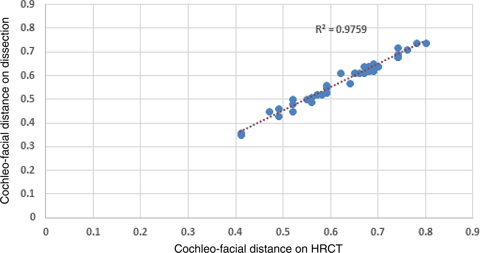
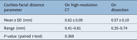
The present study comprised 40 temporal bones. On high-resolution CT, the mean (± standard deviation (SD)) cochleo-facial distance was 0.62 ± 0.09 mm, ranging from 0.41 to 0.81 mm, while on dissection it was 0.57 ± 0.10 mm, ranging from 0.35 to 0.74 mm. The p-value for paired t-test was 0.368, indicating that the difference between the two measurements was not significant. The results are summarised in Table 1. Figure 3 summarises the cochleo-facial distance of all 40 human temporal bones and compares this measurement on high-resolution CT and on dissection. The comparison can be best portrayed on a linear regression plot, which can be seen in Figure 4.
Comparison of cochleo-facial distance on high-resolution computed tomography (HRCT) and on dissection.

Linear regression plot of cochleo-facial distance on high-resolution computed tomography (HRCT) and on dissection. Blue dots indicate temporal bones.

Comparison of cochleo-facial distance on high-resolution CT and dissection

CT = computed tomography; SD = standard deviation
The Pearson correlation co-efficient of r = 0.89 denotes a very strong relationship for the distance between the middle turn of the cochlea and the labyrinthine portion of the facial nerve on high-resolution CT and the same distance on dissection. As seen in Figure 4, a very strong, positive, linear relationship is observed between this cochleo-facial distance on high-resolution CT and the same distance on dissection. The co-efficient of determination is R2 = 0.9759.
Discussion
Cochlear implantation is an advanced surgical treatment for bilateral severe to profound hearing loss. Appropriate interpretation of the radiological findings, and their correlation with intra-operative findings, enhances surgical precision and results in fewer complications. One of the less-discussed complications of implant surgery is stimulation of the facial nerve along with the cochlear nerve, leading to significant problems in a considerable number of patients. This happens because of the close proximity of the labyrinthine portion of the facial nerve with the middle turn of the cochlea, leading to stimulation of the nerve when current passes through the nearby electrodes placed in the cochlea. This distance becomes especially important when cochlear implantation is performed in otosclerosis cases, as otospongiotic bone conducts current more easily and can therefore increase the likelihood of facial nerve stimulation.
This cochleo-facial distance, although very small, varies considerably amongst individuals as well as between bony labyrinths of the two sides in the same person. A pre-operative high-resolution CT temporal bone scan can determine this distance and aid decisions regarding the side of surgery for implantation, to decrease the likelihood of facial nerve stimulation. It also helps us to counsel patients regarding the possibility of facial nerve stimulation following implantation. This study was performed with these objectives in mind.
On reviewing the literature, very few studies were found that measured the distance between the facial nerve and the cochlea. There was no study comparing the cochleo-facial distance on cadaveric dissection and radiology. In our study, this mean (± SD) distance on high-resolution CT was 0.62 ± 0.09 mm, ranging from 0.41 to 0.81 mm, and on dissection it was 0.57 ± 0.10 mm, ranging from 0.35 to 0.74 mm.
Kruschinski et al.,Reference Kruschinski, Weber and Pabst5 in their 2003 study, found the average distance to be 0.33 ± 0.14 mm, which is much shorter than our finding. Another study, by Redleaf et al.,Reference Redleaf and Blough9 conducted in 1996, found that this distance ranged between 0.06 and 0.8 mm.
The comparison between the distances measured on CT scans and those measured on dissection gave a mean difference of only 0.05 mm. This was not found to be statistically significant, with a p-value of 0.368. Therefore, a reasonably good idea of the proximity of the facial nerve labyrinthine segment to the middle turn of the cochlea (cochleo-facial distance) can be obtained pre-operatively through a high-resolution CT scan of the temporal bone.
It should also be noted that this distance varied considerably amongst a few specimens (range, 0.35–0.74 mm), being even shorter than 0.4 mm in some cases. Usually, the smallest burrs used in otology practice are 0.6 mm in diameter, highlighting the need to acknowledge the care and precautions needed while working in this area.
Management of troublesome facial nerve stimulation includes switching off the relevant electrodes, which can significantly hamper the hearing outcome in some cases. Fluoride was used as a treatment in refractory cases by Gold et al., with good results, showing dramatic resolution of the facial nerve stimulation and thus allowing continued successful use of the implants.Reference Gold, Miller, Kamerer and Koconis10
• Cochlear implants have dramatically changed the management of severe to profound hearing loss, with good outcomes
• Facial nerve stimulation is a lesser studied but troublesome complication of cochlear implantation
• The labyrinthine portion of the facial nerve is the segment closest to the cochlea, mainly the middle turn
• Electrical impulses from this middle turn might stimulate the labyrinthine segment, giving facial twitching, more so in otospongiotic bone
• This cochleo-facial distance can be determined pre-operatively by high-resolution computed tomography temporal bone scan
• Such information can aid implant side selection and pre-operative patient counselling
It is advised that we use half-banded, modiolus-hugging electrodes in cases of otosclerosis, so that the spread of current is limited and not towards the facial nerve. This distance from the facial nerve is also important in the middle cranial fossa approach, as in those cases there is a high chance of opening the cochlea while dissecting the labyrinthine portion of the facial nerve, leading to unexplained hearing loss post-operatively.Reference Kruschinski, Weber and Pabst5,Reference Redleaf and Blough9
Thus, a variety of factors, including implant design, side of implantation and local tissue impedances, may interact to produce incidental facial nerve stimulation.Reference Niparko, Oviatt, Coker, Sutton, Waltzman and Cohen11 The distance between the labyrinthine segment of the facial nerve and the middle turn of the cochlea may deserve increased recognition as a factor in cochlear implant performance, as this distance – which can be accurately predicted pre-operatively – can significantly alter the outcome with regard to this complication.
Theoretically, we can conclude that the distance between the labyrinthine segment of the facial nerve and the middle turn of the cochlea may influence the development of facial nerve stimulation, and cochleo-facial distance measurements can be used to prevent this complication. These findings can also be applied to otospongiotic bones, in which this complication might occur more frequently.
Conclusion
The labyrinthine segment of the facial nerve is the most likely area of stimulation in most patients suffering from facial nerve stimulation post-cochlear implantation. Pre-operative high-resolution CT temporal bone scans (coronal, double oblique) can aid examination of the bone separating the labyrinthine segment from the middle turn of the cochlea. This may have clinical significance regarding implant side selection (if other audiological and radiological factors are not very different between the two ears) and may benefit pre-operative counselling of the patient. Prevention of facial nerve stimulation could therefore be achieved by cochlear implant designs and surgical planning that consider this minimal distance between the labyrinthine segment of the facial nerve and the middle turn of the cochlea.
Competing interests
None declared