Introduction
Cricopharyngeal dysfunction may result from failure of the cricopharyngeal muscular portion of the cricopharyngeal or upper ooesophageal sphincter to relax adequately, thereby: preventing the larynx from lifting and moving forward; reducing laryngeal motion up and forward; and/or causing poor pressure to drive the food bolus through the sphincter and to widen the opening.Reference Logemann1 When the dysfunction is due to a cerebrovascular or nervous system disorder or a myopathy, it is defined as secondary-related cricopharyngeal dysfunction. Those cases with no apparent underlying cause are defined as idiopathic cricopharyngeal dysfunction.
There are various procedures for treating cricopharyngeal dysfunction. Of these, cricopharyngeal myotomy is reported to be the most effective in cases of advanced dysfunction.
In the current case, a cricopharyngeal myotomy was used to treat cricopharyngeal dysfunction with associated significant piriform sinus expansion.
Patient and methods
An 80-year-old man had experienced a sensation of obstructed swallowing for three years, and had presented to an otolaryngology clinic in 2000. The initial diagnosis had been cervical oesophageal stenosis. The patient's swallowing difficulty had gradually worsened. Over the next two years, he had lost more than 20 kg (his height was 160.5 cm) because of aspiration during mealtimes (two or three times per meal), and he had eventually required more than an hour to complete a meal. Finally, the patient presented at our hospital in September 2002 with severe dysphagia.
A general physical examination showed the patient to be lucid with no abnormal findings, including appendicular peripheral visual fields and muscular strength. The tongue showed good mobility with no fasciculation. The patient's soft palatal and pharyngeal reflexes were moderately decreased.
Laryngofibrescopy revealed good movement of the bilateral vocal folds, significant salivary effusion in the bilateral piriform sinuses and submucous swelling of the inferior pharyngeal wall. Magnetic resonance imaging of the head revealed diffuse cerebral atrophy, but the brainstem was normal. Cervical computed tomography scanning revealed no organic abnormality. Oesophagoscopy could not be employed due to stenosis of the cricopharyngeal region. The patient's iron, creatinine phosphokinase and c-reactive protein levels were normal, and serological tests for rheumatoid factor and antinuclear antibody were negative.
Videofluorography
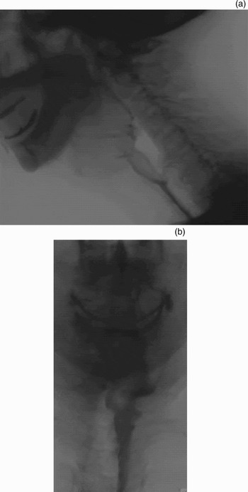
Videofluorography with barium swallow was performed. Although the patient could hold 5 ml of ingested barium in his oral cavity, as seen on lateral view, this bolus could not be delivered normally into the pharynx with the rolling movement of the tongue. In addition, the triggering of the pharyngeal swallowing reflex was moderately poor at the end of the oral phase. In other words, the pharyngeal reflex was not easily triggered. When a small quantity of barium flowed into the bilateral piriform sinus, a pharyngeal reflex occurred after stimulation of the piriform sinus where barium had accumulated. Laryngeal elevation was reduced and pharyngeal wall contractions were weak bilaterally. Because the cricopharyngeal opening was not significant, the barium did not pass easily through the upper oesophageal sphincter.
A cricopharyngeal bar was observed at the inferior wall of the cricopharyngeal region. Most of the barium remained behind in the piriform sinuses, but there was no aspiration. An anterior projection from the inferior hypopharyngeal wall was identified, involving bone growth from the fifth and sixth cervical vertebrae (Figure 1a). In the anteroposterior view, the remaining barium was pooled in the significantly expanded piriform sinuses (Figure 1b).
Videofluorography views. (a) Lateral view; 5 ml of ingested barium did not pass easily into the upper oesophageal sphincter, because the cricopharyngeal opening was not significant. Two cricopharyngeal bars were found (arrows). (b) Anterior view, showing the remaining barium pooled in the significantly expanded piriform sinuses.

Electromyography
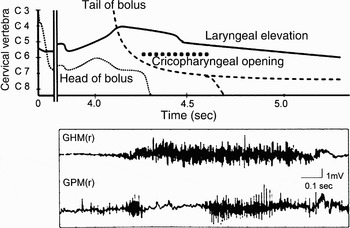
Electromyography was recorded under videofluorography guidance (Figure 2).Reference Yoshida2 The activity waves of the right thyropharyngeal and cricopharyngeal muscles were expressed with the duration of the barium passage in pharyngeal swallowing by deglutition. As a result, the duration of cricopharyngeal relaxation coincided with cricopharyngeal opening. Just after the start of geniohyoid contraction, the onset of barium passage to the cricopharyngeal region occurred simultaneously with the start of cricopharyngeal relaxation. The opening time of the cricopharyngeal region was approximately 0.35 seconds. The restart of cricopharyngeal contraction coincided with completion of barium passage to the cricopharyngeal region.
Electromyographic examination (lower panel) under videofluorography (upper panel) guidance. The right thyropharyngeal and cricopharyngeal action potential were identified in pharyngeal swallow. The no firing time from 4.3 to 4.6 sec of the cricopharyngeal muscle coincided with the duration of cricopharyngeal opening. The opening time of the cricopharyngeal region was approximately 0.35 sec. Laryngeal elevation was based on the level of the vocal fold. GHM = geniohyoid muscle; CPM = cricopharyngeal muscle; r = right; sec = seconds

Pharyngeal manometry
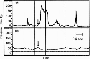
Manometry was conducted under videofluorography guidence.Reference Yokoyama, Mitomi, Tetsuka and Tayama3 The baroceptor had two pressure sensors, one located in the oropharynx (i.e. at the tongue base) and the another in the cricopharyngeal region. The pressure of the cricopharyngeal region was remarkably elevated when the patient swallowed 5 ml of barium, and required approximately 1.0 second to drop to zero. On the other hand, the oropharyngeal pressure was not elevated at all during that time (Figure 3).
Manofluorography results for cricopharyngeal region (upper panel) and tongue base (lower panel). The pressure in the cricopharyngeal region was remarkably elevated during swallowing of 5 ml barium and required approximately 1.0 seconds to drop to zero. In contrast, the oropharyngeal pressure was not elevated at all during this time. Arrow indicate the point at which 5 ml of barium was ingested.

The patient's dysphagia was diagnosed as idiopathic cricopharyngeal dysfunction. A Mendelsohn manoeuvre and head rotation were administered as rehabilitation. However, although the patient had adequate time (over six months) to recover, his dysphagia did not resolve.
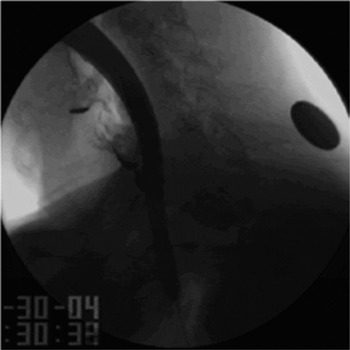
Therefore, surgical widening of the cricopharyngeal region was performed using a bougie (Figure 4); however, the patient gained only minor relief from his dysphagia as a result.
Widening of cricopharyngeal region using a bougie.

Operation
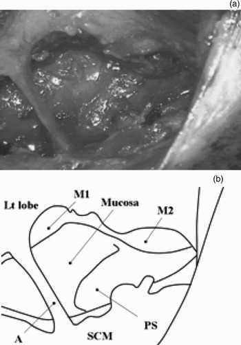
A bilateral cricopharyngeal myotomy was thus performed under general anaesthesia (Figure 5). A balloon catheter was pre-operatively inserted until the oesophagus extended the cricopharyngeal region. An external horizontal incision was made above the cricothyroideus ligament. The sternothyroid and sternohyoid muscles were amputated bilaterally on the incision line. The cricopharyngeal muscles were identified bilaterally from outside each thyroid gland and were cicatricial. The cricopharyngeal muscles were resected posteriorly as far as possible.
(a) Surgical view of cricopharyngeal myotomy; (b) a diagram of the same surgical view. The cricopharyngeal muscle resection was extended upward to include some of the inferior constrictor muscle, and also into the oesophageal muscle. After the resections, the mucosa of the piriform sinuses showed wide expansion bilaterally. M1 = cricopharyngeal muscle; M2 = inferior constrictor muscle; SCM = sternocleidomastoid muscle; Lt lobe = left lobe of thyroid gland; A = inferior thyroid artery; mucosa = mucosa of oesophageal aditus; PS = mucosa of left piriform sinus

The resection was extended upward to include some of the inferior constrictor muscle, and also into the oesophageal muscle. The bilateral inferior constrictor muscles were thinly extended over the frame of the thyroid cartilage. After the resections, the mucosa of the bilateral piriform sinuses showed wide expansion.
Histopathological findings
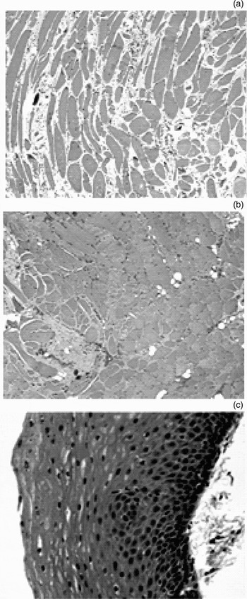
The histopathological findings are shown in Figure 6. Cricopharyngeal muscular fibres (of various sizes) and stromal fibrosis were identified. There was no infiltration of inflammatory cells. Although mild fatty degeneration of the thyropharyngeal muscle was detected, the muscular fibres were almost normal.
Photomicrographs. (a) Cricopharyngeal muscle. Cricopharyngeal muscular fibres of various sizes and stromal fibrosis were observed; there was no infiltration of inflammatory cells. (b) Thyropharyngeal muscle. Mild fatty degeneration was detected; the muscular fibres were almost normal. (c) Cricopharyngeal epithelium. There was no epithelial hyperplasia or infiltration of inflammatory cells in the submucosa. (H&E; ×100)

A post-operative oesophagoscopy was able to be performed through the cricopharyngeal region. As a result, a biopsy of the mucosa of the cricopharyngeal region was obtained. There was no epithelial hyperplasia or infiltration of inflammatory cells in the submucosa.
A post-operative videofluorography was conducted. The triggering of pharyngeal swallowing was found to have considerably improved at the end of the oral phase. The weak pharyngeal contraction had not changed, but the passage disorder of the cricopharyngeal region had improved (Figure 7). The barium effusion into the piriform sinuses, observed pre-operatively, was found to have decreased; in other words, pharyngeal clearance after deglutition had improved.
Videofluorogram performed after the cricopharyngeal myotomy. (a) Lateral view; (b) anteroposterior view. The passage disorder of the cricopharyngeal region was observed to have improved.

Three years later, this patient's dysphagia had resolved.
Discussion
Cricopharyngeal dysfunction accompanies various kinds of neuromuscular disorder. Cases not associated with other diseases are termed idiopathic cricopharyngeal dysfunction.Reference Reichert, Bluestone, Stool, Sieber and Sieber4 Hirano et al. Reference Hirano, Ohkubo, Yoshida, Esaki, Yoshida and Shin5 reported that, in secondary-related cricopharyngeal dysfunction, a passage disorder at the cricopharyngeal region occurs, causing cricopharyngeal muscle relaxation to be completed before the bolus reaches the oesophageal aditus. In the current patient, because the relaxation time of the cricopharyngeal muscle on the electromyogram coincided with the cricopharyngeal opening time on the videofluorography, the cricopharyngeal region appeared to open during the pharyngeal phase but this opening was insufficient. Therefore, it was thought that the current dysphagia had been caused by insufficient relaxation of the cricopharyngeal muscle.
Cruse et al.,Reference Cruse, Edwards, Smith and Wyllie6 Hirano et al. Reference Hirano, Ohkubo, Yoshida, Esaki, Yoshida and Shin5 and Murofushi et al. Reference Murofushi, Nagao, Semba, Mizuno, Niimi and Hirose7 reported that, in cases of idiopathic cricopharyngeal dysfunction, the cricopharyngeal muscle shows diffuse atrophy of muscle cells and/or stromal fibrosis, and has a histological appearance peculiar to myogenic disease. In contrast, Mori et al. Reference Mori, Fukui, Hyodo, Kawakita, Kadota and Inagi8 reported that the findings in similar cases, in spite of advanced age, show more hypertrophy of muscle cells than atrophy, together with fibrosis of muscle cells. Currently, there is no consensus concerning aetiology of idiopathic cricopharyngeal dysfunction because the dysfunction is not caused by a single disease. In the current case, the presence of an inflammatory scar on the cricopharyngeal mucosa suggested a cause for the relaxation deficiency of the cricopharyngeal muscle, but there was no pathological abnormality. The pathological findings of diffuse atrophy of muscle cells and stromal fibrosis suggested the presence of cricopharyngeal myopathy. In other words, it was thought that the significant piriform sinus expansion in the current case developed as a result of the remarkably elevated pressure of the cricopharyngeal sphincter, due to the cricopharyngeal relaxation deficiency caused by cricopharyngeal myopathy. However, further examination would have been required to determine whether the current case was caused by either systemic or local cricopharyngeal myopathy.
Cricopharyngeal dysfunction is generally treated conservatively initially, for example using rehabilitation therapy. Some authors have also suggested that a bougie and a balloon catheter can be used successfully to expand the oesophageal aditus.Reference Fujishima, Hojo, Ohkuma, Shibamoto, Kojima and Tanaka9 However, in advanced cases of cricopharyngeal dysfunction, a cricopharyngeal myotomy is reported to be the most effective treatment.
Conclusion
This report described a case of idiopathic cricopharyngeal dysfunction with significant piriform sinus expansion. Findings from electromyography and manometry, performed under videofluorographic guidance, were indispensable in differentiating cricopharyngeal dysfunction from other diseases. In addition, histopathological examination of the cricopharyngeal muscle, the neighboring muscles and the cricopharyngeal mucosa was necessary to make an accurate diagnosis. Our patient's significant piriform sinus expansion could have resulted from the remarkably elevated cricopharyngeal sphincter pressure, due to the cricopharyngeal relaxation deficiency caused by cricopharyngeal myopathy.