Introduction
The fig leaf roller (FLR), Phycodes radiata (Ochsenheimer, 1808) (Lepidoptera: Brachodidae) is a significant pest of the fig tree (Ficus carica L.). This pest has been reported in various regions, including Nepal, Sri Lanka, India, Afghanistan, and Pakistan. It was first reported from central Iran in 2010 (Ramezani Karim et al., Reference Ramezani Karim, Imani, Basirian and Kumar2010). Recently, FLR has been observed in Semnan province of northern Iran with a high level of damage (Lotfalizadeh et al., Reference Lotfalizadeh, Ajamhassani, Abdos and Mohammadi Khoramabadi2024). The larvae of P. radiata initiate damage to fig leaves from early May to early June, depending on the climatic conditions. As larval feeding progresses, the leaves become crumpled and covered with black webbing and frass, clear damage indicators. In subsequent generations, the larvae extend their feeding to the fruits. By spinning webs from the leaves to the fruits, the larvae establish themselves on the fig fruit, creating channels through which they enter and feed (Dabhi et al., Reference Dabhi, Sisodiya, Trivedi and Patel2021; Kumar and Ramamurthy, Reference Kumar and Ramamurthy2010).
One of the critical approaches to studying the feeding characteristics of various insects is the evaluation of feeding index parameters. These parameters reveal the amount of food consumed and digested by the insects at specific time points, and digestive enzyme role in larval midgut is crucial (Abasabadi et al., Reference Abasabadi, Ajamhassani and Mehrabadi2024; El-Refaie et al., Reference El-Refaie, El-Sayed, Abd-Allah, Ebeid and Abouelnaga2024). The efficiency of digested food is correlated with the insect’s weight and helps to identify the insect’s preferred food sources. The quality of the insect’s diet is determined by the amount of food consumed and digested by the larvae, with feeding index components serving as the primary indicators of the quality and quantity of the insect’s food resources (Hemmati et al., Reference Hemmati, Shishehbor and Stelinski2022; Silva et al., Reference Silva, Bueno, Andrade, Stecca, Neves and Oliveira2017). Insects that feed on nutrient-rich diets exhibit differences in molting frequencies, developmental durations, pupal weights, and fecundity rates compared to those feeding on suboptimal diets. It has been established that insects with optimal nutrition possess stronger immune systems against contaminants and microorganisms (Alaux et al., Reference Alaux, Dukloze, Craucer and Leconte2016; Manjula et al., Reference Manjula, Lalitha and Shivakumar2020). As we know, the immune reaction of the insects is dependent on the energy level received through feeding that is essential for homeostasis (Banville et al., Reference Banville, Browne and Kavanagh2012). When insects consume diets rich in carbohydrates and proteins, the response of haemocytes and phenoloxidase – the primary components of physiological defense against various stresses, fungal and bacterial spores, and toxins – becomes more pronounced (Vogelweith et al., Reference Vogelweith, Moret, Monceau, Thieri and Moreau2016).
Immune activities are crucial mechanisms within the insect circulatory system, influenced by various factors such as starvation, environmental changes, nutrition, diapause, gender, and the entry of invasive agents. These immune responses are categorised into cellular and humoral immunity (Duarte et al., Reference Duarte, Silva, Ribeiro and Carcamo2020; Lavine and Strand, Reference Lavine and Strand2002; Siva-Jothy and Thompson, Reference Siva-Jothy and Thompson2002; Vengateswari et al., Reference Vengateswari, Arunthirumeri and Shivakumar2020). Cellular immunity involves blood cells and is characterised by changes in cell shape, number, type, phagocytosis, and nodulation around foreign agents (Strand, Reference Strand2008). Humoral immunity, conversely, involves changes in the concentration of phenoloxidase and antimicrobial peptides, typically following cellular immunity. Humoral immunity results in the necrosis and melanisation of foreign agents, which are then excreted by the malpighian tubules (Castillo et al., Reference Castillo, Robertson and Strand2006). In immunological studies, the first step is identifying the insect’s haemocytes and their abundance in the haemolymph, followed by their response to foreign agents. The interaction between insect immunity and pathogenic agents does not always favour the pathogen; if the insect has a robust immune system, it can prevent the progression of the infection and even eradicate it (Borges et al., Reference Borges, Santos, Furtado and Figueiredo2008; Moushumi et al., Reference Moushumi, Hazarika, Barooah, Puzari and Kalita2008). The immune capacity of the insect and the concentration of the invading agent are the critical determinants of this interaction. Numerous studies report changes in haemocyte density in response to the introduction of contaminants such as fungi, bacteria, plant metabolites, toxins, and industrial and environmental pollutants into the insect haemolymph (Zheng et al., Reference Zheng, Zhang, Tan, Ma and Yan2023; Suwannakhon et al., Reference Suwannakhon, Sintusopha, Pawaswna, Tatan and Tungjitwitayakul2024). Plasmatocytes and granulocytes, identified as main cells in the immunity of most insects, have shown increased populations in larvae of Chilo suppressalis Walker (Lepidoptera: Crambidae) and Periplaneta americana (L.) (Blattodea: Ectobiidae) treated with Beauveria bassiana (Balsamo) Vuillemin (Zibaee and Malagoli, Reference Zibaee and Malagoli2014; Mudoi et al., Reference Mudoi, Das, Hazarika, Das and Roy2019). Conversely, the blood cell populations in Ostrinia furnacalis (Guenee) (Lepidoptera: Crambidae) and Conogethes punctiferalis (Guenee) (Lepidoptera: Crambidae) larvae have shown significant decreases when exposed to this fungus (Shen et al., Reference Shen, Li, Chu, Lang and An2016; Li et al., Reference Li, Liu, Kang, Li, Lu, Li, Pang, Zheng and Yin2022).
Chemical compounds are commonly used to control insect pests. Despite their effectiveness, these compounds have several drawbacks, including accumulating residues in water and soil, developing resistance in insect populations, and destroying non-target organisms (Raven and Wagner, Reference Raven and Wagner2021; Sanchez Bayo, Reference Sanchez Bayo2021). In contrast, using predators, parasitoids, and entomopathogenic fungi (EPF) as biocontrol agents offers an environmentally friendly alternative for pest management (Barra et al., Reference Barra, Rosso, Nesci and Etcheverry2013; Kamel et al., Reference Kamel, Eid, Nouh and Adly2024). EPFs such as B. bassiana, Metarhizium anisopliae (Metschnikoff) Sorokin, and Lecanicillium muscarium (Petch) Zare & W. Gams not only cause diseases in Lepidoptera (Erasmus et al., Reference Erasmus, Berg and Plessis2021; Sutanto et al., Reference Sutanto, Husain, Rasool, Al-Qahtani and Aldawood2021; Chouikhi et al., Reference Chouikhi, Assadi, Lebdi and Belkadhi2022) but also affect their physiological processes, including the immune system, digestive activities, and fecundity (Baja et al., Reference Baja, Poitevin, Araujo, Mirás-Avalos, Zawadneak and Pimentel2020; Aghaee Pour et al., Reference Aghaee Pour, Zibaee, Gohar Rostami, Hoda and Shahriari2021).
Understanding the immune responses of insects is crucial for effectively applying fungal biocontrol agents. Phycodes radiata has become highly destructive in fig orchards in the Semnan region of northeastern Iran over the past two to three years. Although this pest was first reported in Iran 14 years ago (in the fig orchards of Kerman province in southern Iran) (Ramezani Karim et al., Reference Ramezani Karim, Imani, Basirian and Kumar2010), no comprehensive studies have been conducted on its biology and control. Given the significance of this pest in the Semnan region and its potential spread to other areas, this study on the biology, feeding indices of P. radiata when feeding on four fig cultivars, and immune challenges of larvae in confronting EPF provides a foundation for further research and the development of management strategies for FLR.
Materials and methods
Rearing of Phycodes radiata
In May 2022, an initial population of P. radiata eggs was collected from the leaves of a wild Ficus sp. in the forested area of Cheshme Ali, Semnan province (36° 16ʹ 30ʺ N; 45° 05ʹ 02ʺ E), as well as from three edible fig (Ficus carica) cultivars: Zard Semnan, Ghermez Semnan, and Zard Garmsar, from commercial orchards in Semnan (35° 33ʹ 56ʺ N; 53° 22ʹ 31ʺ E). The eggs were collected in several batches and transferred to the Semnan Agricultural Research Center laboratory. The laboratory conditions were maintained at 25 ± 1°C, with a relative humidity of 50% and a light: dark ratio of 14:10 hours. The eggs were placed on the leaves of the respective cultivars to provide food for the larvae upon hatching. The rearing containers measured 30 × 30 × 30 cm, with the upper surface covered in white organdy mesh fabric for ventilation. The larvae were examined daily; fresh leaves were supplied if the fig leaves became dry or wrinkled. Larval faeces were cleaned regularly, and dead or parasitised larvae were removed from the colony to maintain a healthy population.
Investigation of morphological and biological characteristics of Phycodes radiata
For laboratory-based biological studies, the experiments were conducted in the same rearing conditions as detailed in the section above. First-instar larvae were individually placed in petri dishes with mesh lids, each provided with a fig leaf. A total of 30 replicates were conducted for each fig cultivar, resulting in 120 petri dishes. Fresh leaves were provided daily to the larvae, and the duration of each developmental stage was carefully observed and recorded until pupation. Once adults emerged, they were gently transferred to cylindrical containers measuring 20 cm in height and 15 cm in diameter. For mating and oviposition studies, a male (reared on the same cultivar as the female) was placed with each female in the container. Adult feeding was facilitated using a 1:10 honey-water solution, and ventilation was provided through a portion of the petri dish covered with white organdy mesh.
A binocular microscope and a graduated mesh plate with 1 mm spacing were employed to determine each developmental stage’s morphological characteristics. Larval instars were identified using Dyar’s law (Dyar, Reference Dyar1890) by measuring body length and head width (table 1). The incubation period, larval and pupal duration, pupal weight, adult emergence rate, mating time, oviposition rate, hatching success, and adult longevity were meticulously recorded. The eggs were observed following laboratory mating, and rearing continued until adult mortality.
Morphometric size of different larval development of Phycodes radiata (n = 20)

To investigate the incubation period under orchard conditions, branch tips containing eggs were covered with a white sleeve-like mesh (fig. 1) on the trees. A total of 30 branch tips were covered for each fig cultivar, and data were recorded concurrently with the laboratory studies. Once the adults emerged, the mesh covers were removed, allowing the adults to fly freely and mate. In the laboratory, 20 pairs were observed to evaluate oviposition rate and hatching success. However, due to the adults’ flight and oviposition across different trees in the orchard, it was not feasible to assess the oviposition rate, hatching, or adult longevity in that environment. Observations of the subsequent generations in the orchard were continued until late November when the fig leaves in the Semnan region shed. This insect’s total number of generations was ultimately determined based on these observations.
Branch tips containing eggs were covered with a white sleeve-like mesh on the trees.

Determination of feeding indices
Fourth-instar larvae feeding on the four fig cultivars were used for the feeding index experiments. Ten larvae were selected for each cultivar. The larvae were individually placed in petri dishes with mesh lids, each containing a fig leaf. Before placement in the petri dish, the fig leaf of the respective cultivar and the larva were weighed using a sensitive balance. After 72 hours, the weights of the larvae, the remaining food, and the faeces of each larva were measured and recorded. The following formulas were used to calculate the feeding indices (Waldbauer, Reference Waldbauer1968):
• Relative Growth Rate (RGR) = (A−B)/(B × Day): This index measures the rate of increase in the organism’s weight.
• Relative Consumption Rate (RCR) = D/(B × Day): This index assesses the insect’s food utilisation rate.
• Efficiency of Conversion of Ingested Food (ECI) = (RGR/RCR) × 100: This index indicates the efficiency of converting consumed food into growth and tissue production.
• Efficiency of Conversion of Digested Food (ECD) = (D × 100)/(C−T): This index determines the proportion of digested food that is converted into the insect’s body tissue.
• Approximate Digestibility: This measures the amount of food digested in the insect’s gut.
• Consumption Index (CI) = E/T: This is the ratio of the amount of food consumed by the insect to the duration of the experiment (3 days).
Identification of haemocytes in Phycodes radiata
To identify the haemocytes, the larval haemolymph was examined. Haemolymph was collected using a sterile needle between the third and fourth abdominal segments. The haemolymph was then placed on a slide, and a smear was prepared using another slide. After air drying, a Giemsa (Merck KGaA, Germany) staining solution (9:1 Giemsa and distilled water) was applied to the smear and left for 5 minutes. The slide was then rinsed with distilled water and placed in a saturated lithium carbonate solution for 5 seconds to fix the staining (Yeager, Reference Yeager1945). After another rinse, the underside of the slide was dried using filter paper. Different types of haemocytes were identified using an Olympus BH2 light microscope, which was confirmed with reliable sources and identification keys (Gupta, Reference Gupta and Gupta1991).
Determination of haemocyte abundance percentages in different biological stages of Phycodes radiata
For this experiment, 20 insects were selected from each developmental stage. The haemocyte abundance percentages in a specific haemolymph volume were determined using established methods (Yeager, Reference Yeager1945; Arnold and Hinks, Reference Arnold and Hinks1976). The haemolymph extraction and Giemsa staining steps were the same as those in the haemocyte identification experiment.
Fungal isolate culture
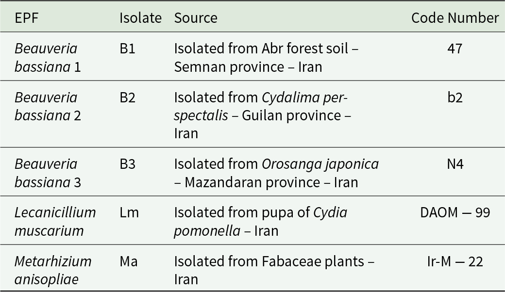
Three isolates of Beauveria bassiana (B1, B2, B3), one isolate of Lecanicillium muscarium (Lm), and one isolate of Metarhizium anisopliae (Ma) were cultured on potato dextrose agar at 25 ± 1°C. After 12 days, fresh conidia were harvested from the petri dishes by adding 10 mL of sterile distilled water containing 0.05% v/v of Tween® 80 as a spreader. Different spore concentrations (spores/mL) were then prepared for testing. The B2 and B3 isolates were obtained from the Research Institute of Forests and Rangelands, while the Lm and Ma isolates were sourced from the Iranian Research Institute of Plant Protection, Tehran, Iran (table 2).
Source of EPF

Bioassay
A 12-day-old Potato Dextrose Agar (PDA) culture of each fungal isolate was surface-scraped using a sterile scalpel. The conidial suspensions were filtered through sterilised filter paper (Whatman No. 01) to remove residual medium and fungal hyphae. Conidia were counted using a Neubauer chamber (HGB, Germany) under optical microscopy, and serial dilutions were prepared to obtain concentrations ranging from 103 to 10⁸ spores/mL for each isolate. Determining spore viability is critical for ensuring the accuracy of the bioassay. To assess viability, 2 µL of the conidial suspension was sprayed onto the centre of a 9-cm petri dish containing PDA medium. After incubation at 25 ± 1°C for 12 hours, the germination percentage was determined by counting 100 conidia per petri dish. Conidia were considered viable if the germ tube length exceeded the diameter of the spores. All isolates demonstrated a viability of approximately 90% and were selected for the bioassays. The control consisted of sterilised distilled water and the adhesive spreader Tween® 80 at 0.05% v/v.
The pathogenicity of the isolates against P. radiata larvae was determined using serial concentrations of the fungal suspensions. Fifth-instar larvae were randomly selected from the rearing cage for the bioassays. For each concentration of the isolates, 40 larvae were used, divided into four groups of 10 larvae. The larvae were directly immersed in the fungal suspension for 6 seconds. Control larvae were treated similarly with Tween 80 (0.05%) and distilled water. After treatment, the larvae were transferred to petri dishes containing fig leaves (var. Zard Semnan) and were observed daily for 7 days. Mortality was recorded (Sevim et al., Reference Sevim, Demir and Demirbağ2010b; Gurlek et al., Reference Gurlek, Sevim, Sezgin and Sevim2018), and larvae that failed to move were considered dead. Data were corrected using Abbott’s formula, and lethal concentration (LC50) values were estimated using POLO-PC software.
Effects of EPF on selected biological characteristics of Phycodes radiata
Third-instar larvae (10 larvae/replicate × 4 replicates/assay) were exposed to LC50 concentrations of the B1, B2, B3, Lm, and Ma isolates of EPF. Sterilised distilled water and Tween-80 (0.05% v/v) served as the control. The larvae from each treatment were individually placed in petri dishes containing fig leaves (var. Zard Semnan) .Fresh leaves were also placed daily until pupation. The conditions of keeping adults until death have already been explained. Fresh fig leaves were placed in adult containers, and the oviposition rate of females was recorded throughout their lifespan. The study examined larval mortality, pupation rate, adult emergence rate, male/female longevity, and fecundity.
Investigation of Haemocyte changes in Fifth-Instar Larvae of Phycodes radiata exposed to Beauveria bassiana, Lecanicillium muscarium, and Metarhizium anisopliae
Two-day-old fifth-instar larvae feeding on Ficus carica var. Zard Semnan were selected for the immunological experiment. Each larva was injected with 1 µL of a Tween 80 (0.05% v/v) solution containing the LC50 concentration of each isolate using a Hamilton syringe. Injections were administered into the larvae’s second and third abdominal leg regions. Treatments included three isolates of B. bassiana (B1, B2, and B3), one isolate of M. anisopliae (Ma), one isolate of L. muscarium (Lm), Tween 80, and distilled water as the control treatments. Post-injection, the total haemocyte count and the counts of plasmatocytes, granulocytes, oenocytoids, and prohaemocytes were recorded at 1, 3, 6, 12, 24, 48, and 72 hours. Each experiment included 5 replicates, with two larvae per replicate. The combined haemolymph from the two larvae (4 µL) was mixed with 8 µL of Tyson’s solution, a diluent and anticoagulant (NaCl2 72 Mm, Na2SO4 9 Mm, Glycerol 43 Mm, Methyl violet 0.06 Mm, Distilled water) (Mahmood and Yusaf, Reference Mahmood and Yousaf1985). Then placed on a Neubauer haemocytometer. Haemocyte changes were observed and recorded under a 40 × magnification light microscope in five squares of the Neubauer chamber at each time interval. The following formula was used to calculate the cell counts (Jones, Reference Jones1962):
 \begin{equation*}\frac{\text{Hemocyte in} \times 1 \text{mm}^2 \times \text{Dilution} \times \text{Depth factor of}}{\text{No. of squares counted}}\end{equation*}
• Dilution: 10 times.
• Depth factor of the chamber: 10.
• Number of squares counted: 5.
Statistical analysis
All experiments were conducted using a completely randomised design, and the data were analysed with SAS 9.1 software. Mean comparisons were performed using Duncan’s multiple range test at a 5% probability level.
Results
Investigation of morphological, biological, and damage characteristics of Phycodes radiata
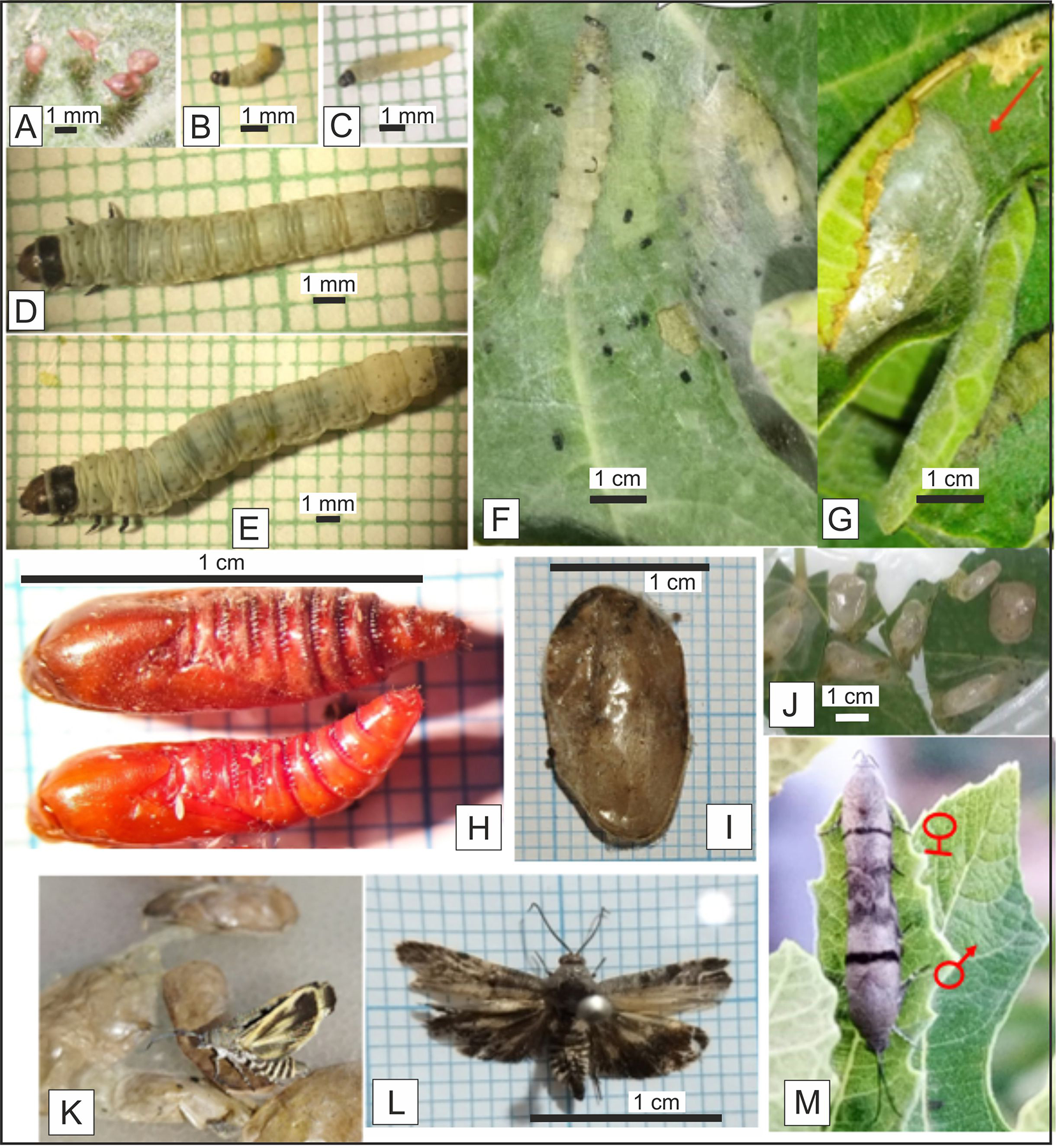
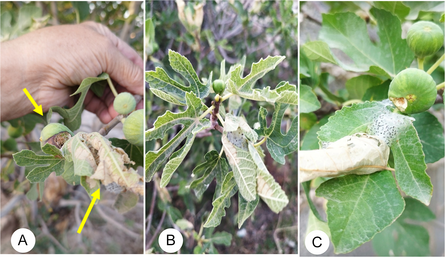
The fig. 2 illustrates the external characteristics of the developmental stages of P. radiata. In early May, red, spindle-shaped eggs with a pedestal were attached to the dorsal surface of leaves from different fig cultivars (fig. 2A). The incubation period on the four fig cultivars ranged from 6.2 to 6.5 days under both orchard and laboratory conditions, with no significant differences observed (table 2). The first-instar larvae were pale green and gradually emerged from the eggs between mid-May and mid-June, depending on the climatic conditions in Semnan. These larvae initially attacked the swollen buds at the beginning of the season, feeding on them and causing the destruction of newly emerged buds. As the larvae progressed through their instars, their body colour intensified (fig. 2B–E). The larvae gradually began feeding on the newly emerged leaves. Early larvae were sometimes observed in groups on both the underside and upperside of the leaves, near the petiole, and on the fruit under woven webs. However, in later larval instars, they were often found individually or in small groups on a single leaf. The late-instar larvae used their salivary secretions to curl the fig leaves and form tubes, leaving black fecal matter within the woven white web (fig. 2F, G). These larvae could hang themselves from the woven web to reach the nearest leaf or fruit. As the larvae feed on the leaf parenchyma in the spring, the epidermal layers dry out, and with the increase in larval age, the entire parenchyma is consumed, causing the leaves to wrinkle and turn brown. In subsequent generations, the larvae extended their feeding to the fruits, chewing one side of the fruit with their mouthparts (fig. 3C). Over time, the damaged part of the fruit dried and turned brown (fig. 3A–C).
Life stages of Phycodes radiata. A, egg; B, first instar larva; C, second instar larva; D, third instar larvae; E, fourth instar larva; F, fifth instar larva; G, prepupa; H, pupa; I, cocoon; J, cocoon on the fig leaves; K and L, adult; M, mating.

A, B, C: damage of larvae of Phycodes radiata on the fig leaf and fruit.

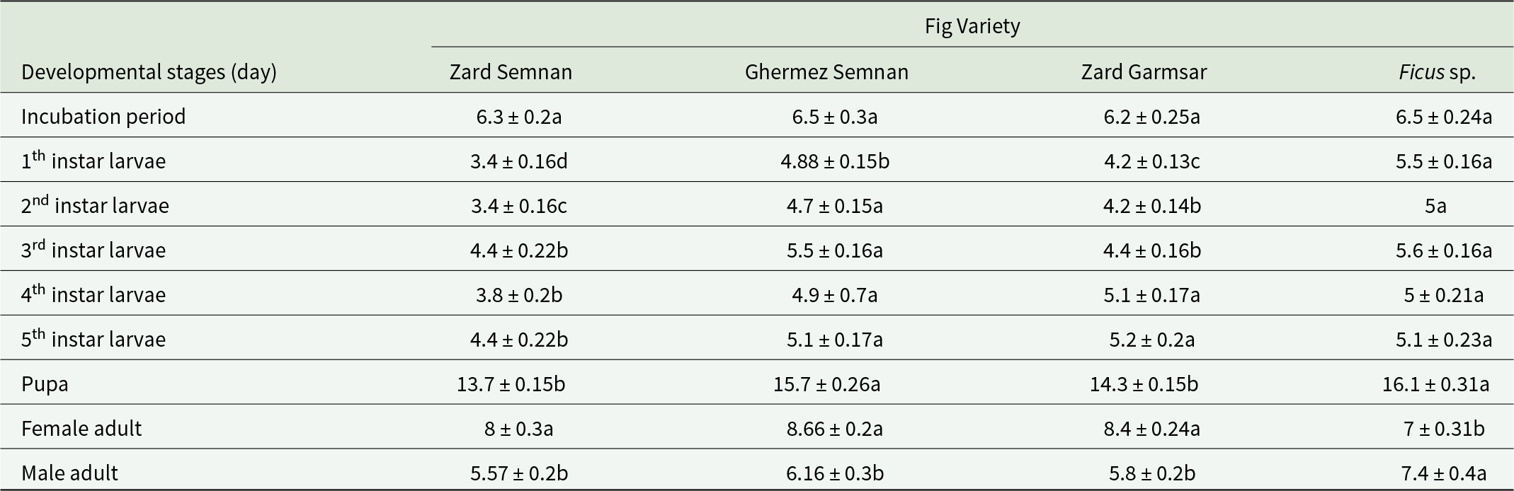
The duration of the first-instar larval stage showed significant differences depending on the fig cultivar (F = 36.2, dft = 3, dfe = 34, p ≤ 0.0001). The most extended duration was observed in larvae fed on wild fig (Ficus sp.), with an average of 5.5 ± 0.16 days, while the shortest duration was recorded for larvae fed on the Zard Semnan and Zard Garmsar varieties (table 3). Significant differences were also noted in the second-instar larval stage duration based on the fig cultivar (F = 22.62, dft = 3, dfe = 34, p ≤ 0.0001). The longest duration occurred in larvae fed on Ficus sp. and Ghermez Semnan, while the shortest duration was observed in larvae fed on Zard Semnan (table 3). The variance analysis for the third-instar larvae duration showed significant differences based on the fig cultivar (F = 14.87, dft = 3, dfe = 34, p ≤ 0.0001). The longest duration was observed in larvae fed on Ficus sp. and Ghermez Semnan, while the shortest was observed in larvae fed on Zard Semnan and Zard Garmsar. The shortest duration of the fourth instar was related to larvae fed on Zard Semnan (F = 10, dft = 3, dfe = 34, p ≤ 0.0001). The fifth-instar larvae fed on Zard Garmsar, Ficus sp., and Ghermez Semnan had the longest duration, while the shortest duration of the fifth instar was recorded for larvae fed on Zard Semnan, with an average of 4.4 ± 0.22 days (F = 2.74, dft = 3, dfe = 34, p ≤ 0.05) (table 3). The red-coloured pupae were 2.1 ± 0.1 cm in length (fig. 2H) and were encased in a cocoon attached to the surface of the fig leaf. The cocoon measured approximately 1.8 ± 0.2 cm in size (fig. 2I, J). The highest mean pupal duration was observed in insects fed on Ficus sp. and Ghermez Semnan (table 3), while the shortest mean duration was recorded for larvae fed on Zard Semnan and Zard Garmsar (F = 18, dft = 3, dfe = 34, p ≤ 0.0001). Significant differences were also found in the pupal weights among the different cultivars, with the lowest pupal weight (0.076 ± 0.02 g) observed in pupae from Ficus sp. (table 4).
Biological period (day) of different stages in Phycodes radiata fed on four fig varieties of Ficus carica

Different letters in each row show significance using Tukey’s test at p < 0.05.
The adult emergence rate from pupae was approximately 99% in the edible varieties and slightly lower, around 97%, in Ficus sp. (table 4). The wingspan of the adult was 1.7 ± 0.1 cm. The dorsal surface of the body with closed wings was grey, while the underside of the wings was yellowish-golden with a dark brown halo. Transverse yellow bands were observed on the abdominal segments (fig. 2K). Male and female moths could be distinguished by their appearance. The filiform antennae were longer in males and darker, whereas they were shorter and whitish in females. Additionally, the dark transverse band seen on the forewing during the resting period was thicker in males than in females (fig. 2M). Females began mating 2–3 days after emergence, and males started mating 3–4 days after emergence from the pupae. Mating typically occurred in the afternoon in the upper parts of fig trees, lasting 3.7–3.88 hours. No significant differences were observed in the mating duration between moths reared on different cultivars (table 4). Laboratory observations indicated that all female moths mated only once during their lifespan, but mated males could mate with other females again. The oviposition rate varied depending on the fig cultivar, with moths whose larvae fed on the three edible varieties exhibiting a higher oviposition rate than those related to Ficus sp. (table 4). The hatching percentage also varied with the fig cultivar, with the highest rates (98-98.8%) observed in Zard Semnan and Ghermez Semnan, while the lowest hatching rate (94.3 ± 0.25%) was associated with insects that had developed on Ficus sp. The analysis of variance for adult longevity data showed significant differences in female (F = 8.22, dft = 3, dfe = 22, p ≤ 0.0007) and male (F = 12.9, dft = 3, dfe = 22, p ≤ 0.0001) based on the fig cultivar. Females had the highest longevity on edible cultivars, while the shortest longevity was observed in females reared on Ficus sp., with an average of 7 ± 0.31 days. Males had the highest longevity on Ficus sp., with an average of 7.4 ± 0.4 days, and the shortest longevity was recorded in males reared on other cultivars (table 3).
Biological characteristics of Phycodes radiata fed on fig varieties (n = 20)

Different letters in each column show significance using Tukey’s test at p < 0.05.
Except for the fourth and fifth larval instars, the developmental stages showed no significant differences between orchard and laboratory conditions. However, the fourth and fifth instar larval durations in the field were 1–1.5 days longer than those observed under laboratory conditions across all fig cultivars. Based on experiments and observations conducted in 2022 and 2023, 3–4 generations of FLR were produced under the climatic conditions of Semnan. The pest persisted until late November (when fig leaves began to fall in the Semnan region), after which the insect entered hibernation as a pupa in the dry leaves at the base of trees or in the surface soils.
Determination of pest feeding indices
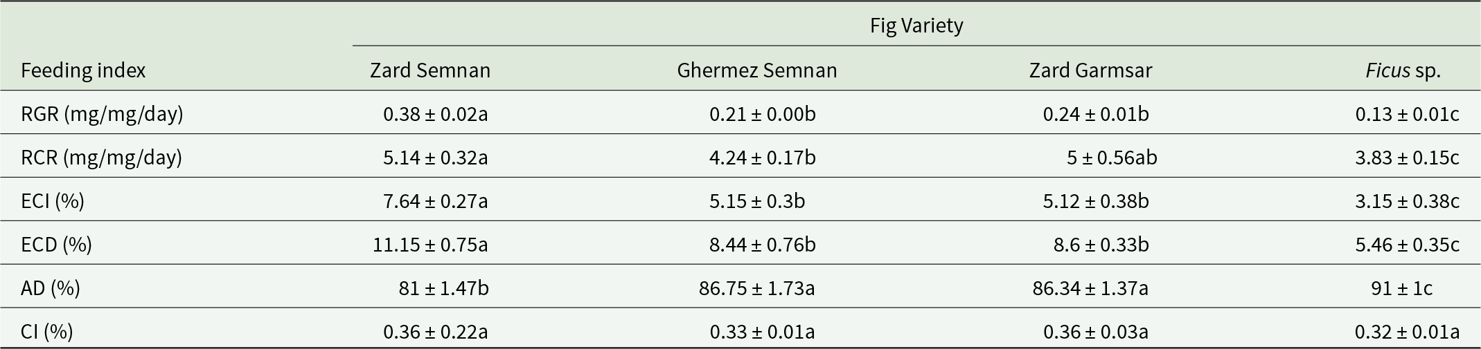
The results showed that the feeding indices of RGR (F = 50, dft,e = 3,34, p ≤ 0.0001), RCR (F = 2.58, dft,e = 3,34, p ≤ 0.0001), ECD (F = 21.37, dft,e = 3,34, p ≤ 0.0001), ECI (F = 24.8, dft,e = 3,34, p ≤ 0.0001), and AD (F = 8, dft,e = 3,34, p ≤ 0.0001) were significantly higher in larvae fed on the Zard Semnan variety compared to the other treatments. The ECD and ECI indices are critical for understanding the relationship between the quality of nutrition and the efficiency of converting nutritional elements into biomass. In this study, the ECD and ECI indices were particularly elevated in larvae fed on the Zard Semnan variety (table 5), indicating that the nutritional elements from this cultivar were of higher quality and had a higher conversion efficiency into biomass compared to the other fig cultivars.
Feeding index of Phycodes radiata fed on fig varieties of Ficus carica

Different letters in each row show significance using Tukey’s test at p < 0.05.
Identification of haemocytes in P. radiata
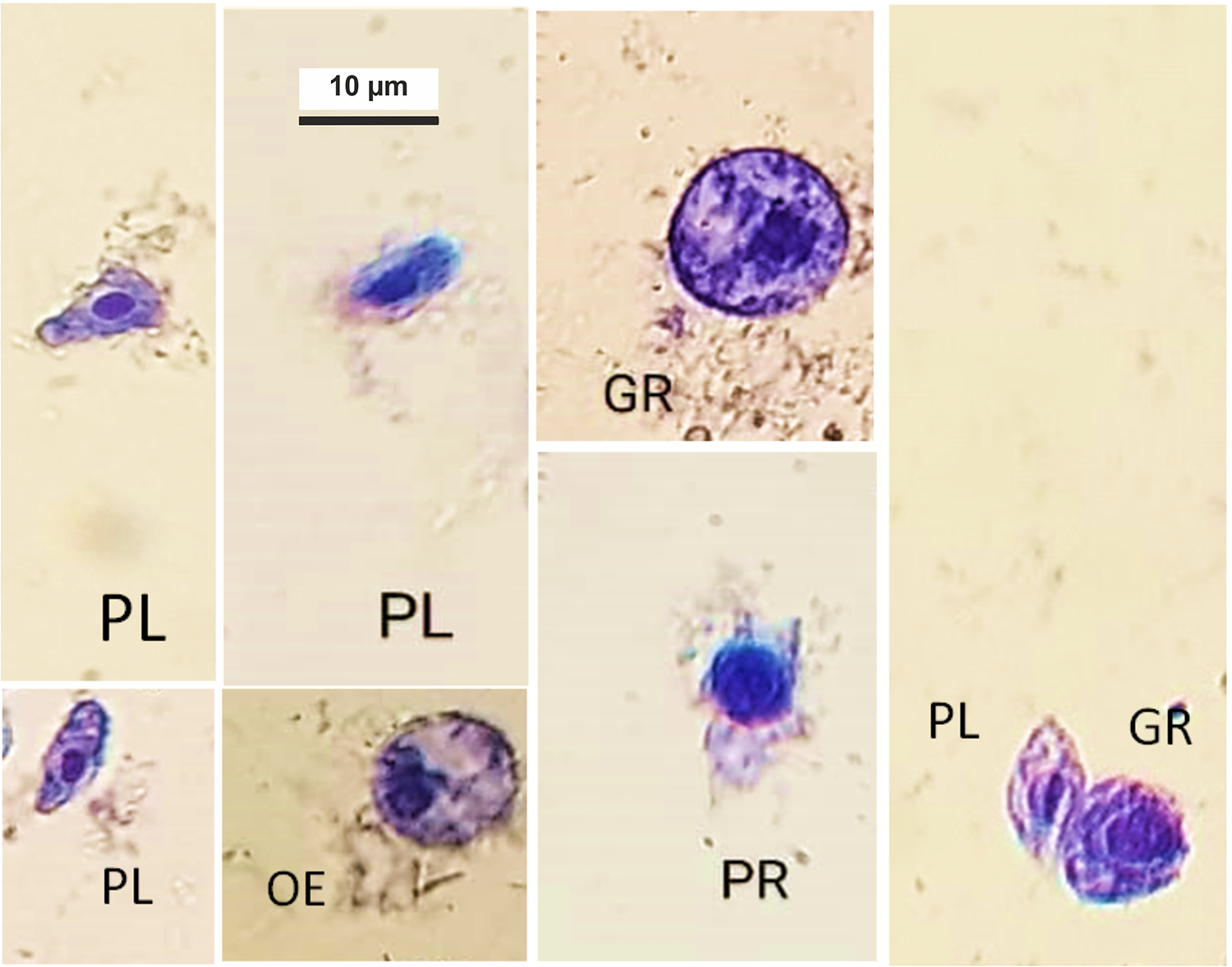
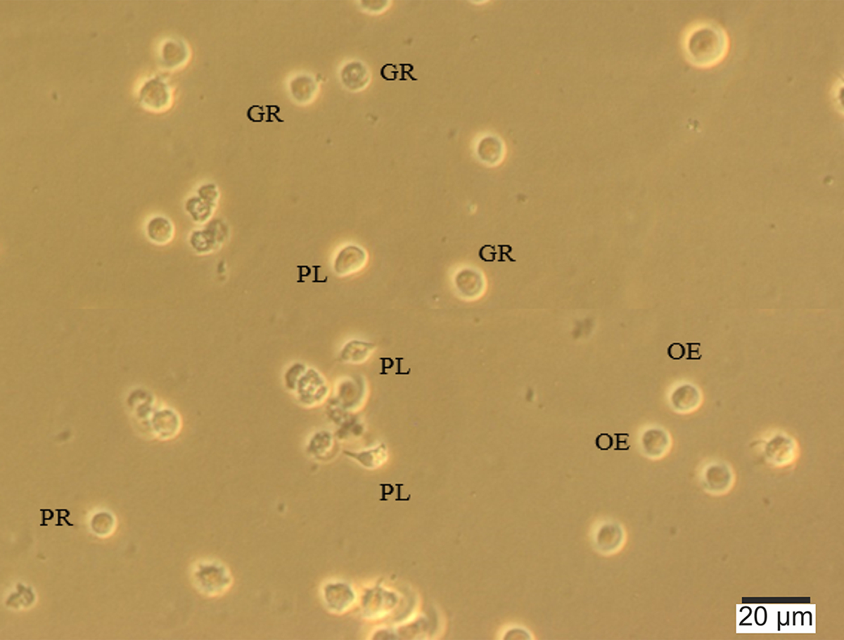
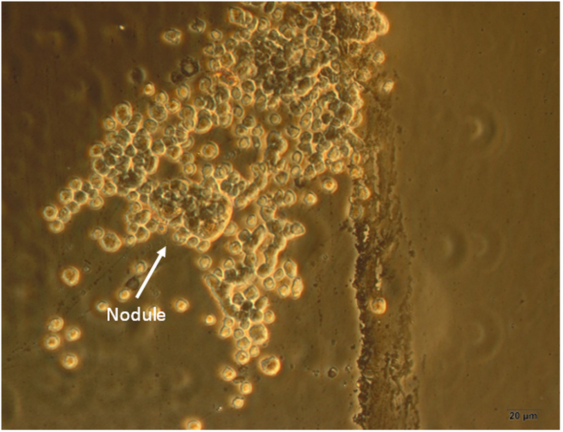
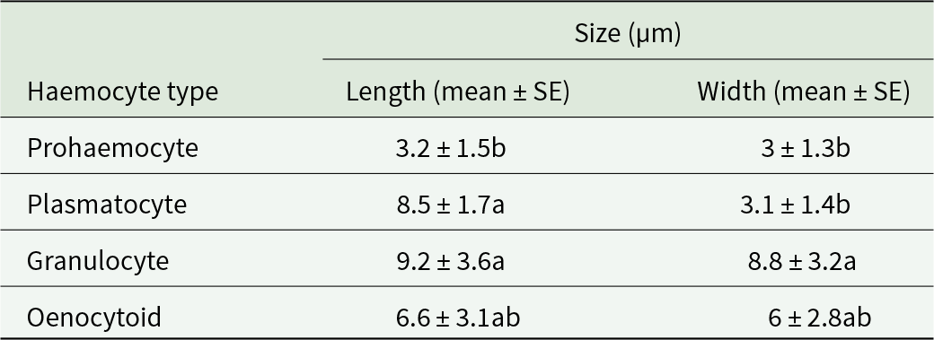
Observations identified four types of haemocytes in the haemolymph of P. radiata larvae: prohaemocytes, plasmatocytes, granulocytes, and oenocytoids (figs. 4, 5). Prohaemocytes were the smallest blood cells observed (table 6). Prohaemocytes were round with a large, round nucleus that occupied most of the cell’s volume. The nucleus was stained dark blue with Giemsa solution, and the cytoplasm appeared as a narrow blue band along the plasma membrane. Plasmatocytes: These cells were spindle-shaped or triangular, with or without cytoplasmic projections, and were observed in varying sizes. The nucleus was large, stained blue with Giemsa, and often centrally located within the cell. The cytoplasm of plasmatocytes was generally smooth and free of granules. Granulocytes: These were the largest haemocytes studied (table 6). Granulocytes were round or oval and varied in size. Their nucleus could be located centrally or laterally, and the cytoplasm was filled with abundant granules (figs. 4, 5). The granular appearance of granulocytes makes them easily distinguishable. Granulocytes and plasmatocytes play a key role in forming nodules around foreign agents, thereby neutralising them (fig. 6). Oenocytoids were larger than prohaemocytes (table 6) and appeared round or oval-shaped. The nucleus, stained dark blue with Giemsa, was located at the periphery of the cytoplasm (figs. 4, 5).
Light microscopy pictures of Phycodes radiata haemocytes stained with giemsa. PR, prohaemocyte; PL, plasmatocyte; OE, oneocytoid; GR, granulocyte. scale bar = 10 µm.

Phase contrast microscopy pictures of Phycodes radiata haemocytes.

Haemocytes of Phycodes radiata in response to Beauveria bassiana forming nodule by phase-contrast microscopy.

Morphometric measurements of haemocytes in larvae of Phycodes radiata (n = 20)

Different letters in each column show significance using Tukey’s test at p < 0.05.
Differential haemocyte count (DHC) of P. radiata
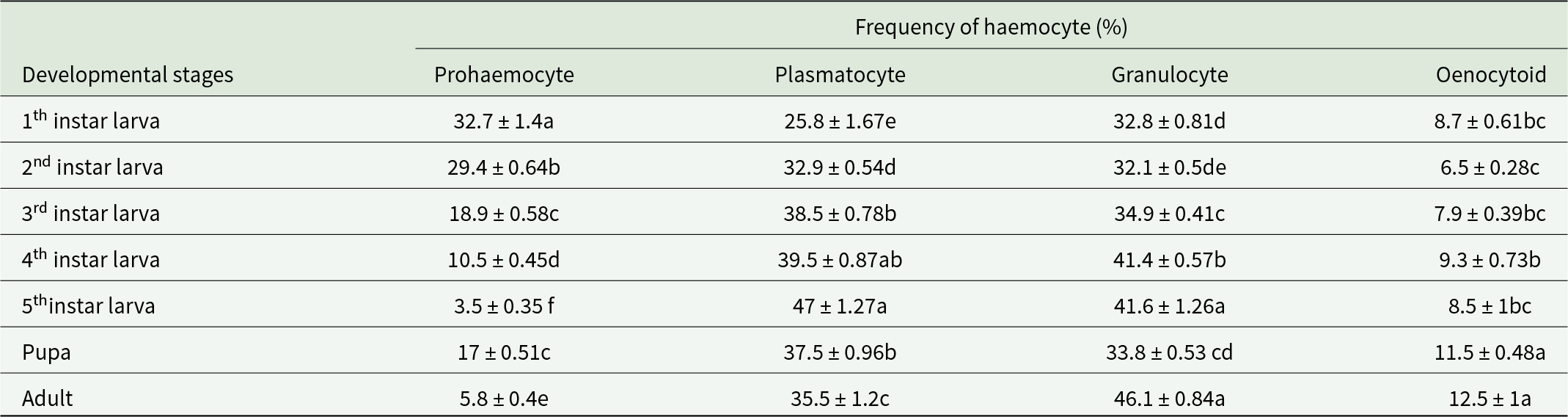
The mean percentage frequency of prohaemocytes at all developmental stages (from first-instar larvae to adulthood) showed significant differences (F = 26, dft = 6, dfe = 77, p ≤ 0.0001). Prohaemocytes exhibited the highest frequency in the early larval instars, with their number gradually decreasing as the larvae developed. In this experiment, the highest percentage frequency of prohaemocytes was observed in the first-instar larvae, with a mean of 32 ± 1.4%, while the lowest frequency was found in the fifth-instar larvae, with a mean of 3.5 ± 0.35% (table 7). The mean percentage frequency of plasmatocytes also showed significant differences across all stages (F = 27, dft = 6, dfe = 77, p ≤ 0.0001). Plasmatocytes were most abundant in the later larval instars, with the highest frequency recorded in the fifth-instar larvae, averaging 47 ± 1.27% cells /mm3 of haemolymph. The first-instar larvae had the lowest frequency, with a mean of 25.8 ± 1.67 cells /mm3 of haemolymph (table 7). Granulocyte frequency significantly varied across all life stages (F = 80, dft = 6, dfe = 77, p ≤ 0.0001). The highest frequency of granulocytes was observed in the higher larval instars, particularly in the fifth-instar larvae, where the mean frequency was 41.6 ± 1.26%. The mean percentage frequency of oenocytoids across all developmental stages showed significant differences (F = 9.04, dft = 6, dfe = 77, p ≤ 0.0001). Oenocytoids had the lowest frequency compared to other haemocytes. The highest percentage frequency of oenocytoids was observed during the adult and pupal stages, while the lowest frequency was recorded in the second instar, with a mean of 6.5 ± 0.31% cells /mm3 of haemolymph. Overall, the frequency of oenocytoids was lower than that of granulocytes and plasmatocytes (table 7).
Differential haemocyte count (DHC) in developmental stages of Phycodes radiata (n = 20)

Different letters in each column show statistical differences among biological stages (Tukey’s test, p ≤ 0.05).
Bioassay
Entomopathogenicity assays
Table 8 reveals the results of the pathogenicity tests for all fungal isolates used in this study on fifth-instar larvae of P. radiata. Larval mortality occurred 5–7 days after treatment with higher concentrations of the isolates. The lowest LC50 value was obtained from larvae treated with B1, followed by B2, B3, and Lm, while the highest LC50 was associated with the Ma isolate (table 8).
LC50 values (spore/mL) of EPF on the fifth instar larvae of Phycodes radiata

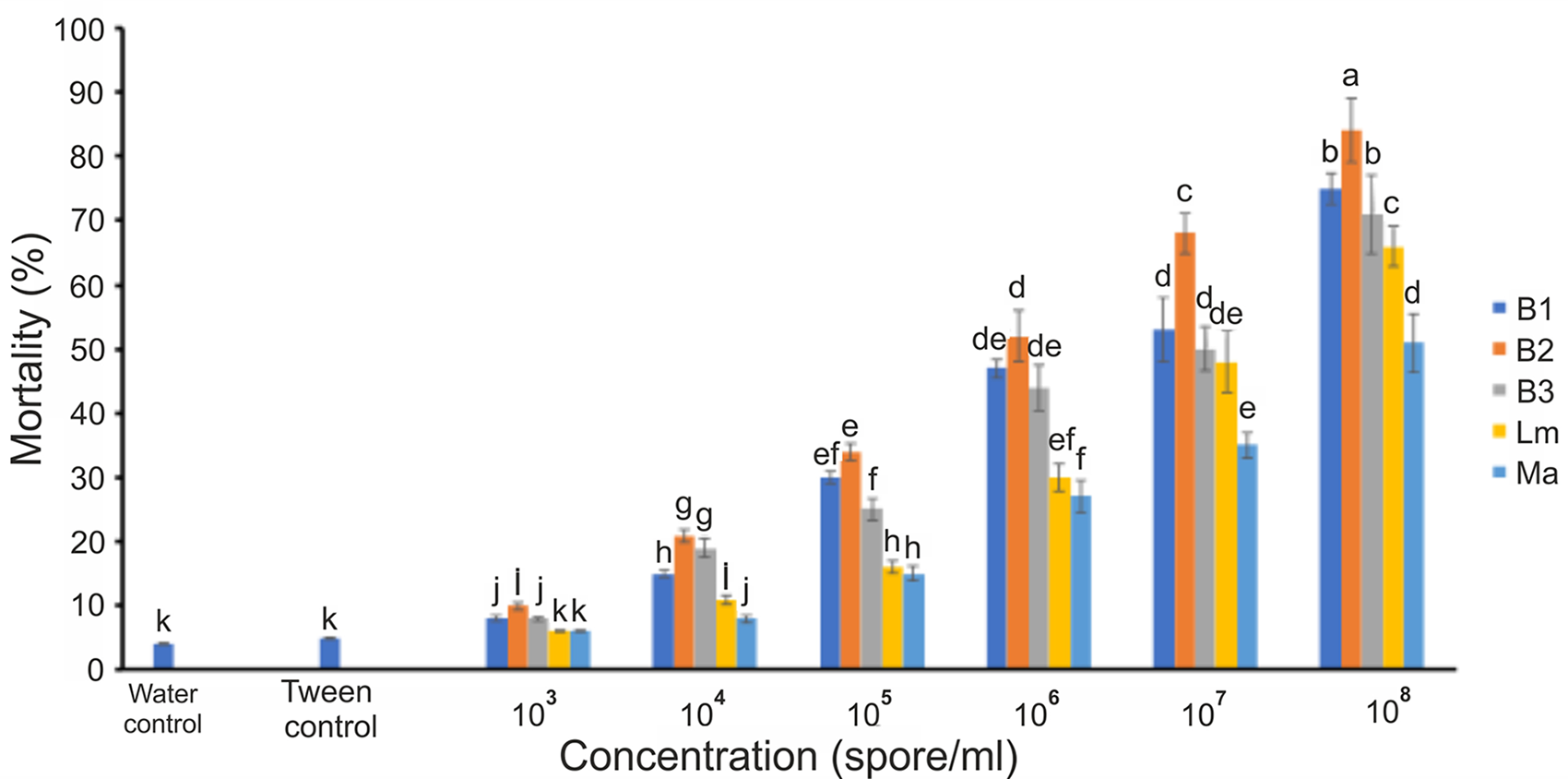
Six concentrations (103, 10⁴, 10⁵, 10⁶, 10⁷, and 10⁸ spores/mL) were tested against P. radiata. The highest mortality was observed at the concentration of 10⁸ spores/mL for all isolates compared to the control treatments using Tween 80 and distilled water. Among the isolates, B2 exhibited the highest effect on larval mortality (84.6%), followed by Lm (66.5%) and Ma (61%) (F = 86.5, df = 7, p ≤ 0.000) compared to the control groups (fig. 7).
Percentage of larval mortality (5th instar) of Phycodes radiata at different concentrations (103, 104, 105, 106, 107, and 108 spore/mL). while two controls are used. Different letters indicate significant differences at p ≤ 0.05.

Effects of EPF on some biological characteristics of P. radiata
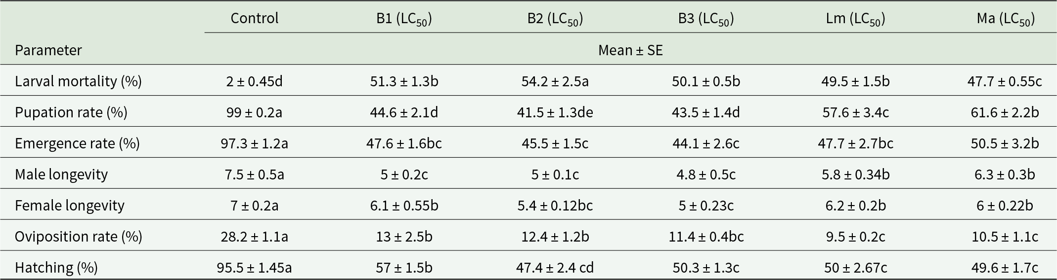
The effects of EPF (LC50) on FLR are presented in table 9. Larval mortality increased significantly in all treatments compared to the control, with isolate B2 having the highest impact. Other parameters, including pupation rate, adult emergence, male/female longevity, oviposition rate, and hatching, decreased sharply in all treatments compared to the control. The reduction in pupation percentage was most pronounced in the B2 treatment. Similarly, the emergence rate was shorter in the B2 and B3 treatments compared to other treatments. Male longevity was shorter in the B1, B2, and B3. Also, female longevity was the lowest when their larvae were treated with B3 treatment. The effects of isolates Lm and Ma were particularly notable in reducing fecundity, while B2 had a pronounced effect on reducing hatching rates (table 9). These findings indicate that EPF induces larval death and modifies developmental stages, reducing male and female longevity, disrupting the reproductive system, and lowering hatching rates.
Effects of EPF on some biological characteristics of Phycodes radiata

Different letters in each row show significance using Tukey’s test at p < 0.05.
Haemocyte changes in larvae exposed to B. bassiana, L. muscarium, and M. anisopliae
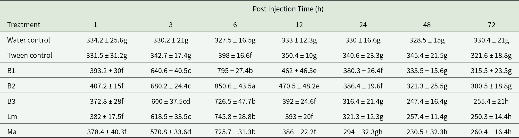
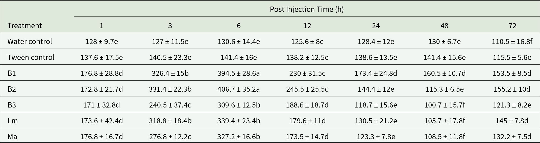
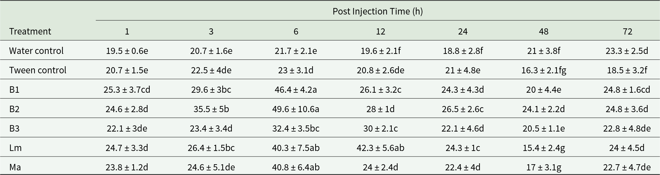
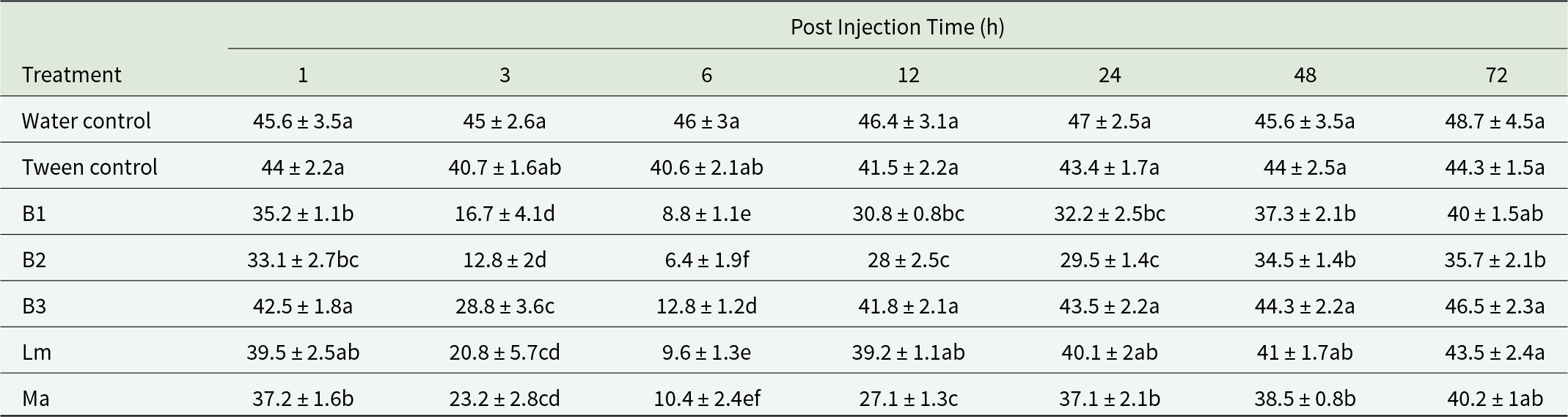
The results demonstrated a significant immune response in larvae treated with EPF. The Total Hemocyte Count (THC) in larvae exposed to fungal spores from all isolates showed a statistically significant increase compared to the control groups (F = 76.7, dft,e = 48,170, p ≤ 0.001). Across all treatments, an increase in the total cell count was observed at 3 and 6 hours post-injection, isolate B2 has a more pronounced effect on haemocyte count increases than the other isolates during this period. Subsequently, the influence of the isolates diminished, and the number of haemocytes displayed a decreasing trend up to 72 hours post-injection (table 10). The pattern of changes in plasmatocytes, the most abundant haemocytes in the haemolymph of fifth-instar larvae, also showed a significant increase at 3 and 6 hours following the introduction of fungal spores from all isolates, compared to the distilled water and Tween 80 (0.05% v/v) controls (F = 50.6, dft,e = 48,170, p ≤ 0.0001). The effect of isolates B2 and B1 was particularly notable 6 hours after injection (table 11). However, after 6 hours, plasmatocyte numbers began to decrease over time. Granulocytes, the second most common haemocytes involved in immune response, increased in all treatments at 3 and 6 hours post-injection (F = 48, dft,e = 48,170, p ≤ 0.002). Again, isolates B2 and B1 had a greater impact than the other isolates at both time intervals (table 12). Oenocytoids, which are potential sources of phenol oxidase production in Lepidoptera, showed variations in their numbers at the 3 and 6-hour intervals (F = 32, dft,e = 48,170, p ≤ 0.001), suggesting changes in immune activities (table 13). Prohaemocytes, as stem cells in the haemolymph that participate in binary division and conversion into granulocytes and other cells during stress and foreign agent entry, exhibited a decreasing trend in response to fungal injection at 3 and 6 hours. Among the isolates, B2, B1, and Lm had the most significant effect on reducing the number of prohaemocytes 6 hours post-injection (F = 24, dft, e = 48,170, p ≤ 0.0001) (table 14).
Changes of total haemocyte numbers in Phycodes radiata larvae injected by different fungal isolates

Different letters show significance using Tukey’s test at p < 0.05.
Changes of plasmatocyte numbers in Phycodes radiata larvae injected by different fungal isolates

Different letters show significance using Tukey’s test at p < 0.05.
Changes of granulocyte numbers in Phycodes radiata larvae injected by different fungal isolates

Different letters show significance using Tukey’s test at p < 0.05.
Changes of oenocytoid numbers in Phycodes radiata larvae injected by different fungal isolates

Different letters show significance using Tukey’s test at p < 0.05.
Changes of prohaemocyte numbers in Phycodes radiata larvae injected by different fungal isolates

Different letters show significance using Tukey’s test at p < 0.05.
Discussion
In the present study, we investigated the morphological and biological characteristics of P. radiata. Previous studies, such as those by Kumar and Ramamurthy in 2010 in India, have also examined the morphology of different stages of this pest, revealing differences in appearance between populations from India and Iran. For example, Iranian larvae are completely green, while the Indian larvae are whitish with paired spots on the dorsal surface of the abdominal segments. The activity of this insect in Semnan, Iran, extends from May to November, which is consistent with the findings of Datta in India (Datta, Reference Datta2024). We observed that the larvae are active on the leaves and fruits of various fig varieties, including Zard Semnan, Ghermez Semnan, Zard Garmsar, and Ficus sp, causing damage by curling and rolling the leaves. In India, P. radiata larvae have been reported to damage various fig cultivars, such as Ficus benjamina, Ficus benghalensis, Ficus religiosa, and Ficus glomerata (Datta, Reference Datta2022, Reference Datta2024). The duration of developmental stages varies between the two climates, with the larval and pupal periods being longer in Iran compared to India, while the incubation period and longevity of males and females are approximately similar. These differences could be attributed to climatic conditions such as temperature, humidity, altitude, and differences in the fig cultivar compositions in each region (Harrington et al., Reference Harrington, Fleming and Woiwod2001; Kang et al., Reference Kang, Cai, Yue and Zhang2021).
Our results indicated that the larval and pupal periods were longer in insects reared on Ficus sp. than in other cultivars. Additionally, the pupal weight, fecundity of females, and hatching rate of insects that fed on the edible cultivars Zard Semnan, Ghermez Semnan, and Zard Garmsar were significantly higher than those that fed on wild Ficus sp. leaves. Similar findings have been reported, where the hatching rate of P. radiata larvae fed on F. religiosa was significantly higher than those fed on F. benghalensis (Datta, Reference Datta2022). It appears that there are factors involved in host selection by insects that are somehow related to their habitat, survival, and evolution (Poore and Hill, Reference Poore and Hill2006). Normally, females choose the best resources (host plants) to provide a more suitable habitat for their offspring (Scheirs et al., Reference Scheirs, Zoebisch, Schuster and de Bruyn2004; Noriyuki, Reference Noriyuki2015). The physicochemical factors of host plants have a prominent role in the larval feeding behaviour. The plants composition of different cultivars varies, affecting the duration of the life cycle and feeding rate. It is known that larvae fed on diet of higher nutritional value have a faster growth and a shorter life cycle compared to larvae fed on lower quality diet (Bala et al., Reference Bala, Sood, Pathania and Thakur2018). Moreover, our observations suggest that the leaf thickness of wild fig trees is much less than that of other cultivars, likely contributing to the lower nutritional quality of these leaves. Wild fig trees, which grow in dense forests, receive less sunlight than orchard trees and produce less chlorophyll. Research by Barzegar and Zare (Reference Barzegar and Zare2020) indicates that younger fig leaves, such as those of the Shahe-Anjir cultivar, have the highest chlorophyll content, likely due to the rapid chlorophyll synthesis in the leaf chloroplasts of younger plants. In contrast, mature leaves have higher anthocyanin content, which leads to strong absorption of blue-green light and reduces the amount of light available for photosynthesis. This shading reduces growth and development and keeps chlorophyll levels low. Leaves grown in shade are thinner and have lower concentrations of xanthophyll compounds, starch, sugars, and fibre. According to Barzegar and Zare (Reference Barzegar and Zare2020), the Shahe-Anjir and Siah cultivars have higher sugar and chloroplast content, making them more suitable for pest feeding. In addition, a similar study has demonstrated that the characteristics of the plant, which are effective in the food selection of the insect, along with larval age and prior feeding experience from the host plant tissue play a significant role in host preference. Different instar larvae of Perina nuda (F.) (Lepidoptera: Lymantriidae) exhibited varying nutritional levels from different fig varieties, indicating a shift in host preference due to developmental stage and the nature of the larval diet being either selective or non-selective (Liao et al., Reference Liao, Lin, Wang, Wang, Wei and Chen2024). In similar case, the preference of Helicoverpa armigera (Hubner) (Lepidoptera: Noctuidae) for tobacco was significantly stronger than cotton after 6 hours of induction of tobacco leaf feeding (Li et al., Reference Li, Yuan, Hu, Li, Guo and Li2014).
Based on these findings, it appears that plant physicochemical characteristics such as nutritional compounds, allelochemicals, metabolites, hardness, size, and shape patterns, along with phytophagous insect characteristics including larval instar and feeding experience, may determine the host preference behaviour and play a crucial role in the bioecology of the target insect (Dodds et al., Reference Dodds, Clancy, Leyva, Greenberg and Price1996; Bruce et al., Reference Bruce, Wadhams and Woodcock2005). Other reports clarified that when larvae of Spodoptera littoralis (Biosduval) (Lepidoptera: Noctuidae), Manduca sexta (Lepidoptera: Noctuidae), and Megastigmus pistaciae (Hymenoptera: Torymidae) feed on various diets, they exhibit different biological future and immune responses (Adamo et al., Reference Adamo, Dvies, Easy, Kovalko and Turnbull2016; Gopalakrishnan and Kalia, Reference Gopalakrishnan and Kalia2022; Ajamhassani et al., Reference Ajamhassani, Ebrahimizadeh, Abdos and Ahangi Rashti2023; Nandhini and Deshmukh, Reference Nandhini and Deshmukh2023).
The differences in food preferences are also reflected in the feeding index parameters. The RGR, RCR, ECI, ECD, and AD values were higher in larvae fed on the edible cultivars, particularly Zard Semnan than those fed on Ficus sp. The RGR is a function of the increase in the body weight of the organism (Waldbauer, Reference Waldbauer1968), and a decrease in food consumption often reflects a decrease in the nutritional value of the host (Naseri et al., Reference Naseri, Fathipour, Moharramipour and Hosseininaveh2010). Therefore, the higher relative consumption observed in the Zard Semnan, Ghermez Semnan, and Zard Garmsar cultivars is likely due to the higher nutritional quality of these cultivars and their effective conversion to larval biomass. The lower RGR in larvae fed on the forest cultivar (Ficus sp.) may indicate the unsuitability of the nutritional elements of this cultivar for pest growth. The results of this study suggest that the leaves of the forest cultivar may be less desirable for P. radiata larvae due to the presence of physical and biochemical characteristics, including compounds that inhibit digestion and absorption or the presence of digestive enzyme inhibitors.
Further research is needed to explore these aspects in more detail. The ECD index reflects the amount of food absorbed from the gut wall, and differences in this index among different hosts may be due to variations in nutrient, water, and fibre content in the host leaf tissue (Srinivasan and Uthamasamy, Reference Srinivasan and Uthamasamy2005). The ECI indicates the insect’s ability to use consumed food for growth and development. In contrast, the ECD indicates the conversion of digested food into growth and biomass (Nathan et al., Reference Nathan, Kalaivani, Murugan and Chung2005). The impact of leaf characteristics such as toughness, thickness, and nutritional quality on the feeding index parameters of other insect species, including Bombyx mori L. (Lepidoptera: Bombycidae), Ectomyelois ceratoniae Zeller (Lepidoptera: Pyralidae), and Tortrix viridana (Linnaeus) (Lepidoptera: Tortricidae), has also been documented (Ramesha et al., Reference Ramesha, Anuradha, Lakshmi, Sugnana Kumari, Seshagiri, Goel and Suresh Kumar2010; Zare et al., Reference Zare, Jalali Sendi, Zibaee and Jafari Nodushan2012; Yazdanfar et al., Reference Yazdanfar, Ghodskhah Daryaei, Jalali Sendi and Ghobari2015).
Haemolymph is a major component of an insect’s body and plays a crucial role in the circulatory system, nourishing tissues, transporting hormones and various compounds, and assisting in heartbeat regulation. Haemolymph consists of two phases: plasma and haemocytes. Haemocytes are considered the primary components of the insect’s physiological defense. Therefore, the first step in immunological studies is the identification of the insect’s haemocytes (Jalali and Salehi, Reference Jalali and Salehi2008). In the present study, four types of haemocytes – prohaemocytes, plasmatocytes, granulocytes, and oenocytoids – were identified in the haemolymph of P. radiata. In most Lepidoptera, five types of haemocytes including prohaemocytes, plasmatocytes, granulocytes, oenocytoids, and spherulocytes have been reported (Strand and Pech, Reference Strand and Pech1995; Blanco et al., Reference Blanco, Crispim, Fernandes, de Oliveira, Pereira, Bazzolli and Martins2017; Ajamhassani and Mahmoodzadeh, Reference Ajamhassani and Mahmoodzadeh2020; Ebrahimi and Ajamhassani, Reference Ebrahimi and Ajamhassani2020), but there are various reports on the diversity of haemocyte types in insects. For example, some insects have additional haemocyte types, such as adipohaemocytes and coagulocytes (Cho and Cho, Reference Cho and Cho2019; Silva et al., Reference Silva, Boleli and Simoes2002), while others may lack certain types. In the haemolymph of Pieris rapae (Linnaeus) (Lepidoptera: Pieridae) and M. pistaciae, as in P. radiata, spherulocytes are absent (Zhang et al., Reference Zhang, Huang, Zhu and Ye2012; Ajamhassani et al., Reference Ajamhassani, Ebrahimizadeh, Abdos and Ahangi Rashti2023). Thus, insect haemocytes have no universal pattern (Bruno et al., Reference Bruno, Montali, Gariboldi, Wronska, Kaczmarek, Mohamed, Tian, Casartelli and Tettamanti2022).
The DHC was calculated at various developmental stages of P. radiata, revealing that haemocyte abundance varied across different stages. Prohaemocytes, as the foundational haemocytes, were most abundant in the first-instar larvae and gradually decreased as the larvae aged. Prohaemocytes are synthesised in the haematopoietic organs and can differentiate into cells that play a role in immunity during growth (Yamashita and Iwabuchi, Reference Yamashita and Iwabuchi2001). As larvae grow older, their haemolymph volume increases, possibly enhancing their immune capacity. Consequently, prohaemocytes differentiate into immunocytes, leading to a decrease in their numbers in larger larvae. Plasmatocytes and granulocytes were most abundant in the late larval instars. The higher susceptibility of older larvae to pathogens compared to younger ones may explain the greater abundance of PL and GR in later instars, which increases resistance (Valades-Lira et al., Reference Valades-Lira, Gonzalez, Damas, Meja, Oppert, Padilla and Guerra2011). In the haemolymph of most Lepidoptera, the combined abundance of these two cell types in later larval instars exceeds 50% (Lavine and Strand, Reference Lavine and Strand2002; Strand, Reference Strand2008). In the fifth-instar larvae of P. radiata, plasmatocytes constituted 47% and granulocytes 41.6% of the total cell population. In Conogethes punctiferalis (Lepidoptera: Crambidae), the abundance of plasmatocytes was 21.4%, and granulocytes 64.8% (Li et al., Reference Li, Liu, Kang, Li, Lu, Li, Pang, Zheng and Yin2022). Oenocytoids were less abundant during the larval stage of P. radiata, but their population increased during the pupal and adult stages. These cells are potential sources of phenol oxidase production in Lepidoptera (Ling et al., Reference Ling, Shirai, Kanekatsu and Kiguchi2005; González-Santoyo and Córdoba-Aguilar, Reference González-Santoyo and Córdoba-Aguilar2012), and due to their lower involvement in immune processes against pathogens, their abundance is typically lower than that of immunocytes. The density of oenocytoids is approximately 1% in Cydalima perspectalis Walker (Lepidoptera: Crambidae) and about 5-10% in Anticarsia gemmatalis Hub. (Lepidoptera: Noctuidae) (Kholghahmadi et al., Reference Kholghahmadi, Karimi-Malati and Jalali Sendi2025; Andrade et al., Reference Andrade, Negreiro, Levy, Fonseca, Moscardi and Falleiros2010).
EPF are considered effective options for pest control because they penetrate the insect’s integument and reach the haemolymph, causing disease and often death of the target insect (Abood et al., Reference Abood, Bajwa, Ibrahim and Sajap2010; Zafar et al., Reference Zafar, Shoukat, Zhu, Fu, Xu and Jin2024). In our results, the B2, B1, and B3 isolate were more virulent and ultimately caused higher mortality in the larvae of P. radiata, respectively. Beauveria bassiana is known as an important biological control agent of various insect orders (Baron et al., Reference Baron, Rigobelo and Zied2019). Testing the effectiveness of EPF requires several conidia densities as a treatment to test their ability to infect (Hasibuan et al., Reference Hasibuan, Simbolon and Candra2024). Therefore, concentrations from 103 to 108 spores/mL were treated to the larvae and the findings showed that the mortality of the larvae is directly related to the increase in concentration in all treatments. However, the effect of isolates of B. bassiana in all concentrations was the highest compared to L. muscarium and M. anisoliae. Different fungal isolates exhibit varying levels of virulence against Lepidoptera (Wraight et al., Reference Wraight, Ramos, Avery, Jaronski and Vandenberg2010; Kirubakaran et al., Reference Kirubakaran, Sathish-Narayanan, Revathi, Chandrasekaran and Senthil-Nathan2014). Factors such as the physiological origin of the fungi, the penetration ability of fungi to larval cuticle, susceptibility of the host insects and developmental stage, gender, and immune system of them influence the effectiveness of pathogenic agents (Holder et al., Reference Holder, Kirkland, Lewis and Keyhani2007; Garcia et al., Reference Garcia, Gonzalez and Bautista2011; Zhang et al., Reference Zhang, Guo, Zou and Jin2018; Zamani et al., Reference Zamani, Gholami Ghavamabad and Kazerani2023). We also observed that the percentage of pupation, adult emergence, lifespan, and oviposition were affected by EPF treatments compared to the control. Similar effects have been reported in other studies (Wang et al., Reference Wang, Yu, Chu, Korai and Liu2018; Xia et al., Reference Xia, Yu, Yang, Shang, He, Song, Li and Jiang2023).
Insect immunity is a critical indicator of contaminants such as fungal spores, poison particles, or other stressors in the body. Upon entering a foreign agent, granulocytes are the first to identify it. The changes in haemocyte types, shapes, and abundances in response to EPF are highly variable. Sometimes, the number of haemocytes decreases (Mishra et al., Reference Mishra, Kumar and Malik2015; Li et al., Reference Li, Liu, Kang, Li, Lu, Li, Pang, Zheng and Yin2022; Bitencourt et al., Reference Bitencourt, Chorrea, Mallet, Santos, Lowenberger, Moreira, Golo, Bitenciurt and Angelo2023; Perumal et al., Reference Perumal, Kannan, Alford, Pittarate and Krutmuang2024), and sometimes it increases (Mudoi et al., Reference Mudoi, Das, Hazarika, Das and Roy2019; Aghaee Pour et al., Reference Aghaee Pour, Zibaee, Gohar Rostami, Hoda and Shahriari2021) during immune challenges. In this study, we observed an increase in THC, PLS, GRS, and OES and a decrease in PRS in the early hours following fungal injection (3 and 6 hours). Haemocyte numbers began to decrease from 12 to 72 hours post-injection. Cellular immunity typically occurs in the initial hours following the entry of a foreign agent or stress into the haemolymph, and it diminishes over time (Stanley and Miller, Reference Stanley and Miller2006). As stem cells, prohaemocytes undergo mitotic division in the initial hours following a foreign agent attack, which explains the significant decrease in prohaemocytes and the increase in granulocytes and plasmatocytes at 3 and 6 hours.
Additionally, granulocytes can increase their population through binary division. Among the treatments, isolates B2 and B1 had the greatest impact on the haemocyte population, although all isolates caused a significant increase in haemocytes compared to the control groups at 3 and 6 hours post-infection. It appears that the physiological properties of each isolate, in terms of toxicity and metabolite production, differ and lead to varied immune responses. The B2 isolate was isolated from the box tree moth Cydalima perspectalis (Walker) (Lepidoptera: Crambidae), while the B1 isolate was sourced from the forest soil of Shahrood Abr. If the physiological origin of the fungi affects their pathogenicity, it can certainly be expected to influence the immune responses of the target insects as well. Given that the B2 isolate was obtained from a lepidopteran species and the target insect in this study is also a lepidopteran, the similarity between these two insects may account for the strong effect of B2 on the immune response of P. radiata. Additionally, since the Abr forest soil is geographically close to Semnan, and immunological experiments were conducted on an insect active in this region, this proximity may partly explain the cellular defense response of the FLR against the B1 isolate (sourced from the Abr forest soil).
When the concentration of foreign agents in the haemolymph becomes very high, haemocytes respond by confronting the invaders through a process known as nodulation (Douglas and Siva-jothy, Reference Douglas, Siva-Jothy, Chapman, Simpson and Douglas2013). In our study, granulocytes, plasmatocytes, and, to a lesser extent, oenocytoids were observed to participate in nodule formation. This process occurs in one, two, or multiple layers. Nodulation proceeds in two distinct stages. The first stage begins approximately one minute after the entry of the foreign agent into the haemolymph, during which granulocytes actively accumulate around the invader. The second stage typically occurs 2-6 hours post-injection, with plasmatocytes attaching to the melanised spot (Ratcliffe and Rowley, Reference Ratcliffe, Rowley and Gupta1979; Sato, Reference Sato2023). Nodule formation is accompanied by producing phenol oxidase, eicosanoids, and the viscosity protein haemochitin (Arai et al., Reference Arai, Ohta, Suzuki, Tanaka, Yoshizawa and Sato2013; Otuka and Sato, Reference Otuka and Sato2023). Previous studies have confirmed the involvement of haemocytes in nodulation as a defense mechanism against pathogens in various insects. For instance, haemocytes in Rhynchophorus ferrugineus (Olivier) (Coleoptera: Curculionidae) have been shown to form nodules in response to Bacillus thuringiensis, B. mori responds similarly to Escherichia coli; and C. suppressalis shows modulation against B. bassiana (Manachini et al., Reference Manachini, Arizza, Parrinello and Parrinello2011; Arai et al., Reference Arai, Ohta, Suzuki, Tanaka, Yoshizawa and Sato2013; Zibaee and Malagoli, Reference Zibaee and Malagoli2014). However, it is important to note that a more precise understanding of the nodulation process in the defense response of P. radiata against pathogens requires further research.
Conclusion
Phycodes radiata is a significant pest in the Semnan region of Iran. This study contributes to a deeper understanding of the biological characteristics of this insect and its feeding indices on four different fig cultivars, offering new insights into pest management strategies. The impact of EPF on the survival of P. radiata was notable, with results indicating that the larvae possess a considerable immune capacity against the spores of B. bassiana, L. muscarium, and M. anisopliae. However, several important questions remain unanswered. For instance, how does the immune response of younger larval instars compare when exposed to pathogens? What is the influence of dietary factors, such as the specific fig cultivar, and environmental variables, such as temperature, on haemocyte abundance and the nodulation process? Additionally, can fungi be effectively utilised as a viable option for the microbial control of this pest? Further research is essential to explore these physiological characteristics and to provide comprehensive information, potentially leading to more effective and sustainable pest management strategies for P. radiata.
Acknowledgements
The authors express special appreciation to the Shahrood University of Technology for their scientific support and helps. We also thank the editorial office of Bulletin of Entomological Research for handling our manuscript.
Financial support
This research did not receive any specific grant from funding agencies in the public, commercial, or not-for-profit sectors.