Kawasaki disease, first described in Japan by Tomisaku Kawasaki in 1967, is an acute, self-limited vasculitis occurring primarily in children less than 5 years of age.Reference Kawasaki 1 It is the leading cause of acquired heart disease in children, with over 5500 cases of Kawasaki disease diagnosed annually in the United States of America alone.Reference Mandell and Hoffman 2 Japan has the highest annual incidence of Kawasaki disease in the world, with one in every 185 children affected.Reference Nakamura, Yashiro and Uehara 3 Kawasaki disease typically affects medium-caliber arteries, with a particular tropism for the coronary arteries. The characteristic coronary complication is aneurysm, which develops in up to 25% of patients, which may lead to thrombosis, stenosis, and occlusion with consequent myocardial infarction, or, in rare cases, rapture.Reference Lacopo, David and Emanuela 4 Recently, coronary CT angiography can now be performed with relatively low radiation doses to non-invasively image coronary artery anatomy.Reference Earls, Berman and Urban 5 Here, we propose a novel imaging technique to quantify and visualise coronary flow using dynamic coronary CT angiography with 320-detector row CT and motion coherence image processing,Reference Nagao, Yamasaki and Kamitani 6 and investigate the assessment of coronary flow in aneurysmal lesion of Kawasaki disease in adulthood.
Material and methods
Study population
We obtained permission from the Internal Review Board of Kyushu university hospital for this retrospective study. Six patients with Kawasaki disease and coronary aneurysms associated (18–38 years old, median=25 years old) and six patients who had no history of cardiac disease and no coronary lesion on coronary CT angiography as controls (30–74 years old, median=53 years old) were enrolled in this study at our hospital from July, 2013 to April, 2015.
Dynamic coronary CT angiography
CT examinations were performed with a 320-detector row CT scanner (Aquilion One ViSION Edition; Toshiba Medical Systems, Tokyo, Japan). Dynamic coronary CT angiography was scanned at mid-diastole during 15–25 (median=20) cardiac cycles with prospective electrocardiogram gating during 20 seconds following 7 seconds after a bolus injection of contrast media of 12–20 ml (Iopamidol, 370 mgI/ml Iopamiron; Bayer HealthCare, Osaka, Japan). Scan parameters were gantry rotation time of 0.275 seconds, tube voltage of 80 kV, current time product of 28 mAs, and cardiac field of view of 200 mm. All patients received sublingual nitroglycerin before the CT study and did not receive β-blockers. Standard coronary CT angiography was performed following dynamic coronary CT angiography. Scan timing was determined by the peak time of attenuation in the ascending aorta obtained from dynamic coronary CT angiography data. Injection of 50–75 ml of contrast was followed by a mixed bolus of 40 ml of contrast and saline – contrast: saline, 1:9 – at 4–5 ml/second during acquisition with inspiratory breath hold.
Image reconstruction with motion coherence image processing
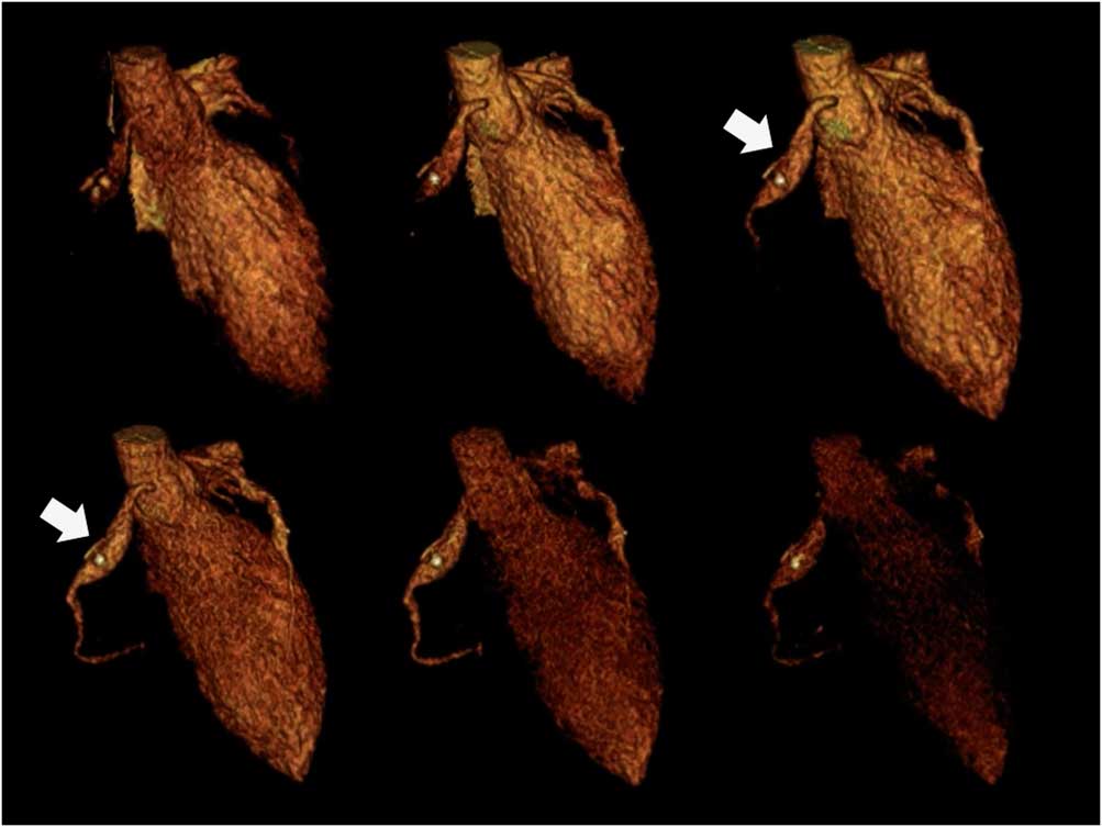
Motion coherence image processing is a novel image processing technique (PhyZiodynamics; Ziosoft Inc., Tokyo, Japan), which performs deformable registration to track all the voxels throughout multiple phases and interpolates images between phases to generate new phases. This imaging technique reduces noise, improves motion coherence, and provides a tool to track specific voxels throughout phases. By using motion coherence image processing, dynamic data sets of 15–25 cycles were interpolated with three additional data sets to generate 90–100 data setsReference Nagao, Yamasaki and Kamitani 7 (Fig 1).
Dynamic coronary CT angiography (CTA) for a 19-year-old man with Kawasaki disease. Volume rendering images of dynamic coronary CTA show a saccular aneurysm with spotty calcifications at the proximal portion of the right coronary artery (arrow).

Coronary flow index
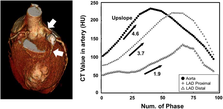
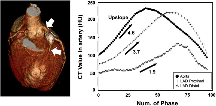
A spherical volume of interest in coronary arteries was automatically tracked and time–density curves optimal for succeeding process were calculated for all phases using motion coherence image processing. On the basis of the maximum slope method, coronary flow index was defined as the ratio of the maximum upslope of the attenuation of coronary arteries to the upslope of the attenuation of ascending aorta on the time–density curve, and was used as a quantification of coronary flow. coronary flow indexes for the proximal and distal sites with a diameter 3–5 mm of right coronary artery and left anterior descending artery, and coronary flow index for intra-aneurysm in patients with Kawasaki disease, were measured (Fig 2).
Calculation of coronary flow ratio. Standard coronary CT angiography shows a saccular aneurysm with calcifications in the proximal site of the left anterior descending (LAD) artery (left, arrow). The sphere volume of interest were set up at the proximal and distal sites of the aneurysm. Graphs (right) show time–density curves of intra-coronary attenuation (Hounsfield units (HU)) for the ascending aorta, and proximal and distal portions of the aneurysm throughout all phases. Maximum upslope (HU/phase) for the three sites was calculated from the linear upslope of the CT value, which represents the first half of the first-pass of contrast media. From the 15th to 25th phase, maximum upslope for the ascending aorta, the proximal site, and the distal site were 4.6 HU/phase, 3.7 HU/phase, and 1.9 HU/phase, respectively. According to the maximum upslope method, the proximal coronary flow index (CFI) and the distal CFI were calculated as 0.80 (3.7/4.6) and 0.41 (1.9/4.6).
Radiation dose
We recorded the dose–length product (DLP) for each examination during dynamic coronary CT angiography and standard coronary CT angiography imaging. We also estimated the effective radiation dose to the chest using the following equation: Effective radiation dose=DLP×0.014.
Statistical analysis
Continuous data are expressed as mean and standard deviation. For comparison of parameters between two patient groups, the Mann–Whitney U-test and paired t test were used. These analyses were performed using JMP statistical program package (version 11.0; JMP Inc., Cary, NC, United States of America).
Results
In six patients with Kawasaki disease, coronary CT angiography showed 10 aneurysms at the proximal site of coronary arteries. Four aneurysms at the right coronary artery and six at the left anterior descending artery were observed. coronary flow index could not be measured for three aneurysms owing to severe calcification of the aneurysmal wall. There was no patient who had significant coronary stenosis or occlusion.
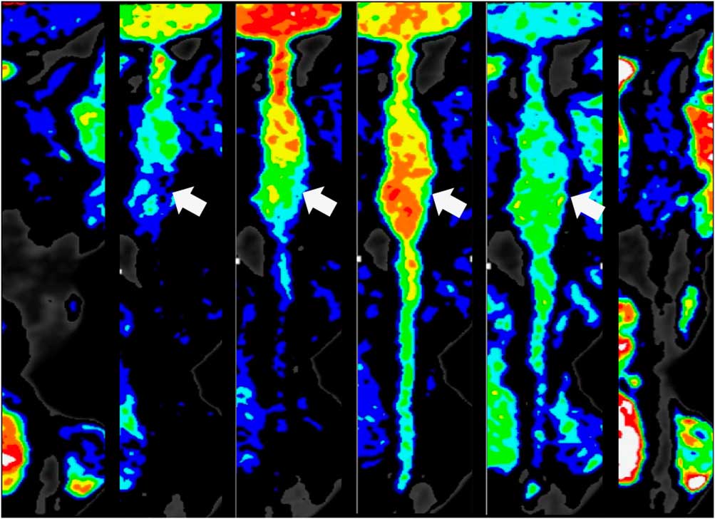
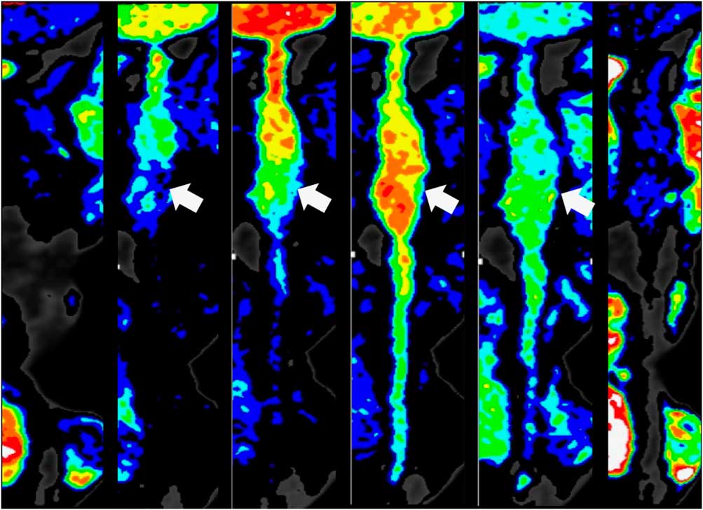
In patients with Kawasaki disease, coronary flow indexes for intra-aneurysm (0.57±0.22) and the distal site (0.44±0.19) were significantly lower than those for the proximal site (0.88±0.34, paired t test p<0.001), respectively. No difference between intra-aneurysm and the distal site was observed. In controls, there was no difference in coronary flow index between the proximal and distal sites (0.81±0.11 versus 0.68±0.33). Distal coronary flow index was significantly lower for Kawasaki disease than for controls (Mann–Whitney U-test, p<0.01), whereas there was no difference in proximal coronary flow index between two groups. Dynamic colour-coding imaging shows a slow and turbulent flow in coronary aneurysms in patients with Kawasaki disease (Fig 3).
Dynamic colour-coding imaging for an aneurysm of the right coronary artery. Dynamic coronary CT angiography data set was reconstructed to the straightened curved planar reformation using multi-planar reformatting. According to intra-coronary attenuation (CT number, Hounsfield unit), coronary artery was delineated to 6 colour scales (warm colour; high CT number, cold colour; low CT number). Cine imaging named for dynamic colour-coding was reconstructed to visualise coronary flow directly. Dynamic colour-coding imaging demonstrates a slow flow in the distal portion of the saccular aneurysm (arrow) in the same patient as in Figure 1.
The effective radiation dose was 5.7±0.6 mSv for dynamic coronary CT angiography and 4.7±1.7 mSv for standard coronary CT angiography. Heart rate for six patients with Kawasaki disease was 68±5 bpm.
Discussion
This study demonstrates a novel imaging technique to quantify and visualise coronary flow using 320-detector row CT with motion coherence image processing, and reveals a slow flow at the distal site of coronary aneurysm associated with Kawasaki disease. A 320-detector row CT system has a wide area detector with a head caudalis direction of 160 mm, allowing all coronary arteries to be covered in a single rotation. This enables to scan whole-heart dynamic angiography every heartbeat for 20 seconds. Motion coherence image processing offers three advantages: dynamic coronary CT angiography shows reduced image noise and high spatial resolution using a hybrid of adaptive iterative dose reduction, although this coronary flow imaging has achieved a relatively low radiation exposure (the mean estimated effective radiation dose, 5.7 mSv); the reconstructed dynamic coronary CT angiography offers a high temporal resolution (100–180 ms/phase); and the volume of interest for the coronary artery on cross-sectional images can be automatically extracted and intra-coronary attenuation can be measured throughout all dynamic images. Motion coherence image processing does not affect the actual CT attenuation. Multiplying data set by interpolation between the original two phases makes the upslope for coronary attenuation to be linear. These advantages allowed accurate creation of time–density curves of coronary arteries with less dispersion. Accordingly, the calculation of coronary flow index derived from maximum slope method is of high reproducibility. We propose the use of coronary flow index on the basis of the maximum slope method as a simple and objective index for coronary flow quantification. Our analytical technique is the first non-invasive means of overcoming the disadvantage of functional assessment of coronary artery flow using CT. The final goal in clinical use is to predict coronary thrombosis and the risk for myocardial infarction in patients with Kawasaki disease.
Distal coronary flow index was significantly lower for Kawasaki disease than for controls. Causes of declines in distal coronary flow index for Kawasaki disease were considered as follows. Proximal coronary flow index did not differ between Kawasaki patients and controls, and it was remarkably reduced in aneurysmal. This can be explained by intracoronary turbulence and increased shear stress in coronary aneurysms in Kawasaki disease.Reference Dibyendu, Andrew and Jane 8 Reduced distal coronary flow index may be simply caused by flow reduction at the aneurysmal site and its impacts on downstream.Reference Hamaoka, Onouchi, Kamiya and Sakata 9 As enrolled patients with Kawasaki disease were less likely to have coronary atherosclerosis, distal coronary arteries have no elevation of shear stress and impaired vascular reactivity.
This study had some limitations that need to be acknowledged. First, the retrospective nature of our analysis in a small patient cohort limits systematic appraisal of our approach to routine, clinical detection of coronary aneurysm in Kawasaki disease. In one patient with Kawasaki disease indicated in Figures 1 and 3, stress/rest myocardial perfusion SPECT showed an ischaemic perfusion pattern in the right coronary artery territories. This suggests that small coronary flow index is related to myocardial ischaemia. However, there were no data of validation by other modalities in other patients. To clarify the relationship between coronary flow index and measurements derived from myocardial perfusion studies is a future challenge. Second, coronary arteries with a diameter >3 mm appear necessary for volume of interest setup, because the volume of interest cannot be automatically extracted in smaller arteries in the motion coherence image processing. Third, volume of interest setup is often difficult in diffusely calcified arteries. Kawasaki disease patients with high calcium volume cannot be reliably evaluated with coronary flow index, because high calcium causes blooming artefact and high brightness shadow. Recently, functional assessment of coronary arteries using adenosine stress dual-energy CT,Reference Nagao, Kido and Watanabe 10 fractional flow reserve CT,Reference Nakazato, Park and Berman 11 and computational fluid dynamicsReference Karmonic, Partovi and Rengier 12 has been proposed. However, those imaging techniques analyse static coronary imaging. Consequently, the dynamic coronary CT angiography analysis described here is closer to the actual coronary haemodynamics.
Conclusion
In conclusion, this is the first analytical technique that can quantify and visualise coronary flow using coronary CT angiography. coronary flow index non-invasively demonstrates a slow coronary flow in intra-aneurysm and the distal portion in adulthood of Kawasaki disease.
Acknowledgement
None.
Financial Support
This research received no specific grant from any funding agency or from commercial or not-for-profit sectors.
Conflicts of Interest
None.
Supplementary material
To view supplementary material for this article, please visit https://doi.org/10.1017/S1047951117002293