In Noonan syndrome, hypertrophic cardiomyopathy is the second most commonly associated cardiac abnormality and tends to affect predominantly the interventricular septum and left ventricle.Reference Burch, Sharland, Shinebourne, Smith, Patton and McKenna1 This report describes a woman with Noonan syndrome presenting with sudden death as a complication of hypertrophic cardiomyopathy.
Case report
A 27-year-old female presented with ventricular fibrillation, which was successfully defibrillated with a single 360 Joule direct current shock. Surface electrocardiogram showed probable left ventricular hypertrophy. She was haemodynamically stable, conscious, and able to give a full history including diagnosis of Noonan syndrome during childhood, which was associated with phenotypic features and chromosomal analysis. At her last visit, at age 16, echocardiography was entirely normal. She was then symptom-free and out of follow-up, until experiencing dyspnoea on exertion and palpitations within the last 6 months.
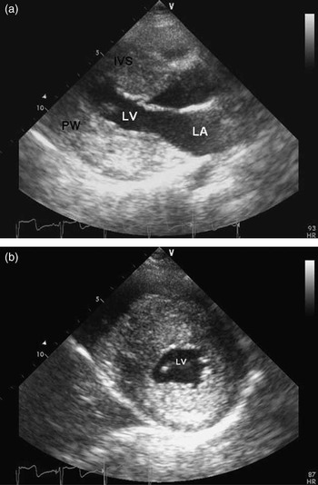
The patient's blood pressure and heart rate were found to be 110/70 millimetres in mercury and 75 beats per minute, respectively. She had a short stature (145 centimetres), a short neck, cubitus valgus, ocular hypertelorism, and a barrel-shaped chest. Auscultation revealed a systolic ejection murmur graded 3 at the mid-left sternal border, and S4 at the apex. Transthoracic echocardiography demonstrated massive left ventricular hypertrophy (septal width 27 millimetres, posterior wall thickness 23 millimetres). Grade I diastolic dysfunction with prolonged isovolumetric relaxation time, the typical “systolic anterior motion” of the anterior mitral leaflet, and a 23 millimetres in mercury peak resting systolic pressure gradient were observed (Fig 1). Colour Doppler imaging displayed mild to moderate mitral regurgitation presumably due to the systolic anterior leaflet motion. Coronary angiography revealed normal coronaries. Metoprolol was started and – because of her documented ventricular fibrillation – an implantable cardioverter defibrillator was implanted successfully.
Parasternal long- (a) and short-axis (b) views demonstrating marked left ventricular hypertrophy (LV = left ventricule; LA = left atrium; IVS = interventricular septum; PW = posterior wall).

Discussion
Noonan syndrome, first described by Noonan and Ehmke in 1963, is a relatively common non-chromosomal syndrome with a Turner-like phenotype and frequent cardiovascular malformations.Reference Noonan and Ehmke2 Despite a prevalence of 1–2 per 1000 persons being reported, the exact rate of occurrence and frequency of cardiac malformations are still obscure.Reference Sreeram, Kitchiner and Smith3
The most common cardiac malformation is dysplasia of the pulmonary valve cusps.Reference Sreeram, Kitchiner and Smith3, Reference Musewe, Robertson and Benson4 Hypertrophic cardiomyopathy is the second most commonly associated abnormality and tends to affect predominantly the interventricular septum and left ventricle.Reference Maron, McKenna and Danielson5 In one of the largest series, left ventricular hypertrophy without significant pulmonary stenosis was noticed in 29 out of 118 (25%) patients; among them, five had severe hypertrophy (greater than 1.7 centimetres) involving the entire septum and the free wall as in the present case.Reference Burch, Sharland, Shinebourne, Smith, Patton and McKenna1 Sudden death is a well-defined catastrophic consequence of hypertrophic cardiomyopathy; proposed risk factors are previous cardiac arrest or a family history of sudden death, non-sustained ventricular tachycardia on Holter or exercise, abnormal exertional blood pressure response, unexplained syncope, and some high-risk genotypes.Reference Maron, McKenna and Danielson5 Several studies have identified a maximal wall thickness greater than 30 millimetres as a risk factor, although its predictive value in isolation is low.Reference Olivotto, Gistri and Petrone6
We suggest that the present case, in which a normal left ventricle at age 16 evolved into a severe hypertrophy at age 27, demonstrates that ventricular hypertrophy might progress even many years after the syndrome is diagnosed, and highlights the importance of regular follow-up of these patients.