Introduction
The occurrence of a hydatid cyst in the infratemporal region is rare, with only a few cases reported in the English language literature.Reference Gangopadhyay, Abuzeid and Kfoury1–Reference Turgut, Ensari, Katirci and Çelikkanat3 Hydatidosis occurs all over the world, but it is especially common in the cattle-raising communities of Africa, Australia, New Zealand and South America, and is endemic in Turkey and the Middle East.Reference Turgut, Ensari, Katirci and Çelikkanat3, Reference Bouckaert, Raubenheimer and Jacobs4
The majority of hydatid cysts appear in the liver (65 per cent) and lungs (25 per cent). The presence of hydatid cysts in other organs is less common, and only 2 per cent of cases involve the maxillofacial region.Reference Amr, Amr, Jitawi and Annab5 Such cases commonly involve cystic lesions located in the mandible, maxillary sinus, orbit, infratemporal fossa, pterygopalatine fossa, parapharyngeal space, tongue, parotid gland or submandibular salivary gland.Reference Gangopadhyay, Abuzeid and Kfoury1, Reference Hotz and Gottstein2, Reference Akyildiz, Ozbilen and Goksu6–Reference Zaidi8
Hydatid disease of the infratemporal fossa is extremely rare; to our knowledge, only a few cases have been reported.Reference Sennaroglu, Önerci, Turan and Sungur7, Reference Kalovidouris, Gouliamos, Andreou, Ioannovits, Levett and Vlahos9–Reference Shuker11 Primary involvement of the maxillary sinuses is very rare, and very few cases have been reported.Reference Mortazavi and Motamedi12
We report a unique case of a 14-year-old girl in whom a hydatid cyst developed in the right infratemporal region, with the only presenting sign being visual loss over a 10-day period; the patient's vision recovered rapidly following surgical removal of cysts.
Case report
A 14-year-old girl presented with right exophthalmos and a two weeks history of visual loss.
Due to pressure on the optic nerve caused by a parapharyngeal cyst pressing through a inferior orbital fissure on the right side, Ophthalmological examination showed limited medial and inferior oblique muscle movement.
The patient's visual acuity of right eye had decreased to detection of hand motion only while the left eye was normal. The anterior, vitreal and fundus examinations of both eyes were normal.
The patient had a history of seizures treated with carbamazepine for two years (from five to seven years of age). During the most recent investigation, multiple hydatid cysts had been noted in the patient's brain. She had been referred to our otolaryngology centre in April 2006 by a neurosurgeon who had performed craniotomies on the patient seven, five and three years earlier. At time of referral, the right frontoparietal lobe had been affected. The patient had been treated intermittently with albendazole.
Examination of the patient's nervous system revealed normal and intact cranial nerves.
While the patient had been receiving treatment with albendazole, a computed tomography (CT) scan of the paranasal sinus had shown multiple cystic masses in the right infratemporal fossa, which were eroding the posterior wall of the maxillary sinus and inferior orbital wall and compressing the orbit (Figure 1).
A multilocular, hypodense lesion in the right infratemporal fossa, with expansion of the fossa and compression of the pterygoid muscles. Erosion of the right maxillary sinus posterior wall is also seen, with some extension within its antrum.

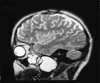
A parasagittal, T2-weighted magnetic resonance imaging (MRI) scan showed a well defined, homogeneous, high signal, multilocular lesion in the right infratemporal fossa, with some extension within the right maxillary sinus antrum. Intracranial extension was not seen in this scan (Figure 2).
Parasagittal, T2-weighted magnetic resonance imaging scan, showing a well defined, homogeneous, multilocular lesion in the right infratemporal fossa, with some extension within the right maxillary sinus antrum. Intracranial extension is not seen in this scan.

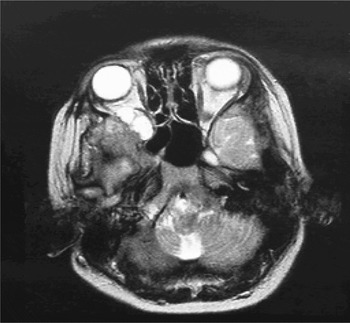
An axial, T2-weighted MRI scan showed a well defined, homogeneous, extraconally located, multilocular lesion in the right infraorbital fissure, which was seen to be expanding and exerting a mass effect (Figure 3). Proptosis was not seen.
Axial, T2-weighted magnetic resonance imaging scan (orbital section), showing a well defined, homogeneous, extraconally located, multilocular lesion in the right infraorbital fissure, which is seen to be expanding and exerting a mass effect on the contents of this anatomical area.

Computed tomography scans of the patient's brain showed the impact of previous craniotomies, together with hypodense defects in the right frontoparietal region.
Laboratory investigations were undertaken, revealing a positive enzyme-linked immunosorbent assay for anti hydatid antibody and 5 per cent eosinophils in the peripheral blood smear. Surprisingly, chest X-rays and liver sonography were normal.
The patient received albendazole 400 mg for three weeks, ceasing one week before surgery.
The patient underwent surgery with a primary diagnosis of a hydatid cyst, on the basis of her previous medical history and radiological findings. Three hydatid cysts were found (2, 1 and 1 cm in diameter); all were encased in a capsule, and extended superiorly from the posterior wall of the maxillary sinus to the infratemporal fossa. These cysts were removed easily and completely from the right maxillary sinus, without any spillage, via a standard Caldwell–Luc approach. During surgery, gauze soaked with 2 per cent formalin was placed around the cysts and iodoform gauze was packed into the antrum and oral cavity. After cyst removal, the surgical area was irrigated with hypertonic saline.
Macroscopically, the yellow-white colour and elastic nature of the cyst walls suggested parasitic disease. Histopathological examination revealed a basophilic cuticular membrane consistent with a hydatid cyst.
Post-operatively, the patient's visual acuity improved considerably over just two weeks.
A six-month course of albendazole (15 mg/kg/day) was prescribed to prevent recurrence of hydatidosis.
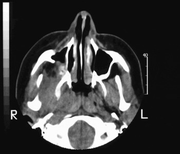
Computed tomography scans were performed four, 12 and 18 months later. No evidence of any lesion in the right infratemporal fossa was seen. Some post-operative inflammation was noted. A residual defect in the right maxillary sinus posterior wall, due to the lesion, was noted (Figure 4).
Axial, paranasal sinus computed tomography scan taken after surgery. No evidence of the lesion is seen in the right infratemporal fossa. Some post-operative inflammation is seen. A residual defect is present in the right maxillary sinus posterior wall, due to the lesion.

At the patient's last follow-up consultation, two years after surgery, she showed no evidence of hydatidosis.
Discussion
Hydatid disease is a parasitic infestation in humans caused by the larval form of Echinococcus granulosus. Infratemporal hydatid disease can only occur when parasitic embryos succeed in passing through the hepatic and pulmonary filtering systems.Reference Kalovidouris, Gouliamos, Andreou, Ioannovits, Levett and Vlahos9, Reference Rudwan and Khaftaji13, Reference Sharma and Ray14
Hydatid cysts in the maxillofacial region are extremely rare, comprising only 2 per cent of reported cases of hydatidosis. They commonly appear as cystic lesions located in the mandible, maxillary sinus, orbit, infratemporal fossa, pterygopalatine fossa, parapharyngeal space, tongue, parotid gland or submandibular salivary gland.Reference Bouckaert, Raubenheimer and Jacobs4, Reference Sennaroglu, Önerci, Turan and Sungur7, Reference Kalovidouris, Gouliamos, Andreou, Ioannovits, Levett and Vlahos9, Reference Sener10, Reference Onerci Turan and Ruacan15
At the time of presentation, our patient showed no evidence of hydatid cysts elsewhere in the body, other than the brain. A maxillofacial hydatid cyst had been present during the period when the patient had received intermittent medical therapy for brain hydatosis.
• Hydatid disease is a parasitic infestation in humans caused by the larval form of Echinococcus granulosus
• This paper describes a case of a hydatid cyst in the infratemporal fossa, with visual loss
• The most effective treatment for hydatidosis is surgical removal
• Immediate surgery is necessary when visual loss occurs
The growth rate of hydatid cysts is highly variable, ranging from 1 to 5 cm a year. The body's tolerance of hydatid cysts, and their effect on function, depends on their growth rate and size.Reference Sennaroglu, Önerci, Turan and Sungur7 In our patient, a slow-growing cyst had displaced the zygomatic arch, mandibular ramus and maxillary bone, causing facial asymmetry. The cysts had also eroded the inferior wall of the orbit, due to a mass effect, leading to visual loss. During surgery, three hydatid cysts (2, 1 and 1 cm in diameter) were removed completely from the right maxillary sinus and infratemporal fossa, without spillage, via a standard Caldwell–Luc approach. Silver nitrate (0.5 per cent), hypertonic saline (20 per cent) and other chemicals are often injected into hydatid cysts at the time of surgery to inactivate the protoscolices. We did not inject any chemicals into the cysts at the time of surgery.
In the present case, the diagnosis of echinococcus infestation was based on the clinical history, physical examination, and radiological and histopathological investigation. The use of serological tests (such as indirect haemagglutination, latex agglutination, enzyme-linked immunosorbent assay and immunoelectrophoresis) in the diagnosis of hydatosis is controversial. Both false positive and false negative results are common.Reference Gangopadhyay, Abuzeid and Kfoury1, Reference Schipper and Kager16 Traditional serological tests are not sufficiently sensitive to establish a diagnosis or to be useful for follow up of patients with hydatid disease. The Casoni intradermal test and the Weinberg complement fixation test frequently give false negative results.Reference Dogăn, Yüksel, Cetin, Süzer, Alp and Kaya17, Reference Bret, Fond, Bretagnolle, Valette, Thiesse and Lambert18
We did not depend on such tests but, rather, undertook CT and MRI scanning, which showed typical features of the hydatid lesion and its location, enabling more accurate diagnosis. The radiologist was able to determine the size and anatomical relations of the cystic lesion, and to inform the otolaryngologist of the possibility of hydatid disease. The most effective treatment for hydatid cysts is surgical removal; therefore, the extent of the lesion must be carefully evaluated to determine the best treatment strategy. Computed tomography and MRI scanning can be used to guide hypertonic saline infusion and cyst fluid aspiration (to decrease cyst size). The surgeon can then decide either to remove the cyst completely or to administer chemotherapy, in cases of inoperable disease due to anatomical factors. Appropriate treatment and ongoing evaluation of the adjacent organs are necessary, due to the risk of recurrence. Immediate surgery is recommended both to prevent disease development and to improve the patient's prognosis.