Introduction
Human toxocariasis is a neglected tissue parasitic disease with worldwide distribution. Caused by the larval migration of Toxocara canis and T. cati, intestinal nematodes are found in dogs and cats, respectively (Chen et al. Reference Chen, Liu, Liu, Wen-Bin, Sung-Jong, Hiromu, Xing-Quan and Hany2018). This disease presents a significant challenge to a global public health problem (Magnaval et al. Reference Magnaval, Bouhsira and Fillaux2022). Humans become infected mainly by ingesting embryonated eggs present in the soil and contaminated vegetables or water. Another route of infection is the consumption of undercooked or raw meat or viscera from paratenic hosts such as chickens, cows, ducks, lambs, pigs, ostriches, or rabbits, which may contain encysted infective larvae in their tissues (Chen et al. Reference Chen, Liu, Liu, Wen-Bin, Sung-Jong, Hiromu, Xing-Quan and Hany2018). In humans, this disease is caused by the migration and residence of T. canis larvae in the liver, lungs, heart, kidneys, brain, and skeletal striated muscles (Fan et al. Reference Fan, Holland, Loxton and Barghouth2015).
Clinical manifestations can range from asymptomatic to severe organ injury and can result in different syndromes. Depending on the type of tissue involved, the infection can be classified as systemic, including visceral larva migrans syndrome (VLM) and common or covert toxocariasis, or compartmentalized, including ocular and neurological toxocariasis (Chen et al. Reference Chen, Liu, Liu, Wen-Bin, Sung-Jong, Hiromu, Xing-Quan and Hany2018). Pathological changes in human toxocariasis include hemorrhage, necrosis, and granuloma formation in somatic tissues (Strube et al. Reference Strube, Heuer and Janecek2013).
Treatment varies according to symptoms and clinical form, starting with an anthelmintic approach. Benzimidazole, albendazole, and mebendazole are the first- and second-choice medications, respectively. Additionally, palliative therapies, such as corticosteroids, antihistamines, and oxygen therapies, may be used (Magnaval et al. Reference Magnaval, Bouhsira and Fillaux2022). The clinical efficacy of these anthelmintics is considered low to moderate, mainly because of their low gastrointestinal absorption and, consequently, low bioavailability after metabolization (Chen et al. Reference Chen, Liu, Liu, Wen-Bin, Sung-Jong, Hiromu, Xing-Quan and Hany2018), indicating the need for research and development of novel drug candidates.
The mouse is an excellent laboratory host to study experimental infections by T. canis, as it tolerates infections for long periods, and the larvae follow the enteric-pulmonary-somatic cycle with signs and symptoms similar to those observed in humans (Nicholas et al. Reference Nicholas, Stewart and Mitchell1984). Thus, murine toxocariasis is to be a suitable model for evaluating chemical compounds, medicines, and probiotics, among others (Mata-Santos et al. Reference Mata-Santos, Pinto, Mata-Santos, De Moura, Carneiro, Carvalho, Del Rio, Pinto, Martins, Fenalti, Da Silva and Scaini2015; Rodrigues et al. Reference Rodrigues, Cunha, Mattos, Martins, Nogueira, De Souza, De Avila, Ramos and Scaini2024; Walcher et al. Reference Walcher, Cruz, de Lima Telmo, Martins, De Avila, Berne and Scaini2017).
A wide range of natural products have been the subject of research in biomedical and biological fields, and camphor and its derivatives have been identified. Camphor is a white crystalline substance that can be obtained from Cinnamomum camphora and other natural sources such as Dryobalanops aromatica and Ocoteaus ambarensis. Camphor can also be synthetically obtained from α-pinene, an abundant natural compound present in turpentine oil (Alves and Victor Reference Alves and Victor2010). This is a secondary metabolite of the plant Artemisia annua, which is known to be active against Plasmodium spp., Toxoplasma gondii, and Leishmania spp. (Feng et al. Reference Feng, Cao, Qiu and Zhang2020). Studies suggest that terpenoids, compounds produced by Artemisia annua, may be active against various helminths such as Haemonchus contortus, Ecchinococcus spp., Schistosoma spp., and Toxocara canis (Lam et al. Reference Lam, Long, Su and Lu2018). These compounds contain special functional groups that allow suitable synthetic transformations, making it possible to prepare several structures of interest within a short period (Silva et al. Reference Silva, Da Silva Santos, De Andrade, Rosa and De Souza2022).
Several extracts and derivatives of camphor have been studied against a wide range of pathogens; for example, Artemisia camphorata has a fungitoxic effect against Bipolaris sorokiniana (Stangarlin et al. Reference Stangarlin, Franzener, Schwan-Estrada and Cruz2003), Alternaria solani (Itako et al. Reference Itako, Schwan-Estrada, Tolentino Júnior, Stangarlin and Cruz2008), and Exserohilum turcicum (Scapin et al. Reference Scapin, Carnelossi, Vieira, Schwan-Estrada and Cruz2010). Furthermore, camphor essential oils showed antibacterial activity against Staphylococcus aureus, Micrococo luteus, Escherichia coli, Salmonella typhimirium, Bacillus cereus, Enterococcus faecalis, Candida albicans, C. glabrata, C. saque, and C. tropicalis (Mighri et al. Reference Mighri, Hajlaoui, Akrout, Najjaa and Neffati2010). This study aimed to select a camphor derivative as a candidate drug for the treatment of visceral toxocariasis using preclinical tests, including in vitro, in silico, and in vivo assays.
Materials and methods
Camphor derivatives
Areneal dehyde and d-camphor were purchased from commercial sources. All solvents and reagents were used as received without further purification. Three camphor derivatives were prepared using an ultrasonic homogenizer model ‘Desruptor Eco Sonics’ (25 KHz, Eco Sonics, Indaiatuba, Brasil), that was used to carry out the reactions at 90 W, with an average yield of 68% (Silve et al. 2016). Camphor derivatives were produced by the Institute of Drug Technology (Farmanguinhos) of the Oswaldo Cruz Foundation (Fiocruz) and were available as lyophilized samples at the New Drug Development Center (NUDEFA/FURG) (Table 1). All the synthesized compounds were solubilized at 2.5% in dimethyl sulfoxide (DMSO) and sterile distilled water to obtain a concentration of 1 mg/mL (Mata-Santos et al. Reference Mata-Santos, Pinto, Mata-Santos, De Moura, Carneiro, Carvalho, Del Rio, Pinto, Martins, Fenalti, Da Silva and Scaini2015).
Chemical structure of the three camphor derivatives used for the study

Parasite
The Toxocara canis eggs were maintained in 2% formalin solution and incubated at 28°C, humidity above 90%, and oxygenation for 30 days (Avila et al. Reference Avila, Conceição, Telmo, Dutra, De Los Santos, Martins, Berne, Da Silva and Scaini2012). The infective larvae (third-stage) (Bowman Reference Bowman2020) were extracted using a modified Baermann apparatus and maintained in sterile tubes for cultivation with RPMI-1640 medium (Gibco®), supplemented with 25 mM HEPES (Sigma®), 1% glucose, PSF antibiotic-antimycotic (Gibco®) at 37°C with an atmosphere of 5% CO2 (Savigny, Reference Savigny1975).
In vitro tests
The in vitro screening tests were performed in duplicate at a concentration of 1 mg/mL in a 96-well flat bottom plate, using 100 T. canis larvae (third-stage) per well and three controls: one live larva, one dead larva (thermal shock), and one DMSO (2.5%). The microculture plate was maintained for 48 h at 37°C with 5% CO2. Subsequently, trypan blue viability indicator was added, and the plate was incubated for an additional 30 min. The samples were examined under an optical microscopy (10× objective). Viability was assessed based on the motility of the larvae and the absence of impregnation using a cell viability indicator. The compounds that showed 100% of larvicidal activity in the screening test (1.0 mg/mL) were evaluated at concentrations of 0.5 mg/mL to 0.05 mg/mL to determine their minimum larvicidal concentration (MLC) (Mata-Santos et al. Reference Mata-Santos, Pinto, Mata-Santos, De Moura, Carneiro, Carvalho, Del Rio, Pinto, Martins, Fenalti, Da Silva and Scaini2015).
To confirm the non-viability of the larvae observed in the in vitro tests of the compounds that presented 100% MLC, we followed the methodology described by Mata-Santos et al. (Reference Mata-Santos, Pinto, Mata-Santos, De Moura, Carneiro, Carvalho, Del Rio, Pinto, Martins, Fenalti, Da Silva and Scaini2015). Briefly, the contents of the wells of a microculture plate containing larvae exposed to the compounds and unexposed larvae (control) were intraperitoneally inoculated in duplicate into female Swiss mice intraperitoneally (IP). After 48 h of inoculation, the mice were euthanized, and tissue digestion of the carcasses, brains, livers, lungs, kidneys, and hearts was performed in a solution of 1% hydrochloric acid and 1% pepsin (Xi and Jin Reference Xi and Jin1998). The material was then examined under a microscope with a 10× objective.
Cytotoxicity
Compounds that showed larvicidal activity against 100% of the larvae in the MLC test were subjected to cytotoxic assays at the same concentrations of the in vitro screening tests to determine the concentration of the compound capable of maintaining 50% cell viability (IC 50). The test was performed using murine macrophages J774.A1 (1.105 cells/100 μL), placed in DMEM medium supplemented with 10% fetal bovine serum. A compound free cell control was used. To read the results, the resazurin cell viability indicator was added at 0.01% and microculture plate was incubated for 6 h. Cell viability was measured by fluorescence at a 620 nm wavelength through a BioTekTM ELx800TM reader (Ansar Ahmed et al. Reference Ansar Ahmed, Gogal and Walsh1994)
In silico bioavailability
The selected compound and albendazole were drawn using the molecular editor Chem Bio Draw Ultra 12.1 (Halford Reference Halford2014) and analyzed using the Swiss ADME computer model (www.swissadme.ch) (Daina et al. Reference Daina, Michielin and Zoete2017). To predict adequate parameters of absorption, distribution, metabolism, and excretion of compounds by the human body, the analysis was made following the ‘rule of five’ (Lipinski Reference Lipinski2004), which includes four parameters and the reference values accepted are equal to or less than multiples of five, being accepted the following values: molecular weight ≤ 500, miLogP ≤ 5, number of hydrogen donors ≤ 5, and hydrogen acceptors ≤ 10.
Evaluation of murine toxocariasis
Female Swiss mice aged 5–7 weeks of age were used for all in vivo tests. The mice were randomly divided into three groups, and two experiments were conducted: the first was to evaluate the efficacy of the compound in mice (n=5) 10 days post-inoculation with T. canis, and the second was to evaluate the efficacy in mice (n=10) 30 days post-inoculation. The selected compounds and two controls were used for this evaluation. The mice were infected with 500 embryonated eggs of T. canis via intragastric (IG) route (Satou Reference Satou, Horiuchi, Akao, Koike, Fujita and Nikaido2005). The groups were administered for 5 days with the selected compound (10 mg/kg, IG), albendazole (40 mg/kg, IG), and phosphate buffered saline solution (PBS) (0.15 M and pH 7,2) (Mata-Santos et al. Reference Mata-Santos, Pinto, Mata-Santos, De Moura, Carneiro, Carvalho, Del Rio, Pinto, Martins, Fenalti, Da Silva and Scaini2015). After 5 days of treatment, all the mice were euthanized. The recovery of T. canis larvae was performed by tissue digestion of organs (liver, lungs, heart, kidneys, brain, and ocular globes) and carcasses. The larvae were examined and quantified using microscopy (10× objective) (Xi and Jin Reference Xi and Jin1998).
The mice were housed in mini-isolators (Tecniplast®) under a controlled environment of 22±1 °C, with a lightening cycle of 12/12 h light/darkness, provided with food without antibiotics and free access to water. This study was approved by the Ethics Committee in Research of the Federal University of Rio Grande (P034/2020). An invention patent titled ‘Use of synthetic compound derived from camphor as an anthelmintic’ owned by the Federal University of Rio Grande was deposited on April 17, 2024, at the National Institute of Industrial Property – INPI, under the number BR 10 2024 007530 7.
Statistical analysis was performed using analysis of variance (ANOVA) with Tukey’s post-hoc test, with a significance level of p≤0.05.
Results
After the in vitro screening tests, two compounds (C1 and C2) showed activity against 100% of T. canis larvae at a concentration of 1 mg/mL. Both were submitted to the MLC test, and the results showed 100% efficacy at 1 mg/mL for C1 and at 0.25 mg/mL for C2 (Table 2); this way, compound (E)-2-((1,7,7-trimethylbicyclo[2.2.1]heptan-2-ylidene)amino)phenol (C2) was selected for the next steps. In the cytotoxicity test, compound C2 resulted in 100% cell viability at its MLC (0.25 mg/mL) and an estimated cell viability of 77% at the maximum concentration tested (2 mg/mL). Following the described procedures, a confirmatory test was performed. The larvae exposed to C2 were not viable and did not have the ability to infect mice. This result was corroborated by the controls, as on average, nine larvae were found in the liver and lung tissues.
Percentage of non-viable (dead) Toxocara canis larvae after exposure to camphor-derived compounds evaluated at different concentrations

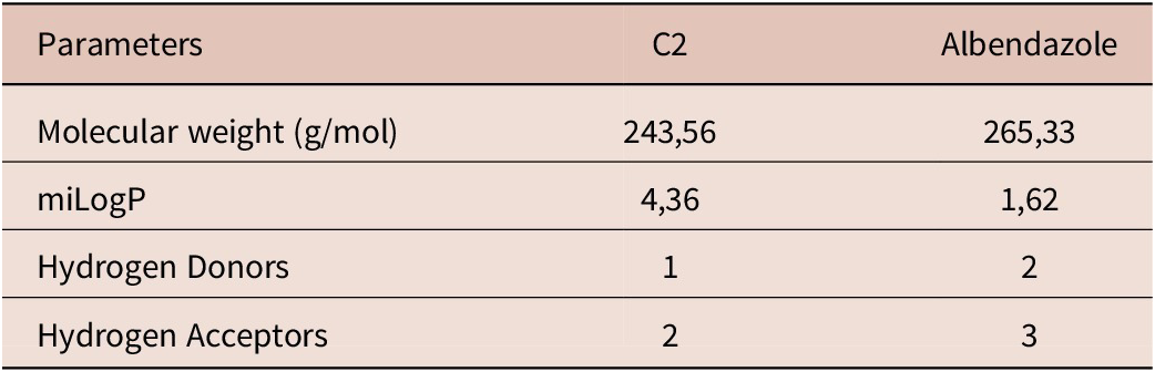
The results presented in Table 3 reveal the characteristics associated with oral bioavailability and indicate acceptable water solubility and gastrointestinal absorption. The physicochemical properties of (E)-2-((1,7,7-trimethylbicyclo[2.2.1]heptan-2-ylidene)amino)phenol (C2) are similar to those of albendazole.
Physicochemical properties of the selected in vitro camphor derived (E)-2-((1,7,7-trimethylbicyclo[2.2.1]heptan-2-ylidene)amino)phenol (C2) and the albendazole

In both in vivo tests, 10 and 30 days post-inoculation with T. canis, C2 promoted a significant reduction in infection intensity compared to PBS and showed similar results to albendazole (Table 4).
Average recovery of the larvae per group 10 and 30 days post-inoculation with 500 eggs of Toxocara canis in Swiss mice

Discussion
Camphor has been used as an herbal medicine for treating several pathogens and diseases. In a study conducted by Kang et al. (Reference Kang, Han, Yoon, Sim, Maeng, Kang and Yoo2019), the leaves of Cinnamomum camphora showed reduction in vitro in the production of inflammatory chemokines and, in vivo, was capable of relieved the symptoms of atopic dermatitis. The same extract has already demonstrated activity against coronavirus (SARS-CoV-2) (Malabadi et al. Reference Malabadi, Kolkar, Meti and Chalannavar2021), while essential oils from Cinnamomum osmophloeum, another species of the genus Cinnamomum, showed antioxidant activity in vivo in Caenorhabditis elegans (Hsu et al. Reference Hsu, Li, Yu, Hsieh, Yang, Liu, Shih, Chu, Yen, Chang and Liao2012). Studies conducted by Fazmiya et al. (Reference Fazmiya, Sultana, Rahman, Heyat, Sumbul, Akhtar, Khan and Appiah2022) demonstrated that C. camphora can also inhibit transient receptor potential A1 (TRPA1) and desensitize the receptor TRPV1, causing an analgesic action with potential use for different clinical conditions.
In this study, we evaluated the efficacy of a synthetic camphor derivative against murine visceral toxocariasis. In the last decade, major studies in vitro have focused on assessing the potential larvicidal activity of many compounds against Toxocara spp., such as Brazilian red propolis essential oil (Sinott et al. Reference Sinott, Sena-Lopes, Leal, Thais, Cardoso, De Moura, Berne and Borsuk2019), natural and synthetic naphthoquinones (Mata-Santos et al. Reference Mata-Santos, Mata-Santos, Carneiro, De Moura, Fenalti, Klafke, Cruz, Martins, Pinto, Pinto, Berne, Da Silva and Scaini2016a; Ortiz-Pérez et al. Reference Ortiz-Pérez, Vázquez, Rivera, Salas, Zarate-Ramos, Trofymchuk, Hernandez-Soberanis and Perales-Flores2021), synthetic fatty acid amide (Mata-Santos et al. Reference Mata-Santos, D’Oca, Mata-Santos, Fenalti, Pinto, Coelho, Berne, Da Silva, D’Oca and Scaini2016b), and synthetic coumarin derivatives (Rodrigues et al. Reference Rodrigues, Cunha, Mattos, Martins, Nogueira, De Souza, De Avila, Ramos and Scaini2024). Several extracts and derivatives of camphor have been studied in fungi (Itako et al. Reference Itako, Schwan-Estrada, Tolentino Júnior, Stangarlin and Cruz2008; Scapin et al. Reference Scapin, Carnelossi, Vieira, Schwan-Estrada and Cruz2010; Stangarlin et al. Reference Stangarlin, Franzener, Schwan-Estrada and Cruz2003) and bacteria (Mighri et al. Reference Mighri, Hajlaoui, Akrout, Najjaa and Neffati2010). This is the first study to demonstrate the potential of camphor for treating visceral toxocariasis.
The results obtained in this study revealed a larvicidal activity in vitro of two camphor derivatives against T. canis, (E)-4-((1,7,7-trimethylbicyclo[2.2.1]heptan-2-ylidene)amino)phenol (C1) at a concentration of 1 mg/mL and (E)-2-((1,7,7-trimethylbicyclo[2.2.1]heptan-2-ylidene)amino)phenol (C2) at 0.25 mg/mL. Considering the low active concentration found for C2, it was selected for subsequent tests and was found to be non-cytotoxic at the concentrations tested. In addition, Silva et al. (Reference Silva, Da Silva Araújo, Moraes, De Souza, Lourenço, De Souza, Wardell and Wardell2016), who synthesized the compounds used in this study, performed in vitro tests against Mycobacterium tuberculosis ATCC 27294 and obtained excellent antimycobacterial activity for compound C2.
Benzimidazoles are widely used to treat infections caused by various intestinal parasites. However, when used in tissue parasitosis, such as human toxocariasis, they are unable to achieve excellent rates of clinical resolution, which may be due to their low solubility in aqueous media (Chen et al. Reference Chen, Liu, Liu, Wen-Bin, Sung-Jong, Hiromu, Xing-Quan and Hany2018). In relation to in silico bioavailability, (E)-2-((1,7,7-trimethylbicyclo[2.2.1]heptan-2-ylidene)amino)phenol (C2) did not violate the Lipinski’s ‘rule of five’ indicating adequate gastrointestinal absorption. In relation to the descriptors evaluated, this compound showed a low molecular weight, indicating its easy transport by proteins, which led to better absorption and metabolization capacity. Moreover, in relation to hydrogen donors and acceptors, this compound may exhibit good permeability through lipophilic membranes. The parameters obtained from the in silico analysis also revealed that C2 showed a miLogP higher than that of albendazole, indicating that this compound may present better results when administered with high-fat diets, as albendazole is poorly absorbed in the gastrointestinal tract and should be taken with a fat diet (Magnaval et al. Reference Magnaval, Bouhsira and Fillaux2022).
The results obtained in vivo in the present study confirmed the efficacy of (E)-2-((1,7,7-trimethylbicyclo[2.2.1]heptan-2-ylidene)amino)phenol (C2) against T. canis infection through a significant reduction in the infection intensity in the experimental models used. When we analyzed and compared the dosages used for the treatment groups, we noticed that despite having similar results in the quantification of larval recovery, the group treated with C2 received a daily dose of 10 mg/kg, which was four times lower than the dose used for albendazole (40 mg/kg).
After infection, T. canis larvae begin to migrate to the liver and lungs via the circulatory system. After the first week, the hepato-pulmonary phase concludes, and the larvae continue with a neurotropic-myotropic phase when the larvae predominate in the brain and muscle (Waindok et al. Reference Waindok, Janecek-Erfurth, Lindenwald, Wilk, Schughart, Geffers, Balas, Durand, Rund, Schebb and Strube2019). In our study, we observed a major reduction in the infection intensity in the group treated 10 days post-inoculation, which revealed that this compound may be more suitable for recent clinical toxocariasis, in which the larvae are still beginning their neurotropic-myotropic phase, and tissues such as the liver, lungs, and heart present a greater number of larvae.
Experiments performed in mice 30 days after T. canis infection have provided consistent data on the efficacy of these compounds in reducing or eradicating the infection (Barrera et al. Reference Barrera, Leonardi, Bolmaro, Echenique, Olivieri, Salomon and Lamas2010; Mata-Santos et al. Reference Mata-Santos, Mata-Santos, Carneiro, De Moura, Fenalti, Klafke, Cruz, Martins, Pinto, Pinto, Berne, Da Silva and Scaini2016a). It is important to note that during this period of infection, the nematode larvae have already completed the initial phase of hepato-pulmonary migration and tend to spread throughout the body of the host. Therefore, the observation of a significant reduction in the intensity of infection is valuable data for a preclinical study. However, it is important to carry out studies on the therapeutic activity of the compound in murine models of chronic toxocariasis. Studies have shown that even mild T. canis infections in mice lead to changes in the general appearance and activity of the animals, causing disturbances in motility and balance, accompanied by the development of necrosis, hemorrhage, and accumulation of foamy cells (Bernardová et al. Reference Bernardová, Jan, Petr, Chia-Kwung and Libuše2024).
These findings, in addition to the lack of cytotoxicity, reinforce the therapeutic benefits of camphor derivatives in treating visceral toxocariasis. The results of this research have also resulted in a patent for the use of this compound as an antiparasitic, which has been filed with the Brazilian National Institute of Industrial Property (INPI) under the number BR 10 2024 007530 7, demonstrating the potential of this compound in the production of a novel antiparasitic drug candidate for use against visceral toxocariasis.
Conclusion
From this research, it can be concluded that the results obtained with synthetic camphor (E)-2-((1,7,7-trimethylbicyclo[2.2.1]heptan-2-ylidene)amino)phenol (C2) demonstrate its high activity against T. canis larvae and in the treatment of murine visceral toxocariasis, in addition to not presenting cytotoxicity and adequate bioavailability. Based on this research and the patent already filed for the use of this compound, it should be considered a candidate antiparasitic therapy for treating visceral toxocariasis.
Data availability statement
Invention patent titled ‘Use of synthetic compound derived from camphor as an anthelmintic’ owned by the Federal University of Rio Grande, deposited on April 17, 2024, at the National Institute of Industrial Property – INPI, under the number BR 10 2024 007530 7.
Financial support
D.F. Ramos is a productivity research fellow from National Council for Scientific and Technological Development (CNPq) (process number PQ 306806/2022-3). The authors acknowledge the financial support from the Brazilian Agency Coordenação de Aperfeiçoamento de Pessoal de Nível Superior and the scholarship granted.
Competing interest
The authors declare that they have no known competing financial interests or personal relationships that could have appeared to influence the work reported in this paper.
Ethical standard
According to Brazilian law no 11.794/2008 (Arouca law), article 2◦ applies to animals of species classified as phylum Chordata, subphylum Vertebrata, subject to environmental legislation. The Federal University of Rio Grande is accredited by the Commission for Ethics in the Use of Animals (CEUA) in accordance with the provisions of article 8◦ of the Arouca law. This study was approved by the Ethics Committee in Research of the Federal University of Rio Grande (P034/2020).