INTRODUCTION
Trypanosoma evansi is a digenetic flagellate which has extensive worldwide distribution, and infects a large number of domestic and wild animals (Levine, Reference Levine1973; Silva et al. Reference Silva, Herrera, Domingos, Ximenes and Dávila1995, Reference Silva, Seidl, Ramirez and Dávila2002). In horses the disease is known as “Mal das cadeiras” or “Surra” (Seidl et al. Reference Seidl, Moraes and Silva1998; Aquino et al. Reference Aquino, Machado, Alessi, Marques, Castro and Malheiros1999; Silva et al. Reference Silva, Seidl, Ramirez and Dávila2002). Humans were considered refractory to infection by T. evansi (Kubiak and Molfi, 1954), but recent reports showed that human infections occurred in India (Joshi et al. Reference Joshi, Shegokar, Power, Herder, Katti, Salkar, Dani and Bhargava2005; Shegokar et al. Reference Shegokar, Powar, Joshi, Bhargava, Dani, Katti, Zare, Khanande, Jannin and Truc2006). The pathogenesis of the anaemia in trypanosome infections is not fully elucidated and the role of enzymes involved in red blood cell and heme synthesis has not been studied.
The cytosolic enzyme delta-aminolevulinate dehydratase (δ-ALA-D) also known as porphobilinogen synthase (PBG- synthase; EC 4.2.1.24) is a metalloenzyme requiring zinc for activation (Bevan et al. Reference Bevan, Bodlaender and Shemin1980). The sulphydryl groups (SH) are essential for the functioning of the enzyme and zinc has the function of maintaining this grouping in the reduced form (Block et al. Reference Block, Lohmann and Beyersmann1990). The main action of this enzyme on the prosthetic groups of proteins such as haemoglobin, myoglobin, cytochrome, catalase and peroxidase (Jaffe, Reference Jaffe1995). δ-ALA-D is sensitive to oxidizing agents in vitro, and may also have reduced activity when associated with situations that occur with oxidative stress in vivo (Folmer et al. Reference Folmer, Soares, Gabriel and Rocha2004). The enzyme, in red blood cells, catalyses the asymmetric condensation of 2 molecules of delta-aminolevulinic acid, with loss of 2 water molecules, forming the composite mono-pyrrole porphobilinogen (Jaffe, Reference Jaffe1995). Inhibition of δ-ALA-D may adversely affect the biosynthesis of heme and iron uptake in erythropoieses. Also, the accumulation of the substrate 5-aminolevulinic may be associated with excessive production of reactive oxygen species (Pereira et al. Reference Pereira, Curi, Kokubun and Bechara1992).
Recent studies showed that T. evansi infection of animals decreased serum levels of iron and zinc (Da Silva et al. Reference Da Silva, Hoehne, Tonin, Zanette, Wolkmer, Costa, Moraes, Flores, Santurio, Lopes and Monteiro2009a) and increased oxidative stress (Da Silva et al. Reference Da Silva, Zanette, Paim, Wolkmer, Costa, Santurio, Lopes and Monteiro2009b; Wolkmer et al. Reference Wolkmer, da Silva, Traesel, Paim, Cargnelutti, Pagnoncelli, Picada, Monteiro and Lopes2009). The activity of δ-ALA-D in the blood may be affected by the serum concentrations of zinc and the levels of oxidative stress in T. evansi-infected animals after onset of anaemia. The aim of this study was to investigate the blood δ-ALA-D activity in rats experimentally infected with T. evansi and to ascertain its correlation with the levels of lipid peroxidation and concentrations of iron and zinc in the serum.
MATERIALS AND METHODS
Experimental animals
Thirty-six male Wistar rats (Rattus norvegicus; 214±25 g) at 90 days of age were kept in cages housed in a room with controlled temperature and humidity (25°C; 70%). They were fed with commercial ration (Supra®) and received water ad libitum. All animals had a period of 15 days for acclimatization and were clinically healthy at the commencement of the experiment (day 0). The procedure was approved by the Committee on Ethics in Animal Experimentation of the Federal University of Santa Maria (protocol number: 52/2010).
Groups and trypanosome infection
The animals were divided into 2 groups (A and B) with 18 animals each. Both groups were again divided in 3 groups (A1, A2 and A3) and (B1, B2 and B3) with 6 animals each in order to evaluate the progression of the disease in different periods of infection. Group A consisted of healthy non-infected rats. Rats in groups B were intraperitoneally inoculated (Day 0) with infective blood from a rat previously infected with a crypreserved strain of T. evansi originally isolated from a naturally infected horse (Da Silva et al. Reference Da Silva, Andrade Neto, Costa, Wolkmer, Mazzantti, Santurio, Lopes and Monteiro2010). The number of inoculated flagellates was estimated using a haemocytometer (Wolkmer et al. Reference Wolkmer, Silva, Cargnelutti, Costa, Traesel, Lopes and Monteiro2007) and the infective dose was 0·1 ml of blood containing 7·5×106 trypomastigotes/animal. Evaluation of the disease was carried out on days 5 (A1 and B1), 15 (A2 and B2) and 30 (A3 and B3) post-infection (p.i.).
Estimation of parasitaemia
Parasitaemia was estimated daily by microscopic examination of smears, stained by the Romanovsky method and visualized at a magnification of 1000× and expressed as number of trypanosomes per field (Da Silva et al. Reference Da Silva, Doyle and Monteiro2006).
Collection of samples
The animals were anaesthetized with isoflurane for collection of blood (6 ml) by cardiac puncture with a 10 ml syringe and needle 25×7. Later, they were decapitated following recommendations of the Ethics Committee. Part of the blood (1 ml) was preserved with anticoagulant (EDTA 10%) for haematology. Another 1 ml was placed in tubes containing heparin for analysis of activity of blood ALA-D. Four ml were allowed to clot to obtain serum for measuring the levels of zinc and iron and lipid peroxidation. Serum was harvested from blood samples after centrifugation of clotted blood at 3500 g for 10 min and stored at −20°C.
Determination of red blood cell parameters
Red blood cell (RBC) count and haemoglobin (Hb) concentration were evaluated with an automated blood cell counter (Vet Auto Hematology Analyzer®). Mean corpuscular volume (MCV) and mean corpuscular haemoglobin concentration (MCHC) were calculated using a standard formula (Feldman et al. Reference Feldman, Zinkl and Jain2000). Haematocrit was measured using the standard microhaematocrit method (Centmicro mod. 1–15-Sigma, Germany) according to the method of Lassen and Weiser (Reference Lassen, Weiser and Thrall2007). Blood smears were prepared and stained for microscopic examination.
Enzyme assay
Blood δ-ALA-D activity was assayed according to the method of Sassa (Reference Sassa1982) using 76 mm sodium phosphate buffer (pH 6·8) and 2·2 mm C-ALA. The reaction product was determined using a modified Ehrlich's reagent at 555 nm, with a molar absorption coefficient of 6·1×104 for the Ehrlich-porphobilinogen salt. The incubation was initiated by adding 200 μl of blood and was carried out for 120 min, at 39°C. The specific activity was expressed as nanomoles (nmol) of porphobilinogen (PBG) formed/h/mg protein. Protein concentrations were determined by the Coomassie blue method (Bradford, Reference Bradford1976) using bovine serum albumin as standard. All samples were run in triplicate.
Determination of serum iron and zinc levels
For determination of iron and zinc levels in serum, samples were denatured according to Flores et al. (Reference Flores, Saidelles, Barin, Mortari and Martins2001). Thus, 1 ml of HNO3 and 250 μl H2O2 were added to 500 μl of serum into 15 ml tubes. The tubes were warmed in a domestic microwave. Milli-Q water was added until a final volume of 10 ml and the solution was analysed by inductively coupled plasma optical emission spectrometry.
Lipid peroxidation
Lipid peroxidation was determined as levels of thiobarbituric acid reactive substances (TBARS) in serum according to the method described by Jentzsch et al. (Reference Jentzsch, Bachmann, Furst and Biesalski1996). Results were obtained by spectrophotometry at 535 nm and expressed in nmol of malondialdehyde/ml serum.
Statistical analysis
The data were summarized as means and standard deviations and were submitted to analysis of variance (ANOVA) followed by the Student t-test (P<0·05). Total erythrocyte count, haematocrit values, haemoglobin concentrations, iron and zinc concentrations, and TBARS levels were correlated with ALA-D activity in blood. The analyses were performed using SAS statistical package (SAS Institute, Cary, NC, USA) with a significance level of 5% (P<0·05).
RESULTS
Parasitaemia and clinical course of infection
The pre-patent period varied between 24 and 48 h. The peak of parasitaemia was observed on day 5 p.i. in the group B (59 trypanosomes per field (t/f)). Thereafter, rats in subgroups B2 and B3 showed a reduction in parasitaemia (0 to 4 t/f) until day 30 p.i. Parasitaemia was 52±11·7 t/f on day 5 p.i. (B1), 3·2±1·6 t/f on day 15 p.i. (B2) and 1·8±1·5 t/f on day 30 p.i. (B3). All B1 rats showed apathy, slow movements and disorientation on days 4 and 5 p.i., while B2 and B3 animals and the control group (A) did not show any clinical signs.
Red blood cell parameters
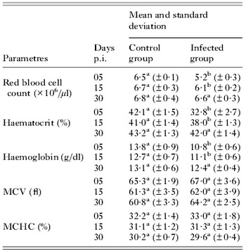
In the infected rats (group B), there was a decrease (P<0·05) in RBC counts, haematocrit and haemoglobin concentrations at days 5 and 15 p.i. (Table 1), but on day 30 p.i. no differences (P>0·05) were observed in these parameters when compared to those of the control group. A mild degree of anisocytosis and polychromasia was observed in blood smears of group B on days 5, 15 and 30 p.i. No difference (P>0·05) occurred between the infected and control group in the MCV and MCHC values (Table 1). The rats showed normocytic normochromic anaemia.
Means (±standard deviation) of haematological parameters of rats experimentally infected with Trypanosoma evansi

Means with different superscript letters on the same line are significantly different (P<0·05) by Student's t-test.
Blood δ-ALA-D activity
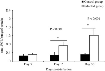
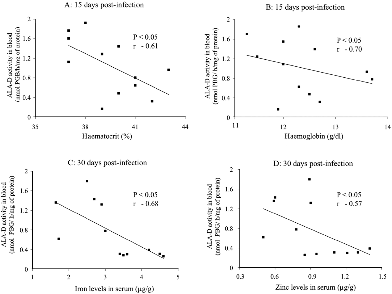
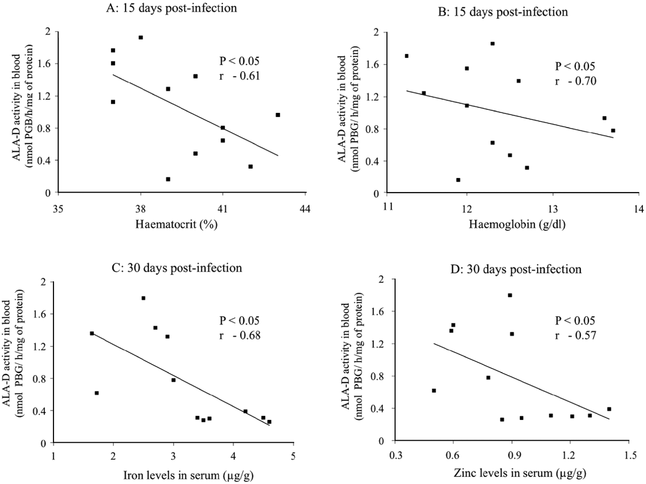
The mean activity of blood δ-ALA-D significantly (P<0·001) increased in rats infected with T. evansi on days 15 and 30 p.i. (Fig. 1). δ-ALA-D activity in blood had a negative correlation (P<0·05) with haematocrit (r=−0·61; Fig. 2A) and haemoglobin (r=−0·70; Fig. 2B) on day 15 p.i., but not on days 5 and 30 p.i.
Rats experimentally infected with Trypanosoma evansi. Activity of δ-ALA-D in the blood of infected rats compared to non-infected rats on days 5, 15 and 30 post-infection.

Correlation of the activity of δ-ALA-D with haematocrit (A) and haemoglobin (B) on day 15 post-infection, and serum iron (C) and zinc (D) on day 30 post-infection (* P<0·05, t-test).
Serum iron and zinc levels
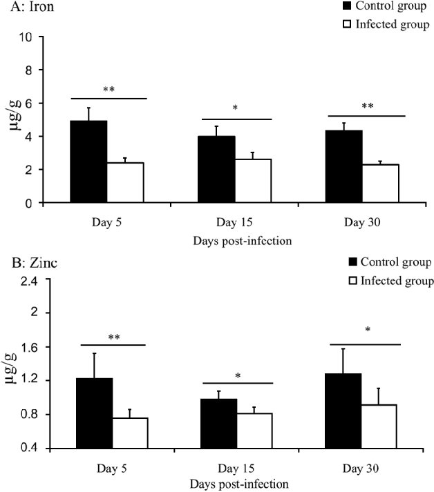
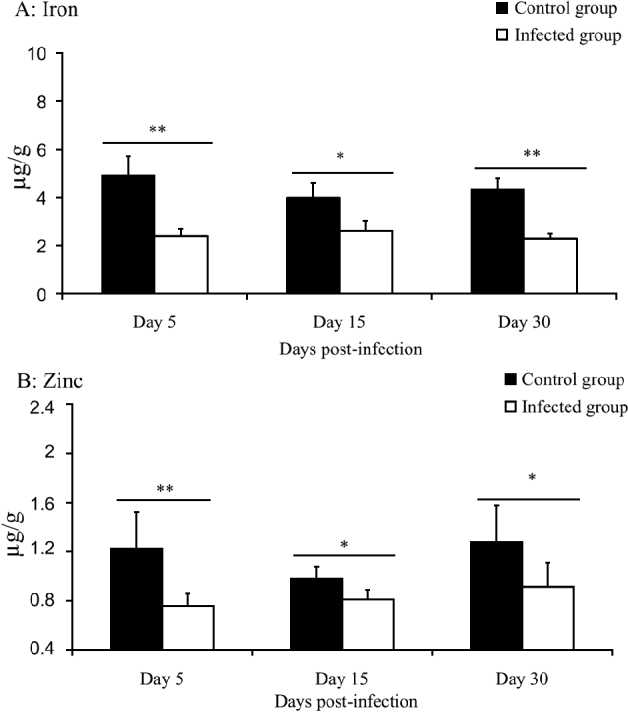
Serum levels of iron and zinc reduced significantly (P<0·05) in rats infected with T. evansi on days 5, 15 and 30 p.i. when compared to the control group (Fig. 3). A negative correlation (P<0·05) between blood δ-ALA-D activity and iron (r=−0·68; Fig. 2C) and zinc (r=−0·57; Fig. 2D) levels in serum was observed on day 30 p.i., but not observed on days 5 and 15 p.i.
Mean values of serum iron (A) and zinc (B) levels in Trypanosoma evansi-infected rats compared with values in non-infected rats. Means differ significantly (* P<0·05; ** P<0·01) between groups evaluated on the same day post-infection (n=6 per group).
Lipid peroxidation
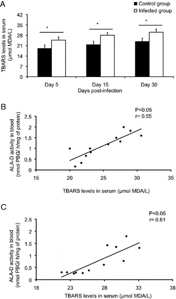
TBARS level in serum, indicated by malondialdehyde MDA concentration, was higher in the infected group than in the non-infected group during the experimental period (P<0·05; Fig. 4A). A positive correlation was observed between the activity of blood δ-ALA-D and levels of circulating TBARS on days 15 (r=0·55) and 30 (r=0·61) p.i. only (Fig 4B,C).
Mean values of TBARS in serum of Trypanosoma evansi-infected rats compared with values in non-infected (A). Correlation of the activity of δ-ALA-D in blood with TBARS levels in serum on days 15 (B) and 30 (C) p.i. * Means differ significantly (P<0·05) among groups evaluated on the same day post-infection (n=6 per group).

DISCUSSION
In the present study, the rats infected with T. evansi showed a reduction of red blood cell counts, haemoglobin concentrations and haematocrit values in anaemia, similarly reported by other authors (Menezes et al. Reference Menezes, Queiroz, Gomes, Marques and Jansen2004; Wolkmer et al. Reference Wolkmer, da Silva, Traesel, Paim, Cargnelutti, Pagnoncelli, Picada, Monteiro and Lopes2009). The anaemia in trypanosomosis is related to several factors some of which are not fully elucidated (Dargie et al. Reference Dargie, Murray, Murray, Grimshaw and Mcintyre1979; Walia et al. Reference Walia, Kalra, Juyal and Ahuja1996; Wolkmer et al. Reference Wolkmer, da Silva, Traesel, Paim, Cargnelutti, Pagnoncelli, Picada, Monteiro and Lopes2009; Da Silva et al. Reference Da Silva, Andrade Neto, Costa, Wolkmer, Mazzantti, Santurio, Lopes and Monteiro2010). The activity of δ-ALA-D in the blood is directly related to the synthesis of the heme fraction and subsequent production of red blood cells (Jaffe, Reference Jaffe1995).
There was activation of blood δ-ALA-D on days 15 and 30 p.i. associated with a negative correlation with haematocrit and haemoglobin, suggesting that the increase in enzymatic activity was in response to anaemia caused by T. evansi infection (Wolkmer et al. Reference Wolkmer, Silva, Cargnelutti, Costa, Traesel, Lopes and Monteiro2007; Da Silva et al. Reference Da Silva, Costa, Wolkmer, Zanette, Faccio, Gressler, Dorneles, Santurio, Lopes and Monteiro2009c). The elevation of this enzyme activity was probably related to an increase in heme synthesis stimulated by the anaemia. On day 5 p.i. the activity of δ-ALA-D had not increased since the erythropoietic response to the anaemia probably had not started. The production of new red blood cells occurs 7 days after the onset of anaemia (Feldman et al. Reference Feldman, Zinkl and Jain2000). An increase in reticulocyte counts occurs in response to anaemia in animals infected with T. evansi (Wolkmer et al. Reference Wolkmer, Silva, Cargnelutti, Costa, Traesel, Lopes and Monteiro2007; Da Silva et al. Reference Da Silva, Costa, Wolkmer, Zanette, Faccio, Gressler, Dorneles, Santurio, Lopes and Monteiro2009c), but may not occur in infections with mild anaemia. The anaemia was normocytic and normochromic, but polychromasia and anisocytosis were indications of some degree erythropoiesis up to day 30 p.i.
A decrease of iron and zinc levels in serum was observed in this study, as well as in cats infected with T. evansi (Da Silva et al. Reference Da Silva, Hoehne, Tonin, Zanette, Wolkmer, Costa, Moraes, Flores, Santurio, Lopes and Monteiro2009a) and rats infected with Trypanosoma cruzi (Matousek de Abel de la Cruz et al. Reference Matousek de Abel de la Cruz, Burguera, Burguera and Anez1993). This reduction of zinc levels coincides with the activation of δ-ALA-D, a metalloenzyme which requires zinc for its activation (Bevan et al. Reference Bevan, Bodlaender and Shemin1980; Fukuda et al. Reference Fukuda, Paredes and Batlle1988). Moreover this element is a nutritional component required for the maintenance of immune functions performed by T lymphocytes (Prasad, Reference Prasad1993; Schlesinger et al. Reference Schlesinger, Arevalo, Arredondo, Lonnerdal and Stekel1993), which are generally activated in T. evansi infection (Da Silva et al. Reference Da Silva, Monteiro, Gonçalves, Spanevello, Oliveira, Costa, Jaques, Schetinger, Mazzanti and Lopes2011).
Iron is a component of metalloproteins and plays an important role in vital biochemical activities of mammals, such as oxygen transport, catalysis and electron transfer (Aisen et al. Reference Aisen, Enns and Wessling-Resnick2001). Possible causes of the reduction of serum iron levels in infection caused by T. evansi are iron sequestration by macrophages (Dargie et al. Reference Dargie, Murray, Murray, Grimshaw and Mcintyre1979), or increased iron use for parasite growth (Lalonde and Holbein, Reference Lalonde and Holbein1984). In this study, the iron reduction had a negative correlation with the activity of δ-ALA-D. This increase in enzyme activity may have led to a higher utilization of iron for the synthesis of heme (Ryter and Tyrrell, Reference Ryter and Tyrrell2000).
Lipid peroxidation is a mechanism by which free radicals can cause tissue damage in several pathological conditions (Halliwell and Chirico, Reference Halliwell and Chirico1993). Studies indicate that oxidative stress causes an inhibition of activity of the enzyme δ-ALA-D due to the high SH group affinity of the enzyme to pro-oxidant agents (Folmer et al. Reference Folmer, Soares, Gabriel and Rocha2004; Gonçalves et al. Reference Gonçalves, Erthal, Corte, Müller, Piovezan, Nogueira and Rocha2005). Wolkmer (Reference Wolkmer, da Silva, Traesel, Paim, Cargnelutti, Pagnoncelli, Picada, Monteiro and Lopes2009) and Da Silva et al. (Reference Da Silva, Andrade Neto, Costa, Wolkmer, Mazzantti, Santurio, Lopes and Monteiro2010) suggested that an increase in lipid peroxidation may be involved in the pathogenesis of anaemia in rats and cats infected with T. evansi. In this study, a positive correlation between serum TBARS levels and blood δ-ALA-D activity indicated that the increase in TBARS was not sufficient to inhibit the enzymatic activity. The increased TBARS probably occurred concomitantly with the activation of antioxidant enzymes, as previously reported in T. evansi infection (Omer et al. Reference Omer, Mousa and Al-Wabel2007).
In conclusion, the activity of blood δ-ALA-D increased in rats infected with T. evansi in response to anaemia. This increased activity correlated with serum TBARS, iron and zinc levels.