Introduction
Oocyte activation has been induced previously by physical and chemical stimuli, such as stimuli by ethanol (Nagai, Reference Nagai1987; Kubiak, Reference Kubiak1989), calcium ionophore A23187 (Meyerhof & Masui, Reference Meyerhof and Masui1977; Ware et al., Reference Ware, Barnes, Maiki-Lauria and First1989), inhibitors of protein synthesis (Siracusa et al., Reference Siracusa, Whittingham, Molinaro and Vivarelli1978; Fulka et al., Reference Fulka, Leibfried-Rutledge and First1991) and electric pulses (Powell & Barnes, Reference Powell and Barnes1992; Prochazka et al., Reference Prochazka, Durnford, Fiser and Marcus1993). It has been reported that the activation rates of mouse oocytes exposed to calcium ionophore and puromycin were approximately 90% and that more than 80% of the activated oocytes showed one pronucleus with extrusion of the second polar body (Nakasaka et al., Reference Nakasaka, Yamano, Hinokio, Nakagawa, Yoshizawa and Aono2000). Nakagawa et al. reported that treatment with calcium ionophore and puromycin activated unfertilized oocytes after intracytoplasmic sperm injection (ICSI; Nakagawa et al., Reference Nakagawa, Yamano, Moride, Yamashita, Yoshizawa and Aono2001a). Murase et al. reported that oocyte activation with calcium ionophore and puromycin enabled successful pregnancy outcome in patients with previous repeated failed fertilization following ICSI (Murase et al., Reference Murase, Araki, Mizuno, Kawaguchi, Naito, Yoshizawa and Araki2004).
On the other hand, oocyte activation is characterized by pulsatile increases in intracellular calcium concentrations and by reduced activities of M-phase promoting factor (MPF) and MPF kinase. In other words, suppression of MPF is considered to be necessary for oocyte activation. Protein synthesis inhibitors such as puromycin are responsible for reducing cytostatic factor synthesis and thus decreasing MPF activity indirectly. However, the mechanism by which puromycin participates in oocyte activation has not been fully clarified. On the other hand, roscovitine is a purine known as a specific inhibitor of cyclin-dependent protein kinase that prevents p34cdc2 dephosphorylation and inhibits MPF kinase activity (Meijer & Kim, Reference Meijer and Kim1997). In addition, roscovitine has been used successfully to arrest meiotic activation without compromising further embryo development in cattle, pigs, goats, cats and dromedary camels (Motlik et al., Reference Motlik, Pavlock, Kubelka, Kalous and Kalab1998; Schoevers et al., Reference Schoevers, Bevers, Roelen and Colenbrander2005; Jimenez-Macedo et al., Reference Jimenez-Macedo, Izquierdo, Urdaneta, Anguita and Paramio2006; Wani, Reference Wani2008; da Silvia Rascado et al., Reference da Silvia Rascado, Martins, Minto, de Sa Lorena and da Cruz Landim-Alvarenga2010). Therefore, oocyte activation in mice may also be shown through the induction of morphological changes as well as the suppression of MPF activity by using roscovitine. However, to the best of our knowledge, the relationship of the effect of roscovitine on oocyte activation with the change in MPF activity in mice has not been clarified.
The aim of the present study was to determine oocyte activation and change in MPF activity induced by calcium ionophore and roscovitine in comparison with those induced by roscovitine alone and those induced by calcium ionophore and puromycin in mice.
Materials and methods
Oocytes
Cumulus-enclosed oocytes were recovered from the oviducts of 6–8-week-old female B6C3F1 mice (Japan SLC Inc, Shizuoka, Japan) that had received intraperitoneal injection of 10 I.U. of pregnant mare's serum gonadotropin (Sigma) followed 48 h later by 10 I.U. of human chorionic gonadotropin (Wako) at 14 h prior to sacrifice by cervical dislocation. The cumulus masses were dispersed in 0.1% hyaluronidase (Sigma) in modified human tubal fluid (mHTF; Nippon Medical & Chemical Instruments Co., Ltd) medium at 37°C in an atmosphere of 5% CO2 in air for 8–10 mins. The cumulus-free oocytes were collected and rinsed twice in fresh mHTF medium prior to use. All animal experiments were conducted in accordance with the ethical standards of the Animal Care and Use Committee of the University of Tokushima Graduate School, Japan.
Chemicals
Stock solutions of 1 mg/ml calcium ionophore A23187 (Sigma-Aldrich), 10 mg/ml puromycin (Sigma) and 10 mg/ml roscovitine (Sigma) were stored at –20°C. Prior to each experiment, these chemicals were diluted with mHTF containing with 0.4% bovine serum albumin (BSA) to the following concentrations: 5 μM calcium ionophore A23187, 10 μg/ml puromycin and 50 μM roscovitine.
Oocyte activation
Oocyte activation was performed in accordance with a previous report (Nakasaka et al., Reference Nakasaka, Yamano, Hinokio, Nakagawa, Yoshizawa and Aono2000). Cumulus-free oocytes were divided into the following five groups: (1) a no activation treatment group, in which oocytes were placed in mHTF medium without calcium ionophore for 5 min and cultured in mHTF medium with 0.4% BSA for 6 h; (2) a calcium ionophore group, in which oocytes were treated with 5 μM calcium ionophore in mHTF medium for 5 min and cultured in mHTF medium with 0.4% BSA containing 5 μM calcium ionophore for 6 h; (3) a roscovitine group, in which oocytes were placed in mHTF medium without calcium ionophore for 5 min and cultured in mHTF medium with 0.4% BSA containing 50 μM roscovitine for 6 h; (4) a calcium ionophore and puromycin group, in which oocytes were treated with 5 μM calcium ionophore in mHTF medium for 5 min and cultured in mHTF medium with 0.4% BSA containing 5 μM calcium ionophore and 10 μg/ml puromycin for 6 h; and (5) a calcium ionophore and roscovitine group, in which oocytes were treated with 5 μM calcium ionophore in mHTF medium for 5 min and cultured in mHTF medium with 0.4% BSA containing 5 μM calcium ionophore and 50 μM roscovitine for 6 h. Oocyte cultures were incubated at 37°C in an atmosphere of 5% CO2 in air. Oocyte activation in the five groups at 0, 2, 4 and 6 h after incubation was observed at a ×400 magnification under an inverted microscope (IMT2–31 Olympus Diaphot) equipped with Nomarski differential interference contrast.
Measurement of MPF activity
Ten cumulus-free oocytes from each of the five groups were transferred into microtubes that each contained 5 μl sample buffer (50 mM Tris–HCl, 0.5 M NaCl, 5 mM ethylene diamine tetraacetic acid (EDTA), 2 mM ethylene glycol tetraacetic acid (EGTA), 0.01% Briji35, 1 mM phenylmethylsulfonyl fluoride (PMSF), 0.05 mg/ml leupeptin, L2884, 50 mM 2-mercaptoethanol, 25 mM beta-glycerophosphate and 1 mM Na-orthovanadate) at 0, 2, 4 and 6 h after incubation. The samples were frozen at –80°C until used for analysis. Oocytes were destroyed completely by repetitions of freezing in liquid nitrogen and ultrasonographic treatment three times for 30 s each time, just before the assay. Assay for MPF activity in the oocytes was carried out using a MESACUP cdc2 kinase enzyme-linked immunosorbent assay (ELISA) kit (R&D Systems). The intra- and inter-assay coefficients of variation were 4–9% and 6–9%, respectively. We set the basal values (0 h after incubation) as 100% and calculated the percentage changes at 2, 4 and 6 h after incubation.
Statistical analysis
Rates of activation in oocytes were evaluated using the chi-squared test. Differences in percentage changes in MPF activity in the five groups were analyzed by analysis of variance (ANOVA). p-values less than 0.05 were considered to be statistically significant.
Results
Appearance of activated oocytes
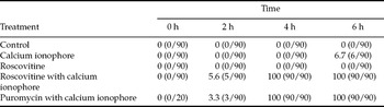
Activated oocytes were defined as oocytes with at least one pronucleus. As can be seen in Table 1, oocytes with no activation treatment and oocytes treated with roscovitine were not activated during the 6-h incubation period. The rate of activation in oocytes with calcium ionophore treatment was 6.7%. All of the oocytes in the calcium ionophore with puromycin group and in the calcium ionophore with roscovitine group were activated during the 6-h incubation period.
Rates of activation in oocytes with various treatments

Values are shown as the percentage rates of activated oocytes.
Oocytes with no activation treatment (control group) and oocytes treated with roscovitine were not activated during the 6-h incubation period. All of the oocytes in the calcium ionophore with puromycin group and in the calcium ionophore with roscovitine group were activated.
MPF activity in oocytes
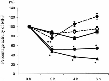
As shown in Figure 1, the percentage activity of MPF in oocytes with no activation treatment did not change significantly during the 6-h incubation period. The percentage activity of MPF in oocytes treated with roscovitine alone was decreased after 2 h of incubation (p = 0.052). However, the suppression of MPF activity was not maintained and the percentage activity of MPF was increased after 4 h of incubation. The percentage activity of MPF in oocytes treated with calcium ionophore alone was not changed significantly during the 6-h incubation period. The percentage activity of MPF in oocytes treated with calcium ionophore and roscovitine was significantly decreased (p < 0.05) and the suppression of MPF activity was maintained during the 6-h incubation period, and this change was similar to that in oocytes treated with calcium ionophore and puromycin.
Percentage changes in M-phase promoting factor (MPF) activity at 2, 4 and 6 h after incubation in the control group, calcium ionophore group, roscovitine group, calcium ionophore and puromycin group, and calcium ionophore and roscovitine group. Mean ± standard deviation. *p < 0.05 vs 0 h, **p = 0.052 vs 0 h. - -○- -: control, - -●- -: roscovitine alone, —■—: calcium ionophore, —●—: calcium ionophore and roscovitine, —▲—: calcium ionophore and puromycin.

Discussion
We found that oocytes treated with both calcium ionophore and roscovitine were activated through the suppression of MPF activity. The suppression of MPF activity was maintained for 6 h. In addition, the pattern of change in MPF activity in the calcium ionophore and roscovitine group was similar to that in the calcium ionophore and puromycin group. In a previous study, it was found that 75.5% of oocytes from cats were activated by treatment with the combination of calcium ionophore and roscovitine and that the rate of activation was as same as the rates of activation of oocytes treated with calcium ionophore and cycloheximide and with calcium ionophore and strontium (da Silvia Rascado et al., Reference da Silvia Rascado, Martins, Minto, de Sa Lorena and da Cruz Landim-Alvarenga2010).
Roscovitine acts specifically on the phosphorylation of MPF and decreases MPF activity. In the present study, suppression of MPF activity continued for only 2 h and oocytes were not activated in the roscovitine alone group, while suppression of MPF activity was maintained for 6 h with oocyte activation in the calcium ionophore and roscovitine group. Therefore, calcium ionophore as well as roscovitine is needed for oocyte activation by maintenance of the suppression of MPF activity. In mammals, meiosis is arrested at metaphase II and this arrest is maintained by high levels of MPF and a cytoplasmic factor that stabilizes MPF activity in metaphase II oocytes. MPF is a heterodimer composed of a cdc2 kinase subunit and a regulatory cyclin B subunit. Intracellular Ca2+ activates calmodulin-dependent kinase II (CaM KII; Sagata, Reference Sagata1996) and mediates cyclin B degradation by increasing the activity of an E3 ubiquitin ligase (Hyslop et al., Reference Hyslop, Nixon, Levasseur, Chapman, Chiba, McDougall, Venables, Elliott and Jones2004) and 26 S proteasome (Jones, Reference Jones2004). Meiotic arrest is broken by a Ca2+ signalling pathway through the inactivation of MPF. Both calcium ionophore and roscovitine may play important roles in transmission of the signal pathway for oocyte activation.
Failure of oocyte activation has been suggested to be the principal cause of fertilization failure in ICSI (Sousa & Tesarik, Reference Sousa and Tesarik1994; Tesarik & Sousa, Reference Tesarik and Sousa1995). Yanagida et al. also reported that in 0.9% of ICSI cases with motile spermatozoa, the motile spermatozoa failed to fertilize the oocytes despite an apparently successful ICSI procedure (Yanagida et al., Reference Yanagida, Katayose, Yazawa, Kimura, Sato, Yanagimachi and Yanagimachi1999). If unfertilized oocytes after ICSI are properly activated, they may form two pronuclei with extrusion of the second polar body and then cleave and develop normally. It has been reported that the method using calcium ionophore and puromycin was effective for oocyte activation in mice (Nakasaka et al., Reference Nakasaka, Yamano, Hinokio, Nakagawa, Yoshizawa and Aono2000). In addition, it has been reported that the combination of calcium ionophore and puromycin proved to be an effective method for producing mouse and human haploid parthenogenones (Nakasaka et al., Reference Nakasaka, Yamano, Hinokio, Nakagawa, Yoshizawa and Aono2000; Nakagawa et al., Reference Nakagawa, Yamano, Nakasaka, Hinokio, Yoshizawa and Aono2001b), suggesting that this activation method might be suitable for rescuing oocytes that have been judged to be unfertilized after ICSI or oocytes that have been injected with a spermatozoon that is unable to activate oocytes. It has been demonstrated that treatment with the combination of calcium ionophore and puromycin could effectively salvage unfertilized oocytes after ICSI (Nakagawa et al., Reference Nakagawa, Yamano, Moride, Yamashita, Yoshizawa and Aono2001a, Lu et al., Reference Lu, Zhao, Gao, Li, Ma, Mullen, Critser and Chen2006). Successful pregnancy outcome has been reported to be achieved by oocyte activation with calcium ionophore and puromycin in patients with previous repeated failed fertilization following ICSI (Murase et al., Reference Murase, Araki, Mizuno, Kawaguchi, Naito, Yoshizawa and Araki2004). However, careful consideration should be given to the use of puromycin for oocyte activation since puromycin is a protein synthesis inhibitor and the mechanism of its action in oocyte activation has not been fully elucidated. Roscovitine displays high efficiency and high selectivity towards cyclin-dependent kinases and reversibly arrests starfish oocytes and sea urchin embryos in late prophase (De Azevedo et al., Reference De Azevedo, Leclerc, Meijer, Havlicek, Strnad and Kim1997; Meijer et al., Reference Meijer, Borgne, Mulner, Chong, Blow, Inagaki, Inagaki, Delcros and Moulinoux1997). Therefore, roscovitine may be a useful drug for oocyte activation in cases of fertilization failure in ICSI.
There are some limitations in the study. First, we could not follow up the parthenotes to blastocyst stage. We defined that activated oocytes were defined as oocytes with at least one pronucleus. As the amplitude of the calcium waves has major consequences for blastocyst formation even if the mouse parthenotes were able to extrude a second polar body and to form a pronucleus, the study for further stages of oocytes may be needed. Second, we used roscovitine at a dose of 50 μM in the present study. It has been reported that the ideal concentration of roscovitine used in mouse oocytes to reduce MPF activity was more than 50 μM and that roscovitine at a concentration of 20 μM did not suppress activity of H1 histone, whereas a dose of 100 μM, although not toxic for oocytes, did not increase activation rates beyond that achieved with a dose of 50 μM (Deng & Shen, Reference Deng and Shen2000). A dose–response experiment may be needed to determine the optimal concentration of roscovitine for oocyte activation in mice.
In conclusion, roscovitine with calcium ionophore is effective for induction of oocyte activation through suppression of MPF activity in mice.