Introduction
Toxocariasis is a disease caused by the larval stage of the dog and cat roundworms, Toxocara canis and T. cati, respectively. Ingestion of embryonated eggs of Toxocara is the primary route of infection. Many epidemiological studies have revealed that the sand in sandpits, such as those in public parks and kindergartens, is often contaminated with these eggs and, thus, that sandpits could serve as a source of infection for young children (Mizgajska, Reference Mizgajska2001; Wiwanitkit & Waenlor, Reference Wiwanitkit and Waenlor2004; Dubna et al., Reference Dubna, Langrova, Jankovska, Vadlejch, Pekar, Napravnik and Fechtner2007). Because the sizes of both species of eggs overlap and the differences in pit structures of egg surface vary (Uga et al., Reference Uga, Matsuo, Kimura, Rai, Koshino and Igarashi2000), it can be difficult to categorize the eggs by morphological observation alone. Therefore, molecular methods for distinguishing between T. canis and T. cati eggs from soil samples (Borecka, Reference Borecka2004; Fogt-Wyrwas et al., Reference Fogt-Wyrwas, Jarosz and Mizgajska-Wiktor2007), as well as for direct detection of Toxocara from sand samples (Borecka & Gawor, Reference Borecka and Gawor2008), have recently been introduced. While T. canis is the most common pathogen of toxocariasis worldwide, some epidemiological studies have shown that T. cati eggs dominate in the environment (Matsuo & Nakashio, Reference Matsuo and Nakashio2005; Talvik et al., Reference Talvik, Moks, Magi, Jarvis and Miller2006; Macuhova et al., Reference Macuhova, Kumagai, Akao and Ohta2010). However, only a few human cases of toxocariasis cati have been reported based on serological diagnostics (Takeda et al., Reference Takeda, Tanabe, Nishi, Tsuji and Iwanaga1975; Shimokawa et al., Reference Shimokawa, Nakashima, Akagi, Omae and Tsuji1982; Hirata, Reference Hirata1987; Mitsugi et al., Reference Mitsugi, Umei, Inoue, Sumida and Hanada1988; Nishikata et al., Reference Nishikata, Hirata, Shimamura, Dohmen, Kudo, Ishibashi, Onizuka and Oda1991; Sakai et al., Reference Sakai, Kawashima, Shibui, Kamata, Kambara and Matsuoka1998). Such findings may suggest either that T. cati eggs are not highly infectious or that our present Toxocara serology could not distinguish toxocariasis cati from toxocariasis canis.
To understand the epidemiological significance of T. cati and T. canis, the presence of eggs in public sandpits in Tokyo, Japan, was carefully investigated. The vertical and horizontal distribution of eggs in two naturally contaminated sandpits was also determined. Additionally, eggs recovered from the sandpits were inoculated to ICR mice to determine their pathogenicity in a paratenic host.
Materials and methods
Sampling and recovery of Toxocara eggs from sandpits
Sand samples were collected from sandpits located in public parks in the metropolitan area of Tokyo, Japan. For the epidemiological study, sand was collected from 34 sandpits (28 parks) in the autumn–winter season (September–January). Sampling was performed at six randomly chosen sites at three different depths in each sandpit. After a thorough mixing, 200 g of sand was examined. For the study on the vertical and horizontal distribution of Toxocara spp. eggs, two additional sandpits (Sandpit A and B) were chosen in one park located in central Tokyo (Macuhova et al., Reference Macuhova, Kumagai, Akao and Ohta2010). Sand samples were collected in the spring season (March) from 13 different sites in each sandpit and at four different depths at each site (0–1 cm, 2–5 cm, 5–10 cm, more than 10 cm). A sand sample of 200 g from each site/depth was examined. Examination was performed according to the method of Uga & Kataoka (Reference Uga and Kataoka1995). Briefly, sand samples were dried and sieved through a set of two meshes (pore diameters: 1 mm and 150 μm). The powdery sand thus obtained was washed with a 0.05% Tween20 water solution. For the floatation method, a sucrose solution with a specific gravity of 1.200 was used. The presence of Toxocara spp. eggs was examined microscopically. Statistical analysis of the egg distributions in the sandpits was performed using a Kruskal–Wallis rank sum test or a one-way ANOVA with a Bartlett test. A P value of < 0.05 was considered statistically significant.
Molecular identification of Toxocara eggs
Recovered eggs microscopically identified as Toxocara spp. were subjected to DNA analysis using the polymerase chain reaction (PCR) technique. Each egg was crushed separately with a fine needle on a glass slide under stereoscopic microscopy. The embryo was then transferred to an Eppendorf tube with a pipette and the DNA was extracted using NaOH, as described previously (Macuhova et al., Reference Macuhova, Kumagai, Akao and Ohta2010) with slight modifications. Briefly, DNA was extracted by incubating samples with 100 μl of 50 mm NaOH in a dry bath at 95°C for 45 min (30 min for eggs examined for the epidemiological study) while mixing thoroughly every 15 min. After centrifugation at 15,000 rpm for 5 min at 4°C, 50 μl of supernatant was taken to a separate tube and neutralized with 50 μl of 1 m Tris HCl (pH 8.0). This solution was stored in a freezer at − 20°C and 1 μl of it was used as a template for the PCR assay. The PCR reaction was performed with amplification of the internal transcribed spacer-2 (ITS-2) gene (Jacobs et al., Reference Jacobs, Zhu, Gasser and Chilton1997; Zhu et al., Reference Zhu, Jacobs, Chilton, Sani, Cheng and Gasser1998; Epe et al., Reference Epe, Meuwissen, Stoye and Schnieder1999; Borecka & Gawor, Reference Borecka and Gawor2008). The forward primer, Tcan (5′-AGTATGATGGGCGCCAAT-3′), was used for T. canis and was also used together with the conserved reverse primer, NC2 (5′-TTA GTTTCTTTTCCTCCGCT-3′), to amplify partial ITS-2. For T. cati, the forward primer, Tcat (5′-GGAGGAAGTAAGATCGTGGCACGCGT-3′), and the reverse primer, NC2, were used to amplify the partial ITS-2 region. The PCR conditions were optimized and performed according to the method described elsewhere (Macuhova et al., Reference Macuhova, Kumagai, Akao and Ohta2010). As a positive control, we employed genomic DNA extracted from T. canis or T. cati larvae. For the extraction of Toxocara DNA from larvae recovered from infected mouse brains, the larva was transferred into an Eppendorf tube under stereoscopic microscopy, together with a small amount of brain tissue. The genomic DNA of the larva was extracted using the above-described method.
Experimental infection of mice
Nine 6-week-old female ICR mice were used to ascertain the pathogenicity of the isolated eggs. The protocol of our experiment was approved by the Institutional Committee for the Animal Experiments of Tokyo Medical and Dental University (Approval number: 0090189). The mice were divided into three groups of three individuals: group 1 comprised mice inoculated with T. canis eggs, group 2 comprised mice infected with the eggs isolated from sandpits, and group 3 comprised the uninfected controls. Each mouse was inoculated with a single dose of 300 eggs in a volume of 0.3 ml distilled water or the same volume of water without eggs. Toxocara canis eggs were obtained from the uteri of adult worms collected from faeces following the administration of anthelmintics to naturally infected puppies. Eggs isolated from sandpits were collected from two heavily contaminated sandpits during examination of the egg distribution. Prior to inoculation, the eggs were treated with 50% sodium hypochlorite for 10 min. Deproteinized eggs were then harvested to a separate tube and washed repeatedly. The fully embryonated eggs were then counted under microscopy. All mice were sacrificed on day 21 after infection. Blood and organs, including tissue from the brain, lung and carcass, were removed. These tissues, except those from the brain, were digested with artificial gastric juice by the method described elsewhere (Jin et al., Reference Jin, Akao, Nobuta and Ohta2008), and the number of migrating larvae was determined. The larvae in brain, which was minced into small pieces on a glass slide and covered with another glass slide, were found and collected one by one under a stereoscopic microscope. Eosinophils in the peripheral blood films were also counted microscopically. Statistical analyses were performed using an unpaired Student's t-test. P values of less than 0.05 were considered to be significant.
Results
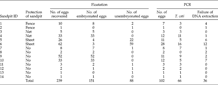
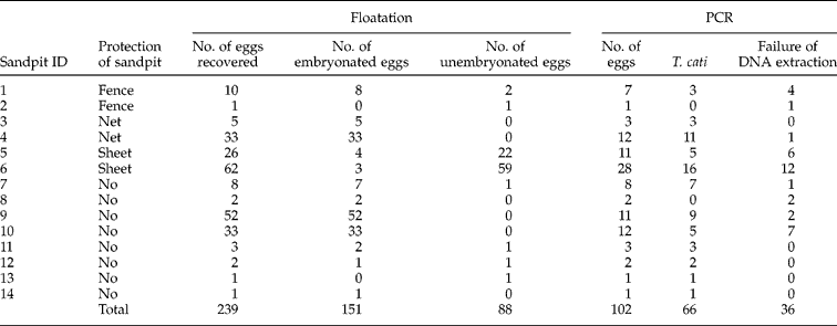
Fourteen of the 34 sandpits (9 from 28 parks) were found to be contaminated with eggs of Toxocara spp. (table 1). In total, 239 eggs were recovered from all sandpits, with an average number of 17.1 eggs per contaminated sample. The recovered eggs were at different stages of development, with 63.2% of eggs being fully embryonated. A fence or net around the sandpit, or a sheet covering, was present in 12 of 34 sandpits. However, six of these sandpits were still found to be contaminated with Toxocara eggs. We examined 102 eggs originating from all infected sandpits by PCR analysis. Sixty-six eggs were determined to be T. cati, while none were T. canis. The remaining 36 samples failed to amplify.
The number of Toxocara eggs recovered by floatation method and identified using PCR analysis from positive sand samples in 14 sandpits in the metropolitan area of Tokyo; no T. canis eggs were identified by PCR.
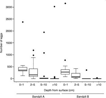
The horizontal and vertical distribution of Toxocara eggs in the sand was evaluated in two additional sandpits (Sandpit A and B) which were previously identified as heavily contaminated with eggs (Macuhova et al., Reference Macuhova, Kumagai, Akao and Ohta2010). In both sandpits, the horizontal distribution of eggs showed no correlation with the location, such as being sampled from a corner, edge or middle of the sandpit (data not shown). In contrast, there were marked differences in the vertical distribution of eggs at four different depth layers. In both sandpits, the median numbers of recovered eggs in each layer showed an inverse relationship with the sand depth, such that the number of eggs decreased as depth increased (fig. 1). Statistically significant differences among the sand layers were observed in both sandpits, based on the Kruskal–Wallis rank sum test (Sandpit A, P= 5.967 × 10− 5) or one-way ANOVA (Sandpit B, F= 0.009064), although outliers were observed in all layers.
Recovery of Toxocara eggs from sandpits A and B, relative to the depth from the soil surface (cm).

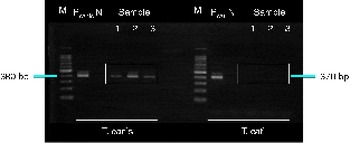
To confirm the pathogenicity of the eggs recovered from sandpits, 300 eggs were orally inoculated into ICR mice. Table 2 shows the numbers of larvae recovered from various organs. In the mice infected with T. canis (group 1), the larvae were distributed almost evenly in the brain and carcass. The average recovery rate of larvae was 19.4 ± 7.2%. In contrast, in the mice infected with the eggs from the sandpit (group 2), almost all the larvae were recovered from the carcasses, with an average recovery rate of 25.8 ± 1.4%. There was no statistically significant difference between the recovery rates of group 1 and group 2 (P= 0.2089). The difference in eosinophil counts between groups 1 and 2 was just below the level of statistical significance (P= 0.0544). A total of three larvae were found in the brain of two mice in group 2. Because the morphological observations did not allow us to distinguish between T. canis and T. cati, these three larvae were subjected to a PCR assay. The results revealed that the PCR product of 380 bp was amplified with T. canis-specific primers but not with T. cati-specific primers, suggesting that all three larvae were T. canis (fig. 2).
Mean larval recovery in the brain, lung and carcasses of ICR mice infected with Toxocara eggs, together with eosinophil counts and optical density values at 21 days post-infection.

a OD, Optical density
b Average ± SD
c ND, not done.
(colour online) Analysis of PCR products amplified from larvae (samples 1 to 3) found in the brain of two mice infected with Toxocara eggs from a sandpit using species-specific primers for T. canis (left) and T. cati (right). The PCR product of 380 bp was amplified by the T. canis-specific primer but not by the T. cati primer (the expected product is 370 bp). M, DNA-size marker; Pcanis and Pcati, positive controls of T. canis and T. cati DNA, respectively; N, no-DNA control.

We also applied PCR analysis to 30 eggs, which were collected from the same samples as the eggs with which the mice were inoculated. Twenty-seven of these eggs were determined to be T. cati, and three failed to amplify. None were determined to be T. canis (data not shown).
Discussion
Contamination of public parks, and especially sandpits, with eggs of Toxocara spp., has been a worldwide health concern. Previous epidemiological surveys have mostly failed to adequately identify the species of Toxocara eggs recovered from the environment (Abe & Yasukawa, Reference Abe and Yasukawa1997; Dubna et al., Reference Dubna, Langrova, Jankovska, Vadlejch, Pekar, Napravnik and Fechtner2007), although a few attempted to distinguish T. cati from T. canis eggs recovered from sandpits based solely on their morphological characteristics (Uga et al., Reference Uga, Matsumura, Aoki and Kataoka1989; Matsuo & Nakashio, Reference Matsuo and Nakashio2005). However, because of their morphological similarities, not all eggs could be clearly identified. According to Matsuo & Nakashio (Reference Matsuo and Nakashio2005), 5% of the recovered eggs were T. canis, 50% were T. cati, and 45% were unidentified. In another study, 6.1% of eggs recovered from sandpits were T. canis, 19.4% were T. cati, and 75.4% were unidentified (Uga et al., Reference Uga, Matsumura, Aoki and Kataoka1989). Recently, more reliable methods of distinguishing T. canis from T. cati eggs has been possible using molecular methods such as PCR and LAMP (loop-mediated isothermal amplification) (Borecka & Gawor, Reference Borecka and Gawor2008; Macuhova et al., Reference Macuhova, Kumagai, Akao and Ohta2010). Borecka & Gawor (Reference Borecka and Gawor2008) found that two of eight positive sand samples were contaminated with eggs of T. canis, three with T. cati, and three with both species. Macuhova et al. (Reference Macuhova, Kumagai, Akao and Ohta2010) found that five sandpits were contaminated only with eggs of T. cati. In the present study, we combined the floatation method, followed by counting of eggs under a microscope, with the PCR method to identify the recovered eggs. This allowed us to quantitatively determine the number of eggs present in sand samples and their developmental stage, as well as to qualitatively estimate which species of Toxocara were present. A total of 239 eggs were recovered from 14 of 34 sandpits. The PCR method applied to 102 eggs determined that 66 were T. cati and none were T. canis, while 36 samples failed to amplify.
It is possible that the failure to amplify was due to complications during transport of the embryo, released from the egg to an Eppendorf tube designed for DNA extraction, since the embryos tend to adhere to the plastic-walled pipette. Alternatively, the failure to amplify may reflect insufficient time allowed for DNA extraction, which we addressed by extending the time from 30 min to 45 min for subsequent experiments.
It was clear that fences, nets and sheets did not protect sandpits from contamination with eggs. Similar findings have been reported previously (Uga & Kataoka, Reference Uga and Kataoka1995; Abe & Yasukawa, Reference Abe and Yasukawa1997).
In the present study, the number of eggs decreased as the depth of the sand increased, such that the number of Toxocara eggs was highest in the superficial layer. This is consistent with a previous study (Uga, Reference Uga1993) which analysed two sandpits (total sites: 24) at depths of 0–3, 15–18 and 35–38 cm and which produced 41, 27 and 15 eggs, respectively, at each depth. This finding may be useful in guiding future environmental studies, because analysis of the superficial layer alone may provide a rapid and easy means of assessment in sandpits. Currently, the depth of sampling varies greatly across studies. In a study by Uga et al. (Reference Uga, Matsumura, Aoki and Kataoka1989), sand samples were taken from a depth of 0–3 cm, while in Dubna et al. (Reference Dubna, Langrova, Jankovska, Vadlejch, Pekar, Napravnik and Fechtner2007) samples were collected to a depth of 5 cm. Abe & Yasukawa (Reference Abe and Yasukawa1997) and Matsuo & Nakashio (Reference Matsuo and Nakashio2005) collected samples from depths of 5–15 and 3–15 cm, respectively.
In the present study, the number of eggs recovered from the two sandpits was surprisingly high. Both sandpits were located in a small park in downtown Tokyo. We did observe stray cats near the sandpits at the time of sample collection. Moreover, since the sand samples were collected in early spring (March), we assumed that the natural vertical distribution could be observed more clearly, because children would not have played in the sand for several months, due to the cold weather.
Although the pathogenic potential of T. cati is usually underestimated (Fisher, Reference Fisher2003), the most important agent of toxocariasis is considered to be T. canis (Smith et al., Reference Smith, Holland, Taylor, Magnaval, Schantz and Maizels2009). This is because many epidemiological studies noted only a few cases involving toxocariasis cati, despite the fact that eggs of T. cati in the infectious stage are commonly found in environments such as public parks and kindergartens. Such findings suggest either that T. cati eggs are not highly infectious or that our present Toxocara serology does not adequately distinguish toxocariasis cati from toxocariasis canis. However, our findings indicate that embryonated eggs in sandpits are capable of invading mouse tissue and can induce viceral larva migrans. Further, the methods used in the present study were able to distinguish T. canis eggs among Toxocara spp. eggs by using an experimental infection to mice and a molecular method. Since T. cati larvae have difficulty reaching the brain during their somatic migration, as compared with T. canis larvae (Prokopic & Figallova, Reference Prokopic and Figallova1982; Okamoto, Reference Okamoto1986; Prociv, Reference Prociv1989), we assumed that the larvae migrating into the brain of mice had a high probability of being T. canis. In addition, we further examined 30 eggs by PCR, which were taken from the same sample of eggs used for infecting the mice. However, we were not able to detect T. canis eggs among them. One reason for this might be that the number of T. canis eggs was far smaller than expected, such that a sample of more than 30 eggs would be required. Infection of mice with eggs from heavily contaminated sandpits not only provided us important information about the pathogenicity of these eggs, but also allowed us to detect the few T. canis eggs present in the sandpit. Moreover, it was a time-consuming and labour-intensive process to extract Toxocara DNA from eggs one by one, and to examine each with PCR.
Experimentally, T. canis infection in mice has been shown to cause neurological disturbance (Cox & Holland, Reference Cox and Holland2001; Holland & Cox, Reference Holland and Cox2001). In contrast, T. cati infection induced ophthalmic toxocariasis but less frequently induced neurological toxocariasis in Mongolian gerbils, which is a good animal model of ophthalmic and neurological toxocariasis (Akao et al., Reference Akao, Tomoda, Hayashi, Suzuki, Shimizu-Suganuma, Shichinohe and Fujita2003). In addition, human cases of neurological toxocariasis are often reported (Duprez et al., Reference Duprez, Bigaignon, Delgrange, Desfontaines, Hermans, Vervoort, Sindic and Buysschaert1996; Finsterer et al., Reference Finsterer, Kallab and Auer2010; Lee et al., Reference Lee, Kim, Oh, Kim, Kim, Jeon and Byun2010), but no confirmed case of neurological toxocariasis cati has been reported. These findings suggest that T. canis remains an important pathogen for human toxocariasis, especially neurological toxocariasis.
The present findings provide further useful epidemiological data on Toxocara eggs as an enviromental contaminant.