Introduction
Hepatic calcifications are rarely found in perinatal autopsies. Most cases have been associated with transplacental infections: TORSCH (Toxoplasma, rubella, syphilis, cytomegalovirus, herpes virus 1 and 2, and others).Reference Simchen, Toi and Bona 1 Another group corresponds to chromosomal disorders, mainly trisomy 13.Reference Simchen, Toi and Bona 1 Vascular causes, such as ischemic thrombosis or other ischemia, have also been identified.
Recently, it has been established that thrombosis leading to loss of functional villous trees may occur in fetal vasculature. This is termed ‘fetal thrombotic vasculopathy (FTV)’ and results in avascular villi distal to thrombosis site.Reference Redline and Pappin 2 It may be associated with cerebral palsy and other neonatal problems.Reference Viero, Chaddha and Alkazaleh 3 FTV is the decrease in the number of fetal blood vessels in terminal chorionic villi. The process of fetal vascular loss in chorionic villi is thought to be secondary to vascular stasis in fetal circulation. Unexpectedly, recent studies found FTV to be most commonly associated with grossly abnormal umbilical cords (UCs) rather than with thrombophilia, due to the fact that such cords often cause cord compression and affect fetal blood flow.
A number of cases describe a clear relation between the vascular patterns of hepatic calcifications with thrombotic events,Reference Williams and Chan 4 ischemic hepatic necrosis,Reference Nguyen and Leonard 5 subcapsular hematomas,Reference Buxton, Maheswaran, Dewbury and Moore 6 as well as with other abnormalities in the perinatal period.Reference Williams and Chan 4
The relation between hepatic calcification and some conditions were observed in our study and are discussed with analysis of the literature.
Methods
We performed an observational analytical study to describe fetal hepatic calcifications and the relation between transplacental infections, isquemic events, UC abnormalities and others main diagnosis from our fetal autopsy database, at the San Ignacio University Hospital (HUSI) between 2007 and 2014. Necropsies had parental consent and all corresponded to natural deaths. Results from necropsies were analyzed looking for description of ‘hepatic calcifications.’ All autopsies also incorporated a complete study of placenta characteristics, including those of the UC: length (which was compared with tables available in relevant literature;Reference Kaplan 7 for short cords it was necessary to have the complete UC; for long cords, fragments of separated cord fragments could be joined), insertion (velamentous, furcate, marginal and paramarginal – inserted within 1 cm from the placental edge – insertions were considered abnormalReference Baergen 8 ); coiled cords (normal coiling index between 0.07 and 0.3 coils/cmReference Predanic, Perni, Chasen, Baergen and Chervenak 9 ), number of cord vessels [two – single umbilical artery (SUA) – or three, or otherReference Rittler, Mazzitelli, Fuksman, de Rosa and Grandi 10 ], entanglements (cord tangles in fetal body parts) and true UC knots.Reference Lehtonen, Markkula, Soidinsalo, Otonkoski and Laine 11 Microscopic abnormalities were also observed in all organs and the placenta. Maternal diseases were searched for in medical records along with information on pregnancy and abortion causes. Once cases were selected, all histological slides were reviewed by the group, emphasizing in calcifications on any tissue. The association between calcifications and interest groups was evaluated by calculating raw odds ratio (OR) analysis with 95% confidence intervals (CI); statistical analysis was performed with Stata 14.2. 12
Results
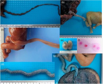
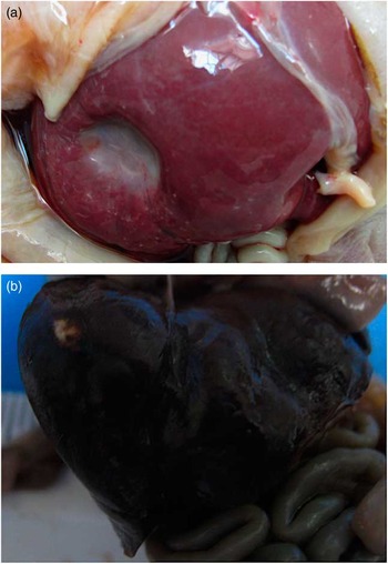
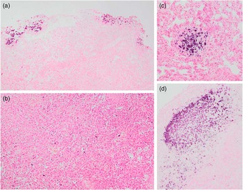
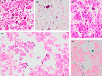
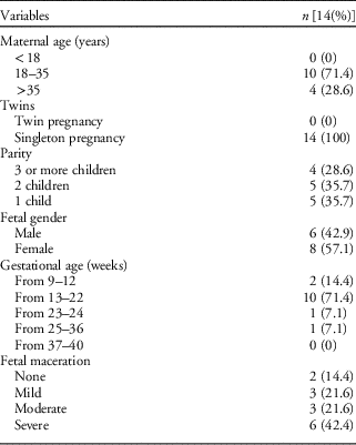
Between 2007 and 2014, there were 591 perinatal cases of fetuses and stillbirths at HUSI. We found 14 cases with the finding ‘hepatic calcification.’ Its distribution and clinical features are shown in Table 1. Medium maternal age (18–35 years) was predominant; parity was very similar among groups. We did not have twin gestations. Female fetuses predominated over males and fetuses on second trimester were more frequent. Severe macerated fetuses were more numerous. UC abnormalities correspond to abnormal insertion, SUA, abnormal length and coiling (Fig. 1). In the gross, sometimes calcification could be observed (Fig. 2). Microscopically, cases were reviewed establishing hepatic calcification’s features like focal or diffuse; intraparenchymatous or subcapsular; polyhedral, punctate or irregular (Figs 3 and 4). We also noted other involved organs in addition to liver, such as brain, thymus and small intestine.
Umbilical cord (UC) abnormalities observed. (a) Long UC, (b) short umbilical cord [(a) and (b), fetuses have similar ages], (c) hypocoiled UC, (d) hypercoiled UC, (e) single umbilical artery (SUA), (f) SUA (histological preparation, photo without zoom), (g) paramarginal insertion of the UC (Photos: M. Olaya-C).

Hepatic calcifications – gross view. (a) Stillbirth with chromosomal disorder; (b) fetus with chromosomal disorder (Photos: M. Olaya-C).

(a) Fetal subcapsular hepatic calcifications with severe maceration, hematoxylin and eosin (H&E) 10×. (b) Diffuse fetal intraparenchymatous hepatic calcifications with severe maceration, H&E 10×. (c) Focal fetal intraparenchymatous hepatic calcifications with moderate maceration, H&E 10×. (d) Peripheral fetal intraparenchymatous hepatic calcifications with moderate maceration, H&E 10× (Photos: M. Olaya-C).

Hepatic calcifications – microscopic features. (a) Oval calcifications, hematoxylin and eosin (H&E) 40×. (b), (c) Irregular calcifications, H&E 40×. (d) Punctata calcifications, H&E 40×. (e) Irregular calcifications. Special staining for calcium (von Kossa) (Photos: M. Olaya-C).

Population features

Among fetuses with some chromosomal disorders (102), 10 (9.8%) had calcifications [OR=13.18; CI (95%) 4.05–42.92]. Some fetuses showed UC abnormalities: 207 (named before), and five of them had hepatic calcifications (2.41%) [OR=2.7; CI (95%) 0.9–8.38]. We did not observed fetuses with TORSCH who exhibited hepatic calcifications.
Unfortunately, the small number of each kind of calcification precluded to analyze the calcification’s morphology or localization related to the etiology. We observed that diffuse were more common than focal (78.57 v. 21.43%, respectively) and intraparenchymatous more than subcapsular in same percentage.
Discussion
We do not know about composition of prenatal calcifications, but some studies in humans have been showed cholesterol, calcium bilirubinates, calcium carbonate polymorphs (calcite, aragonite, vaterite) CaCO3; calcium phosphates carbonated apatite, amorphous carbonated calcium phosphate, calcium palmitate, bile salts.Reference Bazin, Daudon, Combes and Rey 13
Fetal liver calcifications identified by ultrasound, can be divided into three main categories according to location: peritoneal, parenchymal and vascular;Reference Kraus 14 it is related to the etiology. Peritoneal hepatic calcifications can be observed in meconium peritonitis and plastic peritonitis; these are the most common cause of abdominal calcification.Reference Carroll and Maxwell 15 The other type of calcification, parenchymal hepatic calcification, includes the TORSCH complex,Reference Shackelford and Kirks 16 , Reference Brugman, Bjelland, Thomasson, Anderson and Giles 17 and is frequently associated with multiple congenital anomalies and chromosomic disorders;Reference Simchen, Toi and Bona 1 , Reference Taylor, Walkinshaw and Thomson 18 HawassReference Hawass, el Badawi and Fatani 19 found associated anomalies were high (85% of cases). In the Sahlin’sReference Sahlin, Sirotkina, Marnerides, Iwarsson and Papadogiannakis 20 paper almost 50% of patients with liver calcifications had a chromosomal abnormality, and we saw a similar high percentage of 10 out of our 14 patients with hepatic calcifications who had chromosomal anomalies.
Ischemic infarcts, are important cause of calcifications in the liver, those are distributed throughout the liver and show no predilection for peripheral or lobar distribution. The ischemic process could be originated in the UC.Reference Kidron and Sharony 21 Relation between UC abnormalities and hepatic calcifications is not well known; the statistics in this series showed a two-fold higher OR compared with fetuses with UC anatomical abnormalities.
Calcifications could also be present in a number of organs. Primary and metastatic tumors can also have parenchymal calcifications, which usually appear as a compound mass. These tumors could be hepatoblastoma, hemangioma, hamartoma, teratomas, hemangioendothelioma and metastatic neuroblastoma.Reference Lince, Pretorius, Manco-Johnson, Manchester and Clewell 22 The latter have vascular hepatic calcifications that may be seen in portal vein thromboemboli and vascular insufficiency with ischemic infarcts.Reference Nguyen and Leonard 5 Portal vein thromboemboli calcifications are primarily subcapsular in nature and are confined to either the right or left lobe. Other vascular pathologies such as hepatic artery aneurysm, hematoma, calcified thrombi of hepatic veins can also cause hepatic calcification.Reference Kraudel and Williams 23
Rare autosomal recessive disorder such as idiopathic infantile arterial calcinosis, may exhibit calcifications in the ultrasound; those are caused by calcium deposition in the internal elastic lamina of medium and large arteries;Reference Wax, Blackstone, Pinette and Cartin 24 in addition to the liver, it could be also involved cerebral arteries, aorta, pulmonary arteries, coronary arteries, etc.
Some studies on fetal death report more chromosomal disorders, and in some of them, individuals are selected for autopsy if they are abnormal. This study uses consecutive autopsies; in our hospital, the first cause of fetal death is ascending infection; we also included peripartum mortality, where chromosomal abnormalities are less frequent than at the beginning of gestation.
Once chromosome abnormalities, viral infection and fetal circulation obstruction have been ruled out, the prognosis of an isolated fetal liver calcification is good,Reference Butler, Anjum and Koehn 25 because much of them have healthy outcome.Reference Bronshtein and Blazer 26 , Reference Pata, Gündüz and Unlü 27
Our study reveals the need to further investigate the following questions related to dystrophic liver calcifications intimately associated with chromosomal disorders:
-
∙ Are they the result of chronic inflammation process? Which is inflammation etiology?
-
∙ Are there any preponderant chromosome disorders associated with hepatic calcifications?
-
∙ Does this process of hepatic calcifications occur early in gestation?
-
∙ Why is the liver mostly involved, more than other organs?
-
∙ Is there any explanation for patterns of calcifications, or their localization in the liver?
Conclusion
Hepatic calcifications in fetuses are uncommon. Known causes are transplacental infections, chromosomal abnormalities, thrombotic events such as FVT, and ischemic hepatic necrosis and their subsequent subcapsular hematomas. In our fetal population, we found the main risk – chromosomal disorders – associated. Hepatic calcifications diagnosed by ultrasound must alert obstetrician and at autopsies, it could orientate pathologies paying attention to grossly abnormal UCs and placental abnormalities, remarking neonatal vascular dysfunction, infections and chromosomal changes.
Acknowledgments
The authors wish to thank the Pontificia Universidad Javeriana–Hospital Universitario San Ignacio in Bogotá, Colombia for its indispensable support and participation in this study. The authors also like to thank the Staffs of the Departments of Pathology and Obstetrics and Gynecology. The authors extend our thanks to the parents who give their consent for autopsies and to Steven W. Bayless for the English translation. The authors thank Juliana Arenas for her contribution in the bibliographic search at the beginning of this study.